COMMUNICATION The Confusion of Indexing Aspirin Crystals
←
→
Page content transcription
If your browser does not render page correctly, please read the page content below
COMMUNICATION

The Confusion of Indexing Aspirin Crystals
CLARE AUBREY-MEDENDORP,1 SEAN PARKIN,2 TONGLEI LI1
1
    Department of Pharmaceutical Sciences, University of Kentucky, Lexington, Kentucky 40536-0082
2
    Department of Chemistry, University of Kentucky, Lexington, Kentucky 40536-0082

Received 5 March 2007; revised 25 April 2007; accepted 1 May 2007

Published online in Wiley InterScience (www.interscience.wiley.com). DOI 10.1002/jps.21055

                 ABSTRACT: Much of the existing literature dealing with crystalline aspirin is vague or
                 ambiguous with regard to indexing of the crystal faces. The inconsistency with which the
                 indices of the dominant faces have been assigned leads to confusion in analysis of surface
                 properties. To clarify this, we have conducted crystal growth experiments on aspirin,
                 and indexed the crystal faces with X-ray diffraction (XRD), paying special attention to
                 the placement of symmetry elements. The space group was confirmed as P21/c, and the
                 dominant face was (100). Contact angle measurements made on the two major faces of
                 aspirin indicate the (100) face to be more hydrophobic than the (001) face, likely due to
                 the acetyloxy moiety, not the carboxyl, exposed on the (100). ! 2007 Wiley-Liss, Inc. and the
                 American Pharmacists Association J Pharm Sci 97:1361–1367, 2008
                 Keywords:        wettability; contact angle; crystal packing; indexing; aspirin; X-ray
                 diffraction

INTRODUCTION                                                           properties.4 In order to determine and characterize
                                                                       the surface structure of particular crystal faces, the
The majority of pharmaceutical materials are in                        molecular packing and the face indices must be
the solid crystalline form. Understanding the                          assigned properly.
arrangement or packing of molecules in a crystal                          Crystalline aspirin has been studied routinely,
is critical because it helps determine the physi-                      yet despite the persistent investigations since its
cochemical and mechanical properties, including                        discovery in late 1800s,5 confusion remains over
bioavailability.1–3 Depending on the conditions of                     classification of the crystal morphology. In the
preparation, pharmaceutical crystals may exhibit                       1930s, the first attempt was made at defining
variations in habit with the consequence that                          the lattice parameters by X-ray diffraction (XRD)
interfacial properties can be modulated by the                         using oscillation and Weissenberg photographs.
various morphologically important faces. Differ-                       From these results it was deduced that the space
ent habits of the same drug compound can have                          group must be P21/a (a nonstandard setting of
a huge influence on the dissolution kinetics and                       space group number 14).6,7 Using the parameters
consequent bioavailability. A molecular level knowl-                   from this report the predominant face was
edge of the crystal surface structure is therefore                     identified as (001).8,9 However, the first full
crucial to understanding the physical and chemical                     structure determination by single crystal XRD
                                                                       was not until 1964.10 This report and others that
                                                                       followed assigned a different setting of the same
   Correspondence to: Tonglei Li (Telephone: 859-257-1472;
Fax: 859-257-7585; E-mail: tonglei@uky.edu)                            space group, that is, P21/c.11–13 In this setting,
Journal of Pharmaceutical Sciences, Vol. 97, 1361–1367 (2008)          the largest face is (100).14,15 A likely cause of the
! 2007 Wiley-Liss, Inc. and the American Pharmacists Association       confusion over face indexing was then the use of

                                                            JOURNAL OF PHARMACEUTICAL SCIENCES, VOL. 97, NO. 4, APRIL 2008   1361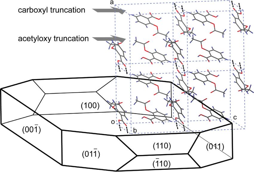
1362      AUBREY-MEDENDORP, PARKIN, AND LI

Table 1. Unit Cell Parameters of Form I of Aspirin               Sigma-Aldrich), acetone (HPLC grade, Sigma-
                                                                 Aldrich), and deionized distilled water (Barnstead
a (Å)               b (Å)         c (Å)          b (8)        Millipore Deionized Water System, Dubuque,
11.242   (7)      6.539   (4)    11.245   (9)    95.90   (3)a    Iowa, 0.2 mm filtered) were used for preparation
11.446   (13)     6.596   (6)    11.388   (9)    95.33   (2)b    of aspirin crystals. Diiodomethane (99%, Sigma-
11.430   (1)      6.591   (1)    11.395   (2)    95.68   (1)c    Aldrich), glycerol (98%, Mallinckrodt, Paris, KY),
11.233   (3)      6.544   (10)   11.231   (3)    95.89   (2)d    and deionized distilled water were used for the
   a                                                             contact angle measurements.
    This work.
   b
    Reference 10.
   c
    Reference 11.
   d
     References 12, 13.                                          Crystal Growth
                                                                 Aspirin single crystals were grown by slow eva-
alternative space group settings, exacerbated by
                                                                 poration from ethanol and acetone. Aspirin was
the almost identical metrics of the a and c axes
                                                                 weighed and dissolved in the liquids, close to its
(Tab. 1), and consequently the functional groups
                                                                 solubility (0.2 gm/mL in ethanol and 0.18 gm/mL
assigned to the indexed faces. Thus, if the face
                                                                 in acetone), by stirring. The beakers were sealed
indices of aspirin single crystals indexed as P21/a
                                                                 with Parafilm1, which had small holes made
are used for a crystal structure indexed as P21/c,
                                                                 using a needle, and stored in ambient conditions.
any conclusions drawn with regard to surface
                                                                 In addition, aspirin crystals were grown in water
properties of a particular face will be in error,
                                                                 with a supersaturation ratio of 225% based on its
particularly from the viewpoint of structure-
                                                                 water solubility of 3.3 mg/mL. The beakers were
property relationships. Indeed, this apparent
                                                                 sealed with Parafilm1 and stored in ambient
confusion has occurred in several reports on
                                                                 conditions. Crystals were harvested after 1 week.
aspirin crystals, including one of ours.16
                                                                 The morphology of aspirin was manually plotted
   Recent studies have probed the chemical nature
                                                                 by the Cerius2 program (Accelrys, Inc., San Diego,
of functional groups exposed on the two major
                                                                 CA) using the single crystal X-ray data that we
faces of aspirin crystals. As a result of incorrect
                                                                 determined to resemble the observed habits of the
face indexing, these studies concluded that the
                                                                 crystals grown from the different solvents.
(100) face was more hydrophilic than the (001)
face due to the presence of the carboxyl group
on the (100). We made a similar mistake when
                                                                 Single Crystal X-Ray Diffraction (XRD)
differentiating the (100) and (001) face in a dis-
cussion of surface wettability.16 The conclusion                 Structure determination and face indexing of
that the smaller contact angle observed with                     aspirin single crystal was done using a Nonius
water was on the supposed (100) face, was justi-                 KappaCCD diffractometer (Madison, WI) with Mo
fied by the presence of the carboxyl group.                      Ka X-rays (l ¼ 0.71073 Å). The crystals were
   Upon close inspection of the face indexing of                 indexed using the COLLECT program.18 Data for
aspirin single crystals with XRD, we believe the                 space group assignment and structure determina-
common assignment of the (100) face as more                      tion were collected in a series of v-scans at 90 K.
hydrophilic (due to the exposure of carboxyl                     These data were integrated, scaled and merged
group) is not correct. The confusion in the lit-                 using the Denzo-SMN package.19 Space group
erature seems to stem from the original assign-                  assignments were made using the maXus and
ment of the space group and indices of the crystal               Shelxtl packages.20,21 Since the a and c axes are
faces.7–9,17 This communication presents experi-                 essentially identical in length, and because of the
mental evidence to clarify the difference between                previous inconsistencies, these assignments were
the two major faces of aspirin crystal, and to revise            checked manually. Structures were solved and
our conclusion of the contact angle data for further             refined using Shelxtl. Crystal face indices were
interpretation of surface wettability.                           assigned relative to the P21/c cell setting with the
                                                                 aid of a video capture utility within the COLLECT
                                                                 program. Video captures of the crystal were taken
MATERIALS AND METHODS                                            along the directions of interest. Miller indices of
                                                                 well-defined faces were deduced by inspection of
Aspirin (acetylsalicylic acid) (>99.0%, Sigma-                   the crystal viewed along specific real and recipro-
Aldrich, St. Louis, MO), ethanol (HPLC grade,                    cal space vectors (for instance, the (100) face was

JOURNAL OF PHARMACEUTICAL SCIENCES, VOL. 97, NO. 4, APRIL 2008                                            DOI 10.1002/jps
THE CONFUSION OF INDEXING ASPIRIN CRYSTALS 1363
Figure 1. Video images used to index aspirin faces. a, b, and c are unit cell axes, while
a", b", and c" are the corresponding reciprocal axes of the P21/c setting.
found with vectors b and c in this plane). This is
shown in Figure 1.
Powder X-Ray Diffraction (PXRD)
The Miller indices of two major faces of aspirin
single crystals were further verified using a
powder X-ray diffractometer (MultiFlex, Rigaku
Co., The Woodlands, TX) with Cu Ka X-rays
(l ¼ 1.54178 Å). The two major faces of a single
crystal were placed face up on a sample holder,
with the faces horizontal with the surface of the
sample holder. The scan rate was set to 58 per
minute and the scan was taken from 28 to 408 of 2u.
Atomic coordinates from the single crystal X-ray
study was used in Materials Studio 3.5 (Accelrys,
Inc., San Diego, CA) to produce simulated powder
patterns for comparison with the experimental
data.
Contact Angle Measurements
Sessile drop contact angles were measured on the
two major faces of aspirin. Crystals were cleaved
to expose fresh faces and secured on a microscope
slide using adhesive tape. The contact angles were
obtained using a video-based contact angle system
(OCA, Future Digital Scientific Co., Bethpage,
NY). Drops (5 mL) of water, diiodomethane, and
glycerol were dispensed and placed on the two
faces using a motor driven syringe, respectively.
Water contact angles were measured in a satu-
rated water vapor environmental chamber to
prevent evaporation during spreading. Each drop-
let was recorded for 240 s. The contact-angle
values recorded for droplets on a surface were
obtained automatically via curve-fitting software.
RESULTS AND DISCUSSION
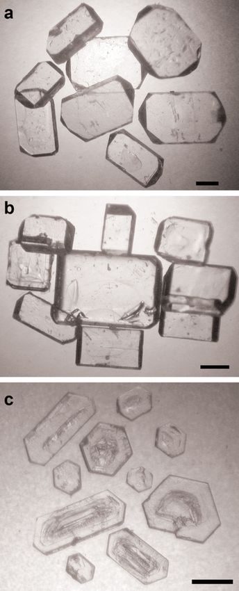
Figure 2. Aspirin crystals grown from (a) ethanol, (b)
Aspirin crystallized in ethanol, water, and acetone acetone, and (c) water. Scale bars denote 1 mm. [Color
results in plate morphology with two major faces figure can be seen in the online version of this article,
and a few different minor faces, shown in Figure 2. available on the website, www.interscience.wiley.com.]
DOI 10.1002/jps JOURNAL OF PHARMACEUTICAL SCIENCES, VOL. 97, NO. 4, APRIL 20081364 AUBREY-MEDENDORP, PARKIN, AND LI
The effects of solvent on crystallization of aspirin Our results demonstrate that for crystals
demonstrate some morphological differences but indexed as P21/c, the largest growth face is
no polymorphism. This was validated in both (100) and the second largest is (001), as shown
the XRD and powder X-ray diffraction (PXRD) in Figure 3. Based on these assignments and an
studies. intimate view of the crystal packing, it is apparent
Aspirin crystals evaluated by single XRD that there are two possible truncations for the
indicated the crystals were of the monoclinic (100) face. One truncation goes through the
space group P21/c with a ¼ 11.242(7), b ¼ 6.539(4), carboxyl group and another passes through
c ¼ 11.245(9) Å, and b ¼ 95.9(3)8. These values are the acetyloxy groups. It is important to make
in agreement with the literature that previously this distinction on the (100) face, especially when
investigated the unit cell parameters of form I probing surface properties in relation to func-
using XRD,10,11 neutron diffraction,12,13 and tional groups on that surface. Although the
oscillation and Weissenberg X-ray photographs.7 standard setting for space group number 14 in
These values are listed in Table 1. More recently modern crystallography is P21/c (thereby placing
an elusive second form (form II) of aspirin has the glide plane along c), it is of course possible
been identified and characterized by single crystal to transform to the nonstandard P21/a setting
XRD. Though some controversy exists,22,23 unit (via the transformation matrix (001, 0#10, 100). If
cell parameters (a ¼ 12.095(7), b ¼ 6.491(4), this transformation is made, then the assigned
c ¼ 11.323(6) Å, and b ¼ 111.509(9)8) are super- face indices are also, of necessity, changed. If the
ficially quite different from those of form I, indexing of major faces from earlier reports is used
although they are likely related. Nonetheless, to correlate the crystal surface structure assigned
both forms exhibit the same space group, P21/c.24 with a different space group setting, and no
Comparison of unit cell parameters and atomic account is made for the particular setting used,
coordinates of the two forms demonstrates a then any results inferred will be in error. As
subtle difference in the molecular packing shown in Figure 3, if a surface property, such as
between forms I and II. In our crystallization wettability, is measured on the (100) face but
studies only form I was produced, though the explained using the functional groups present on
presence of a very small amount of form II cannot the (001) face, conclusions will be misleading. The
be discounted.22,23 crucial point is to recognize which functional
Figure 3. Growth morphology and crystal structure of aspirin. As indicated, there are
two possible truncations of the (100) face, one with carboxyl groups and another with
acetyloxy groups.
JOURNAL OF PHARMACEUTICAL SCIENCES, VOL. 97, NO. 4, APRIL 2008 DOI 10.1002/jpsTHE CONFUSION OF INDEXING ASPIRIN CRYSTALS 1365
groups are exposed on the major faces whether the in the experimental pattern. On the other hand,
space group is assigned as P21/a or P21/c. The axes all four peaks from experimental data for the (100)
could be assigned according to any number of face agree with equivalent peaks from the
conventions, but the functional groups assigned to simulated data: (100) at 7.848 versus simulated
the physical faces are invariant. The dominant 7.808, (200) at 15.768 versus simulated 15.848,
face will always have the acetyloxy group (not the (300) at 23.608 versus simulated 23.808, and (400)
carboxyl group as discussed below) and the other at 31.888 versus simulated 32.08. These results
major face will always have the methyl and phenyl verify the findings from the XRD study, and
groups. This is illustrated in Figure 3. confirm application of the P21/c space group as
Results from the PXRD work confirmed the opposed to using P21/a. The key feature to
XRD studies and verified that the dominant face is distinguish the two faces is the (100) peak at
(100) for the P21/c cell setting. As illustrated in 7.808. If there were no systematic absences
Figure 4, experimental peaks of the single crystal present, the (001) peak would be observed close
(100) and (001) faces correspond well to the to the (100) peak at 7.908.
equivalent peaks in the simulated patterns. Due Contact angles were measured with various
to the systematic absences for P21/c, odd num- solvents on the two major faces of aspirin, and
bered reflections of the (00l) family, such as (001) the results are summarized in Table 2. All of
and (003), are systematic absences when generat- the contact angles measured on the (100) face are
ing XRD peaks; these hypothetical peaks are larger than those on the (001) face. Due to the
colored green in the simulated pattern. The single polar nature of the solvents used, the measure-
peak observed in the experimental pattern of the ments indicate the (100) face is less hydrophilic
(001) surface at 15.688 represents the (002) face, than the (001), emphasizing the importance of
and has an equivalent peak in the simulated knowing the surface crystal structure. Although
pattern. There should also be an observed peak, the (100) face may expose the carboxyl acid group,
similar to the simulated pattern at 328, but the the contact angle measurements indicate other-
peak was too small to detect and was not observed wise. It appears to be the acetyloxy truncation
Figure 4. Experimental and simulated PXRD patterns of the (100) and (001) faces of
aspirin. Note the simulated (001) and (003) are hypothetical due to the systematic
absence.
DOI 10.1002/jps JOURNAL OF PHARMACEUTICAL SCIENCES, VOL. 97, NO. 4, APRIL 20081366 AUBREY-MEDENDORP, PARKIN, AND LI
Table 2. Contact Angles on the (100) and (001) Faces (001), and vice versa. This is the cause of apparent
of Aspirin Measured in Selected Solvents confusion in earlier studies, including one of our
own.16
Water Diiodomethane Glycerol This report also demonstrates the importance of
(100) 66.9 $ 2.9 41.2 $ 1.7 65.1 $ 1.1 X-ray crystallography in solving both the crystal
(001) 56.4 $ 2.4 36.2 $ 1.9 56.5 $ 0.9 structure and the relationship between alterna-
tive cell settings and indices assigned to crystal
faces. Without consistent indexing, erroneous
that is exposed, which leads to the (100) face being conclusions are inevitable. For the case of aspirin,
less hydrophilic than the (001) face. The hydrogen the confusion almost certainly arose from the
bonding between carboxyl groups in the crystal is similarity in length of the a and c axes.
strong; cleavage through this bonding to expose
the carboxyl group is unlikely. Once again, the
relationship between the interfacial properties
and surface structure could be misconstrued when ACKNOWLEDGMENTS
using the improper labeling and thereby identify-
ing the wrong surface functional groups. If the The research was supported by the PhRMA Foun-
more hydrophilic face is mistaken as the (100), dation. The authors would like to thank Ms. Ellen
carboxyl groups would be thought as the dominant Savelli for her work with contact angle measure-
surface group and that it would be involved in ments.
solvent interactions. Our results show that the
carboxyl group is not exposed and therefore a
lower contact angle by a polar liquid is not
expected. REFERENCES
1. Byrn SR, Pfeiffer RR, Stowell JG. 1999. Solid-State
Chemistry of Drugs. 2nd edition. West Lafayette,
CONCLUSIONS IN: SSCI, Inc.
2. Vippagunta SR, Brittain HG, Grant DJW. 2001.
Analysis of the crystal structure of aspirin by Crystalline solids. Adv Drug Deliv Rev 48:3–26.
single X-ray and powder XRD has clarified the 3. Suryanarayanan R, Byrn SR. 2001. Characteriza-
confusion over the face indices of aspirin crystals. tion of the solid state. Adv Drug Deliv Rev 48:1–136.
The dominant face of aspirin crystals grown in 4. Kiang YH, Shi HG, Mathre DJ, Xu W, Zhang D,
ethanol, water, and acetone is (100) when the Panmai S. 2004. Crystal structure and surface
space group P21/c is assigned. The elucidation of properties of an investigational drug—A case
the indices also allowed proper assignment of study. Int J Pharm 280:17–26.
functional groups to the major faces and raised 5. Hoffmann F. Acetyl Salicylic Acid. US Patent
644,077, February 27, 1900.
awareness of two possible truncations on the (100)
6. Kozu S, Takane K. 1935. Cell-dimensions and
face, one revealing the carboxyl group and the
space-group of acetylsalicylic acid. Proc Imper Acad
other the acetyloxy moiety. Our contact angle 11:381–382.
results further show that the (100) is less 7. Nitta I, Watanabe T. 1937. Unit cell and space
hydrophilic than the (001) face when a polar group of acetylsalicylic acid. Sci Pap Inst Phys
liquid is used in the measurement, suggesting Chem Res 31:125–128.
that the carboxyl group truncation is not mea- 8. Watanabe A, Yamaoka Y, Kuroda K. 1980. Study of
sured. The common thought of the (100) face being crystalline drugs by means of polarizing micro-
more hydrophilic in previous studies, including scope. III. Key refractive indices of some crystalline
one of ours,16 is incorrect. The mistake probably drugs and their measurement using an improved
stems from the inconsistent indexing caused by immersion method. Chem Pharm Bull 28:372–378.
9. Watanabe A, Yamaoka Y, Takada K. 1982. Crystal
the use of a nonstandard space group setting. The
habits and dissolution behavior of aspirin. Chem
face indexing of aspirin crystal is not incorrect,8,9
Pharm Bull 30:2958–2963.
because their assignment is relative to their 10. Wheatley PJ. 1964. The crystal and molecular
choice of P21/a. However, if these face indices structure of aspirin. J Chem Soc Suppl 6036–6048.
are used to examine surface moieties based on 11. Kim Y, Machida K, Taga T, Osaki K. 1985. Struc-
a P21/c description without the necessary index ture redetermination and packing analysis of
transformation, the (100) face will be mistaken as aspirin crystal. Chem Pharm Bull 33:2641–2647.
JOURNAL OF PHARMACEUTICAL SCIENCES, VOL. 97, NO. 4, APRIL 2008 DOI 10.1002/jpsTHE CONFUSION OF INDEXING ASPIRIN CRYSTALS 1367
12. Harrison A, Ibberson R, Robb G, Whittaker G, 17. Niini R. 1931. The crystal form and optical con-
Wilson C, Youngson D. 2002. In situ neutron stants of aspirin. Z Kristallogr 79:532–536.
diffraction studies of single crystals and powders 18. Nonius BV. 1997. KappaCCD Server Software.
during microwave irradiation. Faraday Discuss Windows 3.11 edition. The Netherlands: Delft.
122:363–379. 19. Otwinowski Z, Minor W. 1998. Denzo-SMN pro-
13. Wilson CC. 2002. Interesting proton behaviour in gram package. Processing of X-ray diffraction data
molecular structures. Variable temperature neu- collected in oscillation mode. Methods Enzymol
tron diffraction and ab initio study of acetylsalicylic 276:302.
acid: Characterising librational motions and com- 20. Mackay S, Dong W, Edwards C, Henderson A,
paring protons in different hydrogen bonding Gilmore C, Stewart N, Shankland K, Donald A.
potentials. New J Chem 26:1733–1739. 1999. MaXus, Comprehensive crystallography soft-
14. Meenan P. 1997. Crystal morphology predictive ware. 4.0 edition. Glasgow: University of Glasgow.
techniques to characterize crystal habit: Applica- 21. Sheldrick GM. 1990. SHELXTL PC, An integrated
tion to aspirin (C9H8O4). ACS Symp Ser 667:2– system for solving, refining, and displaying crystal
17. structure from diffraction data. 4.1 edition. Madi-
15. Hammond RB, Pencheva K, Roberts KJ. 2006. A son: Siemens Analytical X-ray Instruments.
structural-kinetic approach to model face-specific 22. Bond AD, Boese R, Desiraju GR. 2007. On the
solution/crystal surface energy associated with polymorphism of aspirin. Angew Chem Int Ed
the crystallization of acetyl salicylic acid from 46:615–617.
supersaturated aqueous/ethanol solution. Crystal 23. Bond AD, Boese R, Desiraju GR. 2007. On the
Growth Des 6: 1324–1334. polymorphism of aspirin: Crystalline aspirin as
16. Li T, Liu S, Feng S, Aubrey CE. 2005. Face- intergrowths of two ‘‘polymorphic’’ domains. Angew
integrated Fukui function: Understanding wett- Chem Int Ed 46:618–622.
ability anisotropy of molecular crystals from den- 24. Vishwesher P, McMahon JA, Oliveira M, Peterson
sity functional theory. J Am Chem Soc 127:1364– ML, Zaworotko MJ. 2005. The predictably elusive
1365. form II of aspirin. J Am Chem Soc 127:16802–16803.
DOI 10.1002/jps JOURNAL OF PHARMACEUTICAL SCIENCES, VOL. 97, NO. 4, APRIL 2008You can also read