Increased Mobility of the Atrial Septum in Aortic Root Dilation: An Observational Study on Transesophageal Echocardiography
←
→
Page content transcription
If your browser does not render page correctly, please read the page content below
BRIEF RESEARCH REPORT
                                                                                                                                                  published: 24 August 2021
                                                                                                                                            doi: 10.3389/fphys.2021.701399

                                                Increased Mobility of the Atrial
                                                Septum in Aortic Root Dilation: An
                                                Observational Study on
                                                Transesophageal Echocardiography
                                                Altair Heidemann Jr. 1,2,3 , Lorença Dall’Oglio 3,4 , Eduardo Gehling Bertoldi 1,5 and
                                                Murilo Foppa 1,2,3*
                                                1
                                                 Graduate Studies Program in Cardiology, Universidade Federal do Rio Grande do Sul, Porto Alegre, Brazil, 2 Cardiology
                                                Division, Hospital de Clínicas de Porto Alegre, Porto Alegre, Brazil, 3 NUPIC (Núcleo de Pesquisa em Imagem
                                                Cardiovascular), Hospital de Clínicas de Porto Alegre, Porto Alegre, Brazil, 4 School of Medicine, Universidade Luterana do
                                                Brasil, Porto Alegre, Brazil, 5 School of Medicine, Universidade Federal de Pelotas, Pelotas, Brazil

                                                Background: There is a growing interest in the relationship between atrial septal
                                                anatomy and cardioembolic stroke. Anecdotal reports suggest that the enlargement of
                                                the aortic root could interfere with atrial septal mobility (ASM). We sought to investigate
                                                the association between ASM and aortic root dilation.
                                                Methods and Findings: From all consecutive clinically requested transesophageal
                          Edited by:
                    Jochen Steppan,             echocardiogram (TEE) studies performed during the study period in a single institution,
             Johns Hopkins University,          we were able to review and evaluate the ASM and anteroposterior length, aortic root
                       United States
                                                diameter, and the prevalence of atrial septal aneurysm (ASA) and of patent foramen
                      Reviewed by:
                               Jie Li,
                                                ovale (PFO) in 336 studies. Additional variables, such as left ventricular ejection fraction,
    Augusta University, United States           left atrial diameter, diastolic dysfunction, age, sex, weight, height, previous stroke,
                Erik Josef Behringer,           atrial fibrillation, and TEE indication, were extracted from patient medical records and
 Loma Linda University, United States
                                                echocardiographic clinical reports. In 336 patients, we found a mean ASM of 3.4 mm,
                  *Correspondence:
                        Murilo Foppa            ranging from 0 to 21 mm; 15% had ASA and 14% had PFO. There was a 1.0 mm increase
                mufoppa@hcpa.edu.br             in ASM for every 10-mm increase in aortic root diameter adjusted for age, sex, weight,
                                                height, ejection fraction, and left atrial size (B = 0.1; P = 0.04). Aortic diameter was not
                    Specialty section:
          This article was submitted to         associated with a smaller septal length (B = 0.03; P = 0.7).
                   Vascular Physiology,
                                                Conclusion: An increased motion of the atrial septum can occur in association with
                a section of the journal
                Frontiers in Physiology         aortic dilation. These findings deserve attention for the relevance of aortic root anatomy
            Received: 27 April 2021             in future studies involving atrial septal characteristics and embolic stroke risk.
            Accepted: 20 July 2021
                                                Keywords: atrial septal aneurysm, aortic dilation, atherosclerosis, transesophageal echocardiography, stroke
          Published: 24 August 2021
                               Citation:
         Heidemann A Jr, Dall’Oglio L,
      Bertoldi EG and Foppa M (2021)
                                                INTRODUCTION
Increased Mobility of the Atrial Septum
             in Aortic Root Dilation: An
                                                Embolic stroke has multiple causes, and more than one disease is frequently detected during the
                Observational Study on          assessment of patients (Chatzikonstantinou et al., 2012; Amarenco et al., 2013). There is a growing
  Transesophageal Echocardiography.             knowledge regarding the roles of patent foramen ovale (PFO) and atrial septal aneurysm (ASA)
            Front. Physiol. 12:701399.          as sources of embolism in ischemic stroke (Pearson et al., 1991; Lamy et al., 2002; Ward et al.,
     doi: 10.3389/fphys.2021.701399             2006; Mas et al., 2017; Saver et al., 2017; Søndergaard et al., 2017). Other phenotypic expressions

Frontiers in Physiology | www.frontiersin.org                                          1                                           August 2021 | Volume 12 | Article 701399
Heidemann et al. Increased Mobility of Atrial Septum
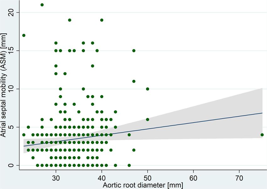
FIGURE 1 | Transesophageal echocardiography. (A) Aortic measurements at Valsalva sinus plane (120◦ , mid-esophageal view). Aorta measured at leading edge echo
signal of posterior wall to the leading edge of anterior wall. (B) Interatrial septal size and oscillation (25–45◦ , mid-esophageal view) measured as the maximal
perpendicular distance from an imaginary line drawn between atrial septal insertion points (dashed line). AO, aorta; LA, left atrium; RA, right atrium; LV, left ventricle;
RV, right ventricle.
of large vessel atherosclerotic diseases, such as aortic enlargement individual informed consent was waived due to the retrospective
and atherosclerotic plaques, are also frequently present in these analysis of data.
patients, and they increase with aging (Reed et al., 1992). Clinical variables, extracted from electronic medical records,
However, the causal role of diffuse advanced atherosclerosis and were age, sex, weight, height, clinical indication for TEE,
senile aortic dilation in stroke as well as their interconnections medical history of previous stroke, and atrial fibrillation.
with other stroke risk factors is less understood. Additional echocardiographic data were extracted from
The anatomic relation between the aortic root and atrial transthoracic reports (performed regularly in all patients who
septum first appeared on case reports of patients with platypnea– had TEE), including left ventricular ejection fraction (LVEF),
orthodeoxia syndrome (Medina et al., 2001; Chopard and left atrial diameter, diastolic dysfunction, and central venous
Meneveau, 2013; Hasegawa et al., 2020). An enlarged aorta pressure estimate.
could geometrically impinge a shortening in the anteroposterior A single investigator (AH) reviewed all archived TEE images,
atrial septal length, consequently augmenting the atrial septal and study-specific measurements were performed, which were
mobility (ASM) (Supplementary Video). This effect, added blinded to the TEE clinical reports. Aortic root size was measured
to the presence of atrial septal defects or PFO, is part of from leading-to-leading edge at mid-esophageal view 120–140◦
the mechanism of hypoxia in this syndrome (Eicher, 2005). at end diastole (Figure 1A). Cine loops at mid-esophageal view
Moreover, differences in the left and right atrial filling pressures 25–45◦ at the plane of Valsalva sinus, showing both the thin
may influence the interatrial septum (IAS) movement dynamics. component of atrial septum and the aortic root, were used to
The potential of aortic root enlargement to increase ASM has quantify atrial septal length and oscillation. ASM was defined as
been previously suggested (Bertaux et al., 2007). This hypothesis the distance between the maximal leftward and rightward atrial
deserves to be tested. Transesophageal echocardiogram (TEE) positions during spontaneous breathing (Figure 1B). Aortic
has high spatial and temporal resolutions, with high sensitivity plaques were computed as simple (Heidemann et al. Increased Mobility of Atrial Septum
variables, respectively. Continuous variables were compared TABLE 1 | Transesophageal echocardiographic measurements and findings of
with Pearson’s correlation coefficients. The impact of clinically patients with clinically indicated studies.
relevant covariates in the main association was investigated in Studied sample (n = 336)
unadjusted and multivariable linear regression models. Intra-
reader reproducibility of the study-specific measurements was Transesophageal images
tested from repeated readings in a randomly selected sample Aortic root diameter (mm) 33.4 ± 5.1
of 20 TEE studies using intraclass correlation coefficients (ICC) Aortic plaques
and Bland–Altman graphs. All tests were two-tailed and P-values Absent 189 (56)
< 0.05 were considered statistically significant. The Statistical Simple 99 (30)
Package for the Social Sciences (SPSS) software program, IBM, Complex 48 (14)
Corp., Armonk, NY, version 23.0 and STATA version 12.0 were Aortic valve morphology
used for analysis. Normal 178 (49)
Aortic valve sclerosis 145 (40)
Aortic stenosis 38 (10)
RESULTS Bicuspid aortic valve 5 (1)
Atrial septal diameter (mm) 24.1 ± 7.2
From the initial 508 studies, we excluded 33 repeated TEE studies Atrial septal mobility (mm) 3.4 ± 3.7
performed in the same patients and 14 patients with congenital Atrial septal aneurysm 49 (15)
atrial septal defects, resulting in 461 patients. We also excluded Patent foramen ovale 46 (14)
64 TEE studies without archived images and 61 studies in which
Transthoracic images
images measurements could not be performed in accordance to
Left atrial diameter (mm) 42.2 ± 8.0
study protocol, resulting in a final sample of 336 patients. It was
Left ventricular ejection fraction
noted that 336 patients included in the analysis did not differ
50% 278 (83)
of atrial fibrillation, or exam indication.
Diastolic function
Included patients were 55 ± 16 years old, 49% were women,
Normal 118 (35)
and 25% were obese (body mass index > 30 kg/m²). LVEF < 40%
Mild dysfunction 113 (34)
was present in 10% of the sample. Previous stroke occurred in
Moderate or severe 25 (7)
124 (37%) patients and 48 (14.3%) of them had atrial fibrillation.
Indeterminate/non-measurable 80 (24)
The main indications for TEE were infective endocarditis in
Dilated inferior vena cava 64 (19)
142 (42.2%), stroke in 103 (30.6%) and pre-cardioversion in 44
(13%) patients. N (%) or mean ± SD.
Table 1 shows the echocardiographic measurements and
relevant findings. The average diameter of the aortic root was
larger in men (35 ± 5 vs. 31 ± 4 mm; P < 0.001) and was
venous pressure (P = 0.37). As expected, patients with ASA
positively associated with age (r = 0.16; P = 0.004), height (r
had a higher prevalence of PFO (43 vs. 10%; P < 0.001). The
= 0.35; P < 0.001), and weight (r = 0.25; P < 0.001). Sixty
prevalence of PFO was thrice as frequent among patients with
(17.9%) patients had aortic root enlargement (≥38 mm), and
leftward bulging of the atrial septum compared with those with
three of them had aortic aneurysm (>50 mm). Forty-four percent
rightward shift (24 vs. 8%; P < 0.001).
of the sample had atherosclerotic plaques in the aorta and 50%
The reproducibility analysis showed an ICC of 0.91 for aortic
had some degree of aortic valve fibrocalcific degeneration. The
diameter, with 95% limits of agreement of −0.9–1.5 mm, and an
prevalences of ASA and of PFO were 15 and 14%, respectively.
ICC of 0.87 for ASM, with 95% limits of agreement of −0.2–
The mean atrial septal anteroposterior length was 24 ± 7 mm
0.6 mm.
and ASM was measured as 3.4 ± 3.7 mm, ranging from 0 to
21 mm. A significant positive correlation was observed between
ASM and aortic root diameter (Figure 2). In multivariable DISCUSSION
analysis (Table 2), the linear association between ASM and aortic
root diameter remained significant when adjusted for age, sex, LV We were able to demonstrate a positive linear association
ejection fraction, and left atrial diameter (adjusted B coefficient between the aortic root diameter and interatrial septal mobility.
= 0.1; P = 0.04). In summary, there was a mean 1 mm increase Determinants of IAS mobility are less understood, but our
in ASM for each 10 mm increase in aortic root diameter. No findings may suggest new connections between two different
significant linear association was found between aortic size and well-known ischemic stroke mechanisms. Bertaux et al.
septal length (P = 0.7). demonstrated this same association but in a smaller and
In addition, we were not able to identify significant stricter sample (Bertaux et al., 2007). Our findings reinforce
associations between ASM and the presence of aortic plaques the hypothesis that, due to their geometry and anatomic
(P = 0.61), diastolic dysfunction (P = 0.17), or elevated central proximity, the aortic root dilation could increase ASM. This
Frontiers in Physiology | www.frontiersin.org 3 August 2021 | Volume 12 | Article 701399Heidemann et al. Increased Mobility of Atrial Septum
FIGURE 2 | Correlation between atrial septal mobility and aortic root diameter measured in transesophageal echocardiography images.
TABLE 2 | Crude and adjusted linear regression coefficients between aortic root diameter and atrial septal mobility (ASM).
B coefficient CI (95%) P
Aortic root diameter
Crude 0.08 0.01–0.16 0.03
Adjusted for:
Age 0.08 0.002–0.16 0.04
Sex 0.11 0.03–0.19 0.01
Weight 0.07 −0.01–0.16 0.10
Height 0.08 −0.01–0.17 0.08
LVEF 0.09 0.01–0.17 0.02
Left atrial diameter 0.10 0.02–0.18 0.01
Multivariable adjusted* 0.10 0.002–0.2 0.04
* Adjusted for age, sex, weight, height, left ventricular ejection fraction (LVEF), and left atrial diameter.
mechanism was already postulated in case reports of patients of aortic size, reinforcing the independent association
with platypnea–orthodeoxia syndrome (Kazawa et al., 2017). between them.
Aortic atherosclerosis and dilation are also associated Although patients with aortic plaques had, in average, a larger
with cardiovascular risk factors and vascular clinical aorta, we could not find a significant direct association between
events (Gardin et al., 2006). Similar to population-based aortic plaques and augmented ASM. It is worth to say that we
studies, we found a positive linear correlation of aortic found the triple prevalence of PFO in patients with leftward IAS
diameter with sex, age, weight, and height. Noteworthy, mobility, suggesting that the movement of IAS toward the left
the association between aortic dimension and IAS mobility atrium could favor the opening of the foramen ovale, probably by
persisted significant after adjusting for the main determinants increasing the distance between septum primum and secundum.
Frontiers in Physiology | www.frontiersin.org 4 August 2021 | Volume 12 | Article 701399Heidemann et al. Increased Mobility of Atrial Septum
Methodological limitations of this study design should participation was not required for this study in accordance with
be considered while evaluating our findings. First, causality the national legislation and the institutional requirements.
could not be inferred due to the cross-sectional design. We
acknowledge that our indirect echocardiographic tools are AUTHOR CONTRIBUTIONS
inaccurate compared with invasive measurements to estimate
the pressure gradients between atria. The echocardiographic AH contributed to the concept/design, data collection, data
images were analyzed retrospectively and, although the aorta analysis/interpretation, drafting of the article. LD’O contributed
can be easily measured, the widest ASM movement might to the data collection and data analysis/interpretation. EB
have been occasionally missed due to the limited set of contributed to the data analysis/interpretation and drafting
cine recording. Missing relevant clinical information from of the article. MF contributed to the concept/design, data
retrospectively collected data could mislead associations. It must analysis/interpretation and drafting of the article. All authors
also be noted that our sample was based on patients of a listed have made a substantial, direct, intellectual contribution to
tertiary hospital, where the prevalence of illness is greater than the work, and approved it for publication.
in population-based studies.
FUNDING
CONCLUSION
This work was supported by the Coordenação de
Our results support the idea that an increased motion of atrial
Aperfeiçoamento de Pessoal de Nível Superior—Brasil (CAPES,
septum can occur due to aortic dilation. There may be a
Coordination of Superior Level Staff Improvement)—Finance
geometric relationship, in which the aortic root manifestations
Code 001, and Hospital de Clinicas de Porto Alegre (No.
of systemic atherosclerotic disease may partially affect the ASM.
FIPE-GPPG: 10-0133). The sponsors had no participation in
These findings raise attention to consider the relevance of
the design and conduct of the study; collection, management,
aortic root anatomy in the associations between atrial septal
analysis, and interpretation of the data; and preparation, review,
characteristics and embolic stroke risk in future clinical studies.
or approval of the manuscript.
DATA AVAILABILITY STATEMENT SUPPLEMENTARY MATERIAL
The raw data supporting the conclusions of this article will be The Supplementary Material for this article can be found
made available by the authors, without undue reservation. online at: https://www.frontiersin.org/articles/10.3389/fphys.
2021.701399/full#supplementary-material
ETHICS STATEMENT Supplementary Video 1 | A 3D transesophageal echocardiography focused in
the aortic valve, illustrating the relationship between the dilated aortic root and the
The studies involving human participants were reviewed and oscillation of the interatrial septum (superior part of the video) during the
approved by GPPG HCPA. Written informed consent for cardiac cycle.
REFERENCES and acute myocardial infarction (from the cardiovascular health study). Am.
J. Cardiol. 97, 270–275. doi: 10.1016/j.amjcard.2005.08.039
Amarenco, P., Bogousslavsky, J., Caplan, L. R., Donnan, G. A., Wolf, M. E., and Hasegawa, M., Nagai, T., Murakami, T., and Ikari, Y. (2020). Platypnoea–
Hennerici, M. G. (2013). The ASCOD phenotyping of ischemic stroke (updated orthodeoxia syndrome due to deformation of the patent foramen ovale caused
ASCO phenotyping). Cerebrovasc. Dis. 36, 1–5. doi: 10.1159/000352050 by a dilated ascending aorta: a case report. Eur. Heart J. Case Rep. 4, 1–4.
Bertaux, G., Eicher, J.-C., Petit, A., Dobšák, P., and Wolf, J.-E. (2007). doi: 10.1093/ehjcr/ytaa045
Anatomic interaction between the aortic root and the atrial septum: a Kazawa, S., Enomoto, T., Suzuki, N., Koshikawa, T., Okubo, Y.,
prospective echocardiographic study. J. Am. Soc. Echocardiogr. 20, 409–414. Yoshii, S., et al. (2017). Platypnea-orthodeoxia syndrome in a
doi: 10.1016/j.echo.2006.09.008 patient with an atrial septal defect: the diagnosis and choice of
Chatzikonstantinou, A., Krissak, R., Schaefer, A., Schoenberg, S. O., Fink, C., treatment. Intern. Med. 56, 169–173. doi: 10.2169/internalmedicine.56.
and Hennerici, M. G. (2012). Coexisting large and small vessel disease in 7728
patients with ischemic stroke of undetermined cause. Eur. Neurol. 68, 162–165. Lamy, C., Giannesini, C., Zuber, M., Arquizan, C., Meder, J. F., Trystram, D.,
doi: 10.1159/000339945 et al. (2002). Clinical and imaging findings in cryptogenic stroke patients with
Chopard, R., and Meneveau, N. (2013). Right-to-left atrial shunting associated with and without patent foramen ovale: the PFO-ASA Study. Stroke 33, 706–711.
aortic root aneurysm: a case report of a rare cause of platypnea–orthodeoxia doi: 10.1161/hs0302.104543
syndrome. Heart Lung Circ. 22, 71–75. doi: 10.1016/j.hlc.2012.08.007 Mas, J.-L., Derumeaux, G., Guillon, B., Massardier, E., Hosseini, H., Mechtouff, L.,
Di Tullio, M. R. (2010). Patent foramen ovale: echocardiographic detection and et al. (2017). Patent foramen ovale closure or anticoagulation vs. antiplatelets
clinical relevance in stroke. J. Am. Soc. Echocardiogr. 23, 144–155; quiz 220. after stroke. N. Engl. J. Med. 377, 1011–1021. doi: 10.1056/NEJMoa1705915
doi: 10.1016/j.echo.2009.12.008 Medina, A., de Lezo, J. S., Caballero, E., and Ortega, J. R. (2001). Platypnea-
Eicher, J.-C. (2005). Hypoxaemia associated with an enlarged aortic root: a new orthodeoxia due to aortic elongation. Circulation 104, 741–741.
syndrome? Heart 91, 1030–1035. doi: 10.1136/hrt.2003.027839 doi: 10.1161/hc3101.093603
Gardin, J. M., Arnold, A. M., Polak, J., Jackson, S., Smith, V., and Gottdiener, J. Pearson, A. C., Nagelhout, D., Castello, R., Gomez, C. R., and Labovitz, A. J. (1991).
(2006). Usefulness of aortic root dimension in persons ≥65 years of age in Atrial septal aneurysm and stroke: a transesophageal echocardiographic study.
predicting heart failure, stroke, cardiovascular mortality, all-cause mortality J. Am. Coll. Cardiol. 18, 1223–1229. doi: 10.1016/0735-1097(91)90539-l
Frontiers in Physiology | www.frontiersin.org 5 August 2021 | Volume 12 | Article 701399Heidemann et al. Increased Mobility of Atrial Septum
Reed, D., Reed, C., Stemmermann, G., and Hayashi, T. Conflict of Interest: The authors declare that the research was conducted in the
(1992). Are aortic aneurysms caused by atherosclerosis? absence of any commercial or financial relationships that could be construed as a
Circulation 85, 205–211. doi: 10.1161/01.cir.85. potential conflict of interest.
1.205
Saver, J. L., Carroll, J. D., Thaler, D. E., Smalling, R. W., MacDonald, Publisher’s Note: All claims expressed in this article are solely those of the authors
L. A., Marks, D. S., et al. (2017). Long-term outcomes of and do not necessarily represent those of their affiliated organizations, or those of
patent foramen ovale closure or medical therapy after stroke. the publisher, the editors and the reviewers. Any product that may be evaluated in
N. Engl. J. Med. 377, 1022–1032. doi: 10.1056/NEJMoa161
this article, or claim that may be made by its manufacturer, is not guaranteed or
0057
endorsed by the publisher.
Søndergaard, L., Kasner, S. E., Rhodes, J. F., Andersen, G., Iversen,
H. K., Nielsen-Kudsk, J. E., et al. (2017). Patent foramen
ovale closure or antiplatelet therapy for cryptogenic stroke. Copyright © 2021 Heidemann, Dall’Oglio, Bertoldi and Foppa. This is an open-
N. Engl. J. Med. 377, 1033–1042. doi: 10.1056/NEJMoa170 access article distributed under the terms of the Creative Commons Attribution
7404 License (CC BY). The use, distribution or reproduction in other forums is permitted,
Ward, R. P., Don, C. W., Furlong, K. T., and Lang, R. M. (2006). Predictors of long- provided the original author(s) and the copyright owner(s) are credited and that the
term mortality in patients with ischemic stroke referred for transesophageal original publication in this journal is cited, in accordance with accepted academic
echocardiography. Stroke 37, 204–208. doi: 10.1161/01.STR.0000196939.123 practice. No use, distribution or reproduction is permitted which does not comply
13.16 with these terms.
Frontiers in Physiology | www.frontiersin.org 6 August 2021 | Volume 12 | Article 701399You can also read