Third Re-Do Surgery in a Young Woman with Massive Early Double-Valve Prosthesis and Thromboembolic Stroke - Romanian Journal of Cardiology
←
→
Page content transcription
If your browser does not render page correctly, please read the page content below
Romanian Journal of Cardiology | Vol. 31, No. 2, 2021

CASE PRESENTATION

Third Re-Do Surgery in a Young Woman with Massive
Early Double-Valve Prosthesis and Thromboembolic
Stroke
Andreea-Antonia NICA1, Andrea-Olivia CIOBANU1,2, Roxana-Cristina RIMBAS1,2, Alexandru VASILESCU1,
Vlad VINTILA1,2, Dragos VINEREANU1,2

         ABSTRACT
         Valvular heart disease affects more that 100 million people worldwide. Valvular replacement remains the only
         definite treatment for most of the patients with severe disease. Careful medical management and periodic follow-
         up of valve function is mandatory in order to prevent or diagnose prosthesis-related complications. We present a
         case of extensive mitral and aortic valve thrombosis and possible recurrent endocarditis in a 44-year-old woman
         non-adherent to anticoagulation therapy, presented with stroke. She also had a history of two mitral and aortic
         valvular heart replacement surgeries. Comprehensive and repeated imaging was used to evaluate and monitor
         the patient progression and outcome. Failure of adequate anticoagulation therapy to improve prosthesis function
         during hospitalization required third re-do surgery for mitral and aortic valve replacement with mechanical
         prosthesis.
         Keywords: mechanical prosthesis, thrombosis, anticoagulant treatment, compliance, re-do.

         REZUMAT
         Bolile cardiace valvulare afectează peste 100 de milioane de oameni din întreaga lume. Înlocuirea valvulară rămâne
         singurul tratament eficient pentru majoritatea pacienților cu afectare valvulară severă. Acești pacienți necesită
         monitorizare atentă și urmărirea periodică pentru a preveni sau diagnostica o complicație legată de proteză.
         Aducem în atenție un caz de tromboză extensivă de valvă mitrală și aortică și posibil endocardită recurentă la o
         pacientă de 44 de ani neaderentă la tratamentul anticoagulant, ce s-a prezentat cu accident vascular cerebral.
         Ea are istoric de două operații de înlocuire valvulara mitrală si aortica. Investigațiile imagistice în dinamică au
         fost utilizate pentru a evalua și monitoriza progresia sub tratament. Eșecul terapiei anticoagulante adecvate de a
         îmbunătăți funcția protezei în timpul spitalizării, a dus la necesitatea unei a treia intervenții chirurgicale de înlocuire
         a valvei aortice și mitrale cu proteză mecanică.
         Cuvinte cheie: proteză mecanică, tromboză, tratament anticoagulant, complianța, reoperație.

INTRODUCTION                                                              Mechanical heart valves (MHV) are durable and offer
Valvular heart disease affects more that 100 million                   good hemodynamic performance. However, MHV are
people worldwide1. Valvular replacement remains the                    more thrombogenic compared to bioprosthetic val-
only definite treatment for most patients with seve-                   ves, requiring lifetime efficient anticoagulation in order
re disease.2 Careful medical management and perio-                     to avoid both thrombotic and bleeding complications3.
dic follow-up of valve function is mandatory in order                     We report a case of a young woman with a history
to prevent or diagnose prosthesis-related complica-                    of two heart valve surgeries, admitted for a recurrent
tions: prosthetic valve obstruction (thrombosis and/                   thromboembolic event caused by mechanical mitral
or pannus formation), thromboembolic or bleeding                       and aortic valve thrombosis, as a result of suboptimal
events, prosthetic valvular or paravalvular regurgita-                 oral anticoagulation. Following the lack of prosthesis
tion, infective endocarditis, prosthetic valve-induced                 function improvement with efficient anticoagulation,
hemolysis2,3.                                                          double-valve replacement with mechanical prosthesis
                                                                       was performed with optimal result.

1
  Department of Cardiology and Cardiovascular Surgery, Emergency          Contact address:
  University Hospital, Bucharest, Romania                              Andrea O. Ciobanu, Department of Cardiology and Cardiovascular
2
  „Carol Davila” University of Medicine and Pharmacy, Bucharest,       Surgery, Emergency University Hospital, Bucharest, Romania.
  Romania                                                              E-mail: andreaciobanu7@yahoo.com

                                                                                                                                    367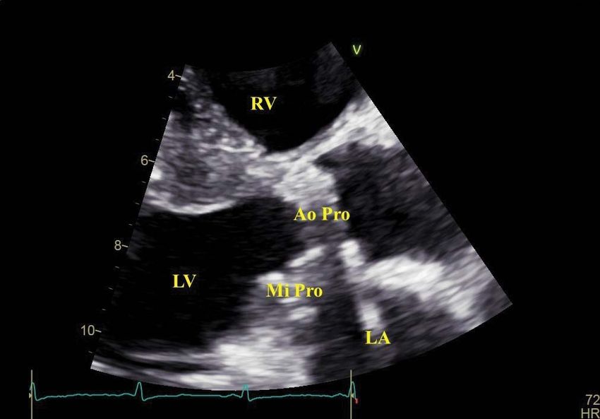
Andreea-Antonia NICA et al.                                                                                        Romanian Journal of Cardiology
Third Re-Do Double Prosthesis Thrombosis                                                                                     Vol. 31, No. 2, 2021

CASE PRESENTATION                                                             and regurgitation, and moderate mitral stenosis and
A 44-year-old woman presented at the emergency                                regurgitation, addressed with double mechanical valve
department with left-sided paresthesia, dizziness and                         replacement during the same index hospitalization. At
diplopia. Symptoms onset was 12 hours prior to pre-                           discharge, mitral size 27 Sorin Carbomedics and aortic
sentation.                                                                    size 18 ATS prosthesis functional parameters were in
   Her cardiovascular history proved to be very                               the normal range, with no perioperative complicati-
complex. In 2009, when she was 33-year-old, she                               ons after the second open-heart surgery. The patient
underwent double-valve replacement with biopros-                              remained asymptomatic during the following months,
thetic mitral valve size 27 St Jude Biocor and aortic                         until June 2020, although she reported stopping anti-
size 21 ATS Stentless valve respectively, for severe                          coagulation treatment over the last 6 months, without
rheumatic heart valve disease. After a 10-year symp-                          seeking medical advice.
tom-free period, she was admitted and treated for                                On admission, the patient was apyrexial, with
blood culture-negative infective endocarditis involv-                         dyspnea at minimal activity, tachycardic at rest, with
ing the bioprosthetic aortic valve, complicated with                          normal blood pressure values. Clinical examination
acute right limb ischemia, followed by surgical arterial                      revealed regular cardiac rhythm, with no significant
thrombectomy. By that time, significant bioprosthe-                           murmur, mechanical aortic and mitral closing click, no
tic degeneration was also identified. The association                         signs of pulmonary congestion, but moderate periphe-
with acute endocarditis resulted in significant valvu-                        ral congestion. Neurological examination revealed
lar dysfunction consistent with severe aortic stenosis                        left-sided paresthesia, with preserved motor functions
                                                                              and left homonymous hemianopsia.

Figure 1. Transthoracic echocardiography, parasternal long axis view, zoom on the aortic and mitral prosthesis. Structural changes of the mitral
prosthesis suggestive of possible thrombosis. No significant changes seen on the aortic prosthesis. Ao Pro, aortic prothesis; LA, left atrium; LV, left
ventricle; Mi Pro, mitral prosthesis; RV, right ventricle.

368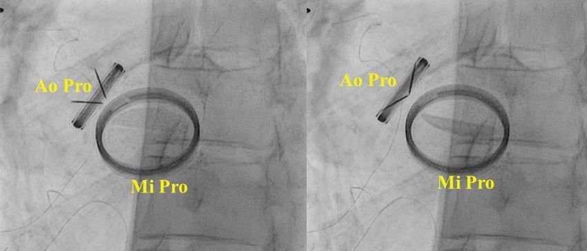
Romanian Journal of Cardiology Andreea-Antonia NICA et al.
Vol. 31, No. 2, 2021 Third Re-Do Double Prosthesis Thrombosis
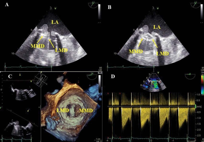
Figure 2. Transesophageal echocardiography evaluation of the mitral prosthesis. Bicommissural views (A- diastole, and B- systole) showing massive
mitral prosthesis thrombosis with a blocked medial disc and incomplete closing of the lateral disc due to extensive thrombosis. C. 3D-TEE en-face
view of the mitral prosthesis showing medial obstructed disc with an organized thrombus. D. Continuous wave Doppler interrogation of the mitral
prosthesis demonstrating elevated transmitral gradients. LA, left atrium; LMD, lateral mitral disc; MMD, medial mitral disc.
Blood tests showed iron deficiency mild anemia, echocardiography (TTE) revealed hypertrophic and
elevated inflammatory markers with high levels of C- non-dilated left ventricle, with apical dyskinesia and
reactive protein up to 10 times the upper limit of nor- ejection fraction of 50%. Left atrium was severely di-
mal and elevated NT-proBNP. Kidney, liver and thyro- lated with dense spontaneous echo-contrast. Prosthe-
id functional were in the normal range. On admission, tic mitral valve disc mobility was impaired by several
INR level was 1.7. The urine test was normal, with no echo-dense masses attached to both discs. Continuo-
signs of infection. us wave Doppler interrogation demonstrated eleva-
12-lead electrocardiogram showed sinus tachycar- ted mean trans-mitral gradient of 9.5 mmHg. Evalua-
dia and left ventricular hypertrophy with strain. tion of prosthetic aortic valve revealed high velocities
Urgent multimodality imaging was performed as and gradients, with a maximum aortic velocity of 3.6
part of the evaluation for a possible cardiac source of m/s and trans prosthetic mean gradient of 33 mmHg,
embolism. Cerebral computed tomography (CT) scan and an effective orifice area of 0.55 cm2. Mild mitral
revealed right occipital hypodensity in the para-hippo- and aortic regurgitations were noted. There was right
campal gyrus, suggesting subacute posterior cerebral heart enlargement, without significant RV dysfunction.
artery ischemic stroke. Doppler ultrasound imaging of Tricuspid valve leaflets were thickened and restricted
carotid and vertebral arteries showed normal arterial consistent with mild rheumatic tricuspid stenosis and
caliber with two small stable left carotid sinus athe- severe regurgitation. Pulmonary artery pressure was
rosclerotic plaques, with no signs of stenosis and good 40 mmHg.
peak systolic and diastolic velocities. Transthoracic
369Andreea-Antonia NICA et al. Romanian Journal of Cardiology Third Re-Do Double Prosthesis Thrombosis Vol. 31, No. 2, 2021 Figure 3. Fluoroscopy showing blocked medial mitral disc. Ao Pro, aortic prosthesis; Mi Pro, mitral prosthesis. Figure 4. Fluoroscopy showing normal opening (left) and closing (right) of both aortic prothesis discs. Ao Pro, aortic prosthesis; Mi Pro, mitral prosthesis. Emergency transesophageal echocardiography cations. Both mitral and aortic prosthesis were seve- (TEE) was performed for a more accurate assessment rely affected by extensive irregular echogenic struc- of prosthesis function and characterization of compli- tures on the atrial and ventricular site of the pros- 370
Romanian Journal of Cardiology                                                                Andreea-Antonia NICA et al.
Vol. 31, No. 2, 2021                                                          Third Re-Do Double Prosthesis Thrombosis

thesis respectively, suggestive of massive thrombosis       was discharged after thorough counseling on the vital
with possibly coexisting vegetations. Moreover, mitral      importance of lifetime anticoagulation. At 6 months
prosthesis medial disc blockage was identified in 2D-       follow-up, the patient remained asymptomatic, with
TEE and confirmed with 3D-TEE and fluoroscopy,              good treatment adherence and therapeutic INR levels.
associated with incomplete lateral disc closing (Figu-
re 2, 3 and 4). Other significant TEE findings included     DISCUSSION
left atrial appendage thrombosis and confirmation of        We present a very challenging case of a young wo-
rheumatic tricuspid valve changes noted on 2D-TTE,          man with a history of rheumatic heart disease and two
associated with severe tricuspid regurgitation, as well     open-heart surgeries for severe aortic and mitral valve
as dilated tricuspid ring. Aortic prosthesis function       disease, with previous infective endocarditis and em-
was also assessed with fluoroscopy and showed nor-          bolic limb ischemia, who presented with early massive
mal discs mobility.                                         thrombosis of both prosthesis due to lack of anticoa-
   Patient history, biological and imaging data alto-       gulation adherence, recurrent embolic event and pos-
gether raised the suspicion of associated infective en-     sibly recurrent endocarditis.
docarditis. Two sets of blood cultures when apyretic           There are several factors associated with prosthetic
were collected and empirical antimicrobial therapy          valve thrombosis such as thrombogenicity of the pros-
with vancomycin and gentamicin was started. The de-         thesis, atrial fibrillation, left atrial geometry, abnormal
cision to continue the antimicrobial therapy with an        prosthetic blood flow and suboptimal anticoagulati-
association of vancomycin, gentamicin and ceftriaxo-        on.4 Inadequate anticoagulation is the most important
ne for at least 4 to 6 weeks was made together with         pathogenetic factor in prosthetic valve thrombosis.
the infectious disease specialist, despite negative blood   Reported incidence of symptomatic prosthetic heart
cultures. Unfractionated heparin (UFH) was also star-       valve thrombosis with obstruction is 0.3% to 1.3% per
ted after careful consideration with the neurologist        year among patients with therapeutical anticoagulation
specialist, to avoid cerebral bleeding complications.       and 6% per year among patients with inadequate anti-
   Clinical course was favorable, with no significant       coagulation. Mortality rate is very high without treat-
heart failure symptoms or fever. Partial remission of       ment, despite emergency surgical intervention5.
paresthesia and dizziness was also attained. Inflamma-         Some studies showed that mitral mechanical pros-
tory markers decreased to normal values. The pati-          thesis thrombosis and embolization might be twice as
ent developed transient acute kidney injury due to          frequent as aortic mechanical prosthesis thrombosis6.
nephrotoxicity of gentamicin, resolved with proper          One explanation could be a higher thrombogenicity of
hydration and stopping the nephrotoxic drugs. Other         the mitral valve. However, data is scarce and with limi-
causes for acute kidney injury such renal embolism,         ted statistical power to make any definite differentiati-
urinary obstructions, hypotension were excluded. Of         ons between the two sites of prosthesis implantation7.
note, blood tests for inherited thrombophilia, lupus           Guidelines recommend chronic anticoagulation
and rheumatic disease were negative.                        treatment with vitamin K antagonist with watchful mo-
   Repeat TEE showed no significant changes in pros-        nitoring of INR. Target INR should be set according to
thesis appearance and function after 11 days of UFH.        the type and thrombogenicity of the mechanical pros-
Hence, VKA treatment with close monitoring of the-          thesis, and individual risk factors8,9. Both American and
rapeutic INR was restarted.                                 European guidelines recommend a median INR value
   Following the Heart Team decision, the patient un-       rather than a range, in order to avoid INR fluctuations
derwent another replacement of the aortic and mitral        associated with increased complications and mortality
dysfunctional prosthesis with aortic size 21 Medtronic      in these patients. Patients should be appropriate trai-
mechanical prosthesis and mitral size 29 Sorin Car-         ned to monitor INR levels and regular check-ups are
bomedics mechanical prosthesis. Tricuspid repair was        recommended. There is evidence that INR self-mana-
also performed. Postoperative outcome was unevent-          gement reduces INR variability and clinical events10.
ful. We achieved and maintained therapeutic INR, and           The clinical history and subtherapeutic anticoagula-
also resumed the antibiotic regimen recommended             tion should alert the physician and take into considera-
by the infectious disease specialist, despite no signi-     tion prosthetic valve thrombosis. Patients usually pre-
ficant intraoperative evidence of infected vegetations      sent with recent dyspnea or an embolic event. Com-
associated with prosthesis thrombosis. The patient          prehensive evaluation by 2D- and 3D transesophageal

                                                                                                                     371
Andreea-Antonia NICA et al. Romanian Journal of Cardiology
Third Re-Do Double Prosthesis Thrombosis Vol. 31, No. 2, 2021
echocardiography and fluoroscopy are recommended TV tricuspid valve
to confirm the diagnosis,8,9 as it was the case with our UFH unfractionated heparin
patient. Association with infective endocarditis must
always be taken in consideration, especially in patients Compliance with ethics requirements:
with previous episodes of endocarditis, as it is related The authors declare no conflict of interest regarding this
to worse prognosis. Management of valve thrombosis article. The authors declare that all the procedures and ex-
depends on the size of the thrombus, associated val- periments of this study respect the ethical standards in the
Helsinki Declaration of 1975, as revised in 2008(5), as well
vular disease or the thromboembolic event and inclu-
as the national law. Informed consent was obtained from all
des anticoagulation, fibrinolysis and surgery8,9. In this the patients included in the study.
case, we opted for unfractionated heparin to optimi-
ze the anticoagulation level, associated with empiric
antibiotic treatment for a possible blood-culture-ne- References
1. Raman K, Mohanraj A, Palanisamy V, Ganesh J, Rawal S, Kurian
gative infective endocarditis. However, there was no VM, Sethuratnam R. Thrombolysis and surgery for mitral prosthetic
significant improvement in thrombus appearance and valve thrombosis: 11-year outcomes. Asian Cardiovasc Thorac Ann
2019;27: 633-40.
prosthesis dysfunction, as demonstrated by repeated 2. Lancellotti P, Pibarot P, Chambers J, Edvardsen T, Delgado V, Dulgh-
TEE. The decision to reoperate was made after ca- eru R, Pepi M, Cosyns B, Dweck MR, Garbi M, Magne J, Nieman K,
reful consideration of the Heart Team regarding the Rosenhek R, Bernard A, Lowenstein J, Vieira MLC, Rabischoffsky A,
Vyhmeister RH, Zhou X, Zhang Y, Zamorano JL, Habib G. Recom-
perceived benefits and possible complications associ- mendations for the imaging assessment of prosthetic heart valves:
ated with a third open-heart surgery by the age of 44 a report from the European Association of Cardiovascular Imaging
endorsed by the Chinese Society of Echocardiography, the Inter-
years old. After a successful re-do surgery followed by American Society of Echocardiography, and the Brazilian Depart-
an uneventful recovery, the patient was trained with ment of Cardiovascular Imaging. Eur Heart J - Cardiovasc Imaging
respect to the crucial importance of strict adherence 2016;17: 589-90.
3. Vesey JM, Otto CM. Complications of Prosthetic Heart Valves. Curr
and close monitoring of the anticoagulation treatment, Cardio Rep 2004;6: 106-11.
in order to achieve and maintain lifetime optimal INR. 4. Magarakis M, Macias AE, Ghodsizad A, Salerno TA. Surgical man-
agement of cardiovascular thrombotic conditions. In Cardiovascular
Thrombus: From Pathology and Clinical Presentations to Imaging,
CONCLUSION Pharmacotherapy and Interventions 2018, Elsevier, p.367-76.
The aim of this challenging clinical case is to raise awa- 5. Durrleman N, Pellerin M, Bouchard D, Hebert Y, Cartier R, Perrault
PL, Basmadjian A, Carrier M. Prosthetic valve thrombosis: twenty-
reness on the life-threatening complications in pati- year experience at the Montreal Heart Institute. J Thorac Cardio-
ents with mechanical valvular prosthesis, the role of vasc Surg 2004;127: 1388-92.
6. Roudaut R, Serri K, Lafitte S. Thrombosis of prosthetic heart valves:
constant counseling to maintain treatment and beha-
diagnosis and therapeutic considerations. Heart 2007;93: 137-42.
vioral adherence, in addition to the real benefit in a 7. Cannegieter SC, Rosendaal FR, Briet E. Thromboembolic and bleed-
third re-do high-risk surgery. ing complications in patients with mechanical heart valve prostheses.
Circulation 1994;89: 635-41.
8. Baumgartner H, Falk V, Bax JJ, De Bonis M, Hamm C, Holm PJ, Iung
Abreviations B, Lancellotti P, Lansac E, Muñoz R, Rosenhek R, Sjögren J, Mas PT,
2D two-dimensional Vahanian A, Walther T, Wendler O, Windecker S, Zamorano JL.
2017 ESC/EACTS Guidelines for the management of valvular heart
3D three-dimensional disease. European Heart Journal 2017;38: 2739-91.
Ao Pro aortic prosthesis 9. Otto CM, Nishimura RA, Bonow RO, Carabello BA, Erwin JP 3rd,
Gentile F, Jneid H, Krieger EV, Mack M, McLeod C, O’Gara PT, Rigo-
CT computed tomography
lin VH, Sundt TM 3rd, Thompson A, Toly C. 2020 ACC/AHA Guide-
INR international normalized ratio line for the Management of Patients With Valvular Heart Disease: A
LA left atrium Report of the American College of Cardiology/American Heart As-
sociation Joint Committee on Clinical Practice Guidelines. Circula-
LMD lateral mitral disc tion 2021;143: e72-e227.
LV left ventricle 10. Heneghan C, Ward A, Perera R; Self-Monitoring Trialist Collabora-
Mi Pro mitral prosthesis tion, Bankhead C, Fuller A, Stevens R, Bradford K, Tyndel S, Alonso-
Coello P, Ansell J, Beyth R, Bernardo A, Christensen TD, Crom-
MMD medial mitral disc heecke ME, Edson RG, Fitzmaurice D, Gadisseur AP, Garcia-Ala-
MHV mechanical heart valve mino JM, Gardiner C, Hasenkam JM, Jacobson A, Kaatz S, Kamali F,
Khan TI, Knight E, Körtke H, Levi M, Matchar D, Menéndez-Jándula
RA right atrium
B, Rakovac I, Schaefer C, Siebenhofer A, Souto JC, Sunderji R, Gin
RV right ventricle K, Shalansky K, Völler H, Wagner O, Zittermann A. Self-monitoring
TEE transesophageal echocardiography of oral anticoagulation: systematic review and meta-analysis of indi-
vidual patient data. Lancet 2012;379:322-34.
TTE transthoracic echocardiography
372You can also read