A case report of a recurrent early and late Bioresorbable vascular scaffold thrombosis: serial angiography and optical coherence tomography ...
←
→
Page content transcription
If your browser does not render page correctly, please read the page content below
Lee et al. BMC Cardiovascular Disorders    (2020) 20:146
https://doi.org/10.1186/s12872-020-01426-z

 CASE REPORT                                                                                                                                       Open Access

A case report of a recurrent early and late
Bioresorbable vascular scaffold thrombosis:
serial angiography and optical coherence
tomography findings
Cheol Hyun Lee, Yun-Kyeong Cho, Hyuck-Jun Yoon and Seung-Ho Hur*

  Abstract
  Background: In bioresorbable vascular scaffolds (BVSs), there is some concern about a possible increase in the rate
  of scaffold thromboses (ScTs). Although several characteristics similarly contribute to the development of both early
  and late ScTs, there are also clearly different pathomechanisms between the two time-dependent types of
  thromboses, especially with BVSs.
  Case presentation: We recently experienced a very rare case of a 69-year-old man who had recurrent early and
  late ScTs with somewhat differing pathomechanisms as assessed by optical coherence tomography (OCT). For the
  late ScT, OCT identified a scaffold dismantling in the same place that a peri-strut low intensity area (PLIA) was
  observed in the previous OCT finding.
  Conclusion: We report the management of an ScT in a case with findings such as a heterogeneous a BVS
  degradation, peri-strut low intensity area (PLIA), intraluminal scaffold dismantling, and under-sizing and/or stent
  malapposition observed in OCT.
  Keywords: Bioresorbable vascular scaffold, Scaffold thrombosis, Optical coherence tomography, Acute coronary
  syndromes

Background                                                                            However, with bioresorbable vascular scaffolds (BVSs),
A stent thrombosis (ST) is an uncommon but devastating                                there is some concern about an increased rate of scaffold
adverse event that can occur after percutaneous coronary                              thromboses (ScTs) [4]. Optical coherence tomography
intervention (PCI) [1]. Various factors can influence the                             (OCT) imaging is instrumental in identifying the mecha-
likelihood of an ST occurrence, including the patient-                                nisms of STs as well as ScTs [5]. Several case reports have
related characteristics, procedural and lesion characteris-                           revealed the mechanism of ScTs using OCT. Here, we
tics, presence of acute coronary syndrome, and premature                              describe and discuss a rare recurrent ScT case with an
discontinuation of antiplatelet therapy [2, 3]. With the ad-                          intraluminal BVS dismantling identified by OCT, which
vent of newer-generation drug-eluting stents (DES) and                                may have led to a very late thrombosis in a patient with a
the continuation of a dual antiplatelet therapy (DAPT),                               history of a subacute ScT, who presented with acute
this phenomenon has come to be seen less frequently.                                  coronary syndrome 23 months after the BVS placement.

* Correspondence: shur@dsmc.or.kr
Division of Cardiology, Keimyung University Dongsan Medical Center, 56
Dalseong-Ro, Jung-Gu, Daegu 700-712, South Korea

                                       © The Author(s). 2020 Open Access This article is licensed under a Creative Commons Attribution 4.0 International License,
                                       which permits use, sharing, adaptation, distribution and reproduction in any medium or format, as long as you give
                                       appropriate credit to the original author(s) and the source, provide a link to the Creative Commons licence, and indicate if
                                       changes were made. The images or other third party material in this article are included in the article's Creative Commons
                                       licence, unless indicated otherwise in a credit line to the material. If material is not included in the article's Creative Commons
                                       licence and your intended use is not permitted by statutory regulation or exceeds the permitted use, you will need to obtain
                                       permission directly from the copyright holder. To view a copy of this licence, visit http://creativecommons.org/licenses/by/4.0/.
                                       The Creative Commons Public Domain Dedication waiver (http://creativecommons.org/publicdomain/zero/1.0/) applies to the
                                       data made available in this article, unless otherwise stated in a credit line to the data.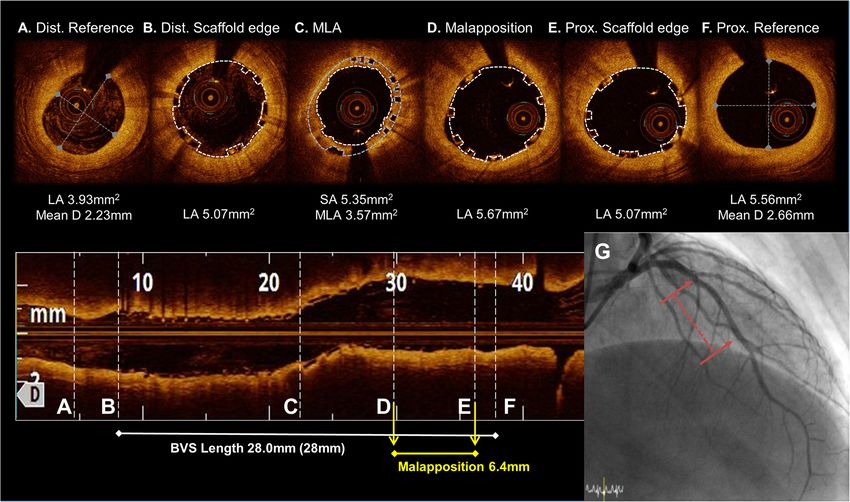
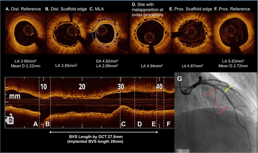
Lee et al. BMC Cardiovascular Disorders     (2020) 20:146                                                                               Page 2 of 7

Case presentation                                                           scaffold using a 2.5 × 20 mm semi-compliant balloon and
A 69-year-old man, a current smoker with hypertension                       angioplasty with a 3.0 × 12 mm NC balloon up to 18 atm
and diabetes mellitus, presented to the emergency de-                       in order to obtain a TIMI 3 flow (Fig. 2). He was pre-
partment with a non-ST-segment elevation myocardial                         scribed aspirin and ticagrelor upon re-discharge. He
infarction (NSTEMI). Coronary angiography revealed a                        showed no abnormalities other than mild stenosis in the
total occlusion of the mid left anterior descending artery                  middle of the scaffold at the one-year follow-up coron-
(LAD). He underwent a placement of a 2.5 × 28 mm Ab-                        ary angiography. The one-year follow up OCT (Dragon-
sorb GT1 BVS (Abbott Vascular, Santa Clara, CA, USA)                        fly, St. Jude Medical, St. Paul, MN, USA) revealed a peri-
under OCT-guidance, pre-dilated with a 2.0 × 20 mm                          strut low intensity area (PLIA) in the proximal part of
TREK (Abbott Vascular, Santa Clara, CA) balloon and                         the BVS. However, since the minimal lumen area
post-dilated with a 2.75 × 15 mm non-compliant (NC)                         (MLA) was 2.66 mm2 and the neointimal hyperplasia
TREK balloon up to 12 atm in his mid-LAD (Fig. 1). The                      (NIH) was 55% without symptoms, we decided to
OCT images after the initial scaffold implantation                          continue medication (Fig. 3). Since an acute myocar-
showed an acute malapposition of 6.4 mm in length at                        dial infarction occurred at 1 year, aspirin and clopido-
the proximal part of the BVS, likely due to the use of an                   grel were prescribed again.
undersized BVS. After high pressure ballooning, it no                          Two years after the BVS implantation, the patient was
longer exhibited a significant value (< 200 μm) on OCT.                     referred to the hospital for chest pain at rest. The patient
He was prescribed aspirin and clopidogrel upon dis-                         underwent coronary angiography because the patient de-
charge, but arbitrarily stopped both for a colonoscopy                      veloped an ST-segment elevation myocardial infarction
16 days later. He was re-admitted to the hospital with                      (STEMI). Coronary angiography revealed a very late ScT
chest pain 20 days after the BVS implantation. An                           in the proximal part of the prior BVS in the LAD artery
NSTEMI was confirmed by a subacute ScT in the mid-                          (Fig. 4). OCT identified a thrombus in the proximal seg-
dle of the scaffold on the coronary angiography examin-                     ment of the scaffold, at the same place where the PLIA
ation. We performed a pre-dilation at the site of the                       was observed in the previous OCT finding. Two layers

 Fig. 1 The OCT images during the BVS implantation from the distal to proximal 2.5 × 28 mm BVS was deployed at a nominal pressure and post-
 dilated with a 2.75 mm noncompliance balloon at 16 atm. (a to f). An under-sizing and minimal malapposition over the cutoff value (< 200 μm)
 were seen at the proximal part of the BVS. (c to e) A tissue prolapse was seen at the MLA site. (c) The final coronary angiogram at baseline
 following the BVS implantation (red dotted line). (g) Abbreviations: Dist., distal; Prox., proximal; MLA, minimal lumen area; LA, lumen area; D,
 diameter; SA, scaffold area; BVS, bioresorbable vascular scaffolds
Lee et al. BMC Cardiovascular Disorders      (2020) 20:146                                                                                 Page 3 of 7

 Fig. 2 OCT image during the subacute scaffold thrombosis event after a 2.5 mm preballoon dilatation up to 4 atm. (a to f). Intraluminal red
 thrombus with stacked struts in the middle to distal part of the BVS. (b to c) No previous under-sizing and malapposition could be seen in the
 proximal part of the BVS. (c to e) Coronary angiogram of a subacute scaffold thrombosis event. A TIMI 0 flow was observed in the distal part of
 the previous BVS (red dotted line). (g) Abbreviations: Dist., distal; Prox., proximal; Pre-, previous; MLA, minimal lumen area; LA, lumen area; D,
 diameter; SA, scaffold area; BVS, bioresorbable vascular scaffolds

of the BVS struts, consisting of a strut supporting the                      metallic DES. Our case had important implications for
vessel wall and a strut protruding into the lumen, were                      the OCT assessment when evaluating the underlying
also detected (Fig. 5). Since the BVS struts were dis-                       pathomechanisms of early and late ScTs.
rupted with an increased risk of a distal embolization by                       The key factor of the early and subacute ScT in the
an aspiration catheter, direct balloon angioplasty was                       present case may have been the discontinuation of the
performed immediately without a thrombus aspiration                          dual antiplatelet therapy (DAPT). Although some mech-
in order to obtain coronary flow. A 2.75 × 33 mm Xience                      anical factors such as a malapposition may have contrib-
Alpine balloon (Abbott Vascular, Santa Clara, CA) was                        uted to the development of the ScT, when the subacute
then deployed at 10 atm in the LAD covering the whole                        ScT event occurred, a significant acute malapposition (≥
segment of the prior scaffold. The stent was then post-                      200 μm) was no longer observed on OCT after an ad-
dilated with a 3.0 × 12 mm non-compliant balloon at 16                       junctive NC balloon inflation. Therefore, we decided to
atm. The entire newly-placed DES showed a good ap-                           use a more potent P2Y12 inhibitor after ensuring coron-
position, as the intra-luminal dismantled BVS struts                         ary flow by balloon angioplasty as opposed to an add-
were compressed toward the vessel wall, according to                         itional DES implantation. Maintaining a DAPT beyond 1
the OCT examination.                                                         year is more important than a metallic DES for the pre-
                                                                             vention of a late and very late ScT with BVSs, because
Discussion and conclusions                                                   the degradation of a BVS strut can heterogenically occur
The occurrence of a stent thrombosis after a metallic                        even over 3 to 4 years [6]. In addition, our case exhibited
DES implantation has been attributed to inflammatory                         a procoagulable status under the NSTEMI condition.
and allergic reactions to specific clinical and stent com-                   The patient had to maintain the DAPT for over a year
ponents (clinical presentation, drug, polymer, or stent).                    according to the guidelines for preventing future events
In contrast, a scaffold thrombosis has been reported to                      [7]. Several reports have identified under-sizing as a fac-
be caused by various factors beyond the factors of a                         tor for an early ScT with both DESs and BVSs [8–11]. In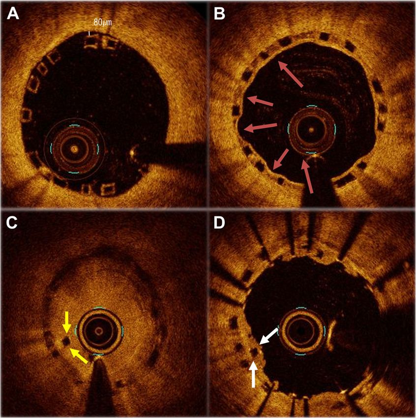
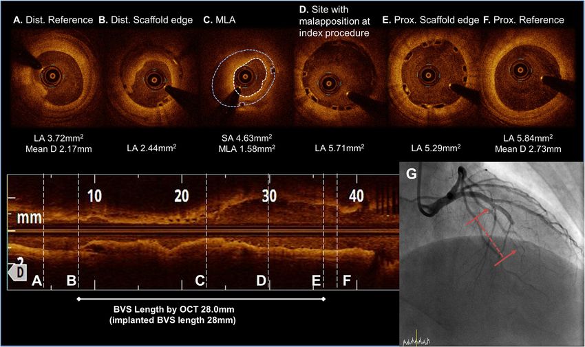
Lee et al. BMC Cardiovascular Disorders     (2020) 20:146                                                                                Page 4 of 7

 Fig. 3 OCT image at the one-year follow up after the BVS implantation. (a to f). The MLA site migrated to the proximal side of the BVS. (c) A
 peristrut low intensity area (PLIA) was observed in the proximal part of the BVS. (d) A coronary angiogram at the one-year follow up after the BVS
 implantation. A discrete moderate stenosis was observed at the MLA site (yellow arrow) of the previous BVS. (red arrow line). (g) Abbreviations:
 Dist., distal; Prox., proximal; Pre-, previous; MLA, minimal lumen area; LA, lumen area; D, diameter; SA, scaffold area; BVS, bioresorbable
 vascular scaffolds

the case shown in Fig. 1, the implantation of an under-                     in the same segment where the PLIA had been seen in
sized BVS may have been another mechanism of the                            the one-year follow-up OCT. Eventually, the remaining
subacute ScT, because a 2.5 mm scaffold was implanted                       minimal malapposition increased the vulnerability
in a vessel with an external elastic lamina (EEL) diameter                  through the inflammation mechanism and vascular
of approximately 3.0 mm. However, unsurprisingly, pa-                       edema in the late reabsorbtion process. The PLIA find-
tients with under-sizing are likely to be treated for an                    ing as an unhealthy cover of the BVS strut conse-
acute myocardial infarction (AMI) during the index pro-                     quently might not be able to prevent a strut disruption.
cedure, because there are elevated potent vasoconstric-                     It became the possible factor of a very late scaffold
tors in the blood level as well as the presence of                          thrombosis (VLScT). Furthermore, a thick strut with a
microcirculatory dysfunction in the setting of an AMI                       minimal malapposition may provide high shear stress
[12, 13]. In our case, despite concern for under-sizing,                    arising from an incompletely apposed and bulky stent
the minimal scaffold area in the index procedure was                        strut within the proximal transition zone. This may
not small (> 5 mm2) after post-NC ballooning up to 12                       have also contributed to the occurrence of a late/very
atm, leading to no need for further intervention.                           late ScT. A recent report of OCT findings in four cases
   A delayed inflammatory reaction has been identified                      of VLScTs suggests that discontinuity of the scaffold
as a possible mechanism associated with inflammation                        may cause a VLScT in advanced stages of a scaffold re-
of late/very late ScTs following the implantation of a                      sorption, although the possibility of scaffold damage by
BVS. A PLIA might be a marker of vascular edema and                         a thrombus aspiration procedure could not be ruled
vascular vulnerability by previous OCT study because                        out [15]. However, our OCT images were obtained be-
of the degradation products of poly-lactic acid crystals,                   fore (Fig. 5c) and after the inflation of a small sized bal-
the substrate for a proteoglycan rich matrix [14]. Con-                     loon with a low pressure (Fig. 4). Thus, it was
sidering our case based on previous research, 23                            important to confirm that the scaffold was dismantled
months after the BVS implantation, the BVS strut                            intraluminally during the late period, not due to scaf-
showed intraluminal dismantling and strut disruption                        fold damage by the thrombectomy.
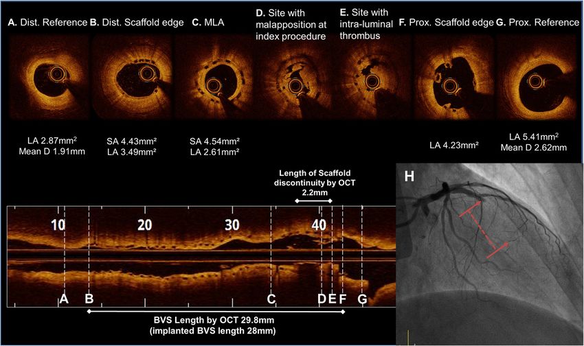
Lee et al. BMC Cardiovascular Disorders       (2020) 20:146                                                                                   Page 5 of 7

 Fig. 4 OCT image during the very late scaffold thrombosis (VLScT) event after a 2.0 mm preballoon dilatation up to 4 atm (a to g). The thrombus
 was observed at the same site where the peri-strut low intensity area (PLIA) had been previously observed, and the strut had been dismantled
 and was floating in the intraluminal area. (d to e) In the coronary angiogram during the VLScT event, a TIMI 0 flow was observed in the proximal
 part of the previous BVS (red dotted line). (h) Abbreviations: Dist., distal; Prox., proximal; Pre-, previous; MLA, minimal lumen area; LA, lumen area;
 D, diameter; SA, scaffold area; BVS, bioresorbable vascular scaffolds

  We found that a disruption of the strut of the VLScT                         malapposition. On the other hand, these findings,
was a possible nidus through the OCT. We treated it                            which can only be seen on OCT, may increase the
with a long DES adequate to cover all previously-                              ScT rate, which may eventually be the reason why it
implanted scaffolds. We changed the P2Y12 inhibitor                            cannot be used in real practice. The limitation of this
back to ticagrelor, a more potent P2Y12 inhibitor, be-                         case report is that OCT findings cannot explain all
cause we observed a higher level of platelet reactivity in                     causes of the 2nd ScT event. For this patient, not
the platelet function test or verify now test when we ex-                      only the occurrence of scaffold discontinuity associ-
perienced the recurrent ScT. In addition, as mentioned                         ated with a PLIA on OCT, but also the flow disturb-
above, the long-term maintenance of the DAPT beyond                            ance of the MLA site and the patient’s clopidogrel
1 year may be helpful, because the degradation of the                          resistance may be involved in the 2nd ScT event.
BVS strut has heterogenicity. In summary, if the occur-                        However, our case report showed a hypothesis gen-
rence of an ScT is caused by a DAPT interruption,                              erating role in which patients with a BVS implant-
under-expansion, or under-sizing during the acute and                          ation should actively analyze the OCT findings in
subacute periods, a thrombectomy and balloon angio-                            order to prevent or identify the cause of the ScT. In
plasty can be considered. If a scaffold fracture and geo-                      addition, further studies are required to define the
graphical miss are observed in the long segment during                         natural course of a PLIA following a BVS implant-
the late and very late periods, DES treatment can be per-                      ation. A next generation bioresorbable scaffold that
formed [16, 17].                                                               can minimize late inflammation may need to be
  Overall, OCT plays a major role in cases of a BVS                            developed.
late thrombosis in identifying associated conditions                              In conclusion, we report a unique case of a sub-
such as a PLIA, accurate evaluation of a luminal                               acute and very late scaffold recurrent thrombosis
stenosis arising from intimal hyperplasia, intraluminal                        likely due to the withdrawal of antiplatelet agents and
scaffold dismantling, under-sizing, and/or stent                               late scaffold dismantling, which led to acute coronary
Lee et al. BMC Cardiovascular Disorders          (2020) 20:146                                                                                     Page 6 of 7

  Fig. 5 An expanded serial OCT image of the same point during the initial procedure, the one-year follow up, and very late scaffold thrombosis
  (VLScT) event. During the initial procedure, a minimal malapposition after high pressure NC ballooning is observed. (a) A PLIA was observed in
  the proximal part of the BVS at the one-year follow up (red arrow). (b) The thrombus was observed at the site where the PLIA was observed, and
  the strut had been dismantled and was floating in the intraluminal area during the VLScT event (yellow arrow). This image obtained before
  balloon predilatation. (c) At the time of the VLScT, the Xience alpine 2.75 × 33 mm stent covered the inside of the disrupted scaffold strut with
  compression toward the vessel wall (white arrow), and a good-expansion was observed in the final OCT image. (d)

syndrome. A serial OCT examination can illustrate                               read and approved the final manuscript. All authors agreed to their
the pathophysiologic process during the development                             contribution.

of a recurrent ScT. This report emphasizes the im-                              Funding
portance of OCT-guided treatment planning in the                                There is no funding for this study.
event of problems following a BVS implantation. It
                                                                                Availability of data and materials
may be used as a reference for the future develop-                              The data analyzed in the case report (medical history of patient) are not
ment of a newer BVS device.                                                     publicly available due to the privacy policy of the hospital but are available
                                                                                from the corresponding author on reasonable request.
Abbreviations
BVS: Bioresorbable vascular scaffolds; DAPT: Dual antiplatelet therapy;         Ethics approval and consent to participate
DES: Drug-eluting stents; EEL: External elastic lamina; LAD: Left anterior      Not applicable.
descending artery; MLA: Minimal lumen area; NC: Non-compliant;
NIH: Neointimal hyperplasia; NSTEMI: Non ST-segment elevation myocardial        Consent for publication
infarction; OCT: Optical coherence tomography; PLIA: Peri-strut low intensity   Informed consent for the publication of the case report was obtained from
area; ScT: Scaffold thrombosis; ST: Stent thrombosis; STEMI: ST-segment         the patient in a written form.
elevation myocardial infarction; VLScT: Very late scaffold thrombosis
                                                                                Competing interests
                                                                                The authors declare that they have no competing interests.
Acknowledgements
Not applicable.                                                                 Received: 8 November 2018 Accepted: 12 March 2020

Authors’ contributions
CHL, the first author, performed the diagnostic coronary angiography in the     References
patient, wrote the manuscript, and made the illustrations. SHH, the             1. Cutlip DE, Windecker S, Mehran R, Boam A, Cohen DJ, et al. Clinical end
corresponding author, performed the percutaneous coronary intervention for          points in coronary stent trials: a case for standardized definitions.
the patient and participated in the OCT imaging interpretation. YKC and HJY         Circulation. 2007;115:2344–51.
participated in the design of the report and helped to draft the manuscript     2. Kirtane AJ, Stone GW. How to minimize stent thrombosis. Circulation. 2011;
reporting the rare case of an early and late scaffold thrombosis. All authors       124:1283–7.
Lee et al. BMC Cardiovascular Disorders (2020) 20:146 Page 7 of 7
3. Lee CW, Kang SJ, Park DW, Lee SH, Kim YH, et al. Intravascular ultrasound
findings in patients with very late stent thrombosis after either drug-eluting
or bare-metal stent implantation. J Am Coll Cardiol. 2010;55:1936–42.
4. Wykrzykowska JJ, Kraak RP, Hofma SH, van der Schaaf RJ, Arkenbout EK,
et al. Bioresorbable scaffolds versus metallic stents in routine PCI. N Engl J
Med. 2017;376:2319–28.
5. Cuculi F, Puricel S, Jamshidi P, Valentin J, Kallinikou Z, et al. Optical
coherence tomography findings in Bioresorbable vascular scaffolds
thrombosis. Circ Cardiovasc Interv. 2015;8:e002518.
6. Serruys PW, Onuma Y, Dudek D, Smits PC, Koolen J, et al. Evaluation of the
second generation of a bioresorbable everolimus-eluting vascular scaffold
for the treatment of de novo coronary artery stenosis: 12-month clinical and
imaging outcomes. J Am Coll Cardiol. 2011;58:1578–88.
7. Roffi M, Patrono C, Collet JP, Mueller C, Valgimigli M, et al. 2015 ESC
guidelines for the management of acute coronary syndromes in patients
presenting without persistent ST-segment elevation: task force for the
Management of Acute Coronary Syndromes in patients presenting without
persistent ST-segment elevation of the European Society of Cardiology
(ESC). Eur Heart J. 2016;37:267–315.
8. Fujii K, Carlier SG, Mintz GS, Yang YM, Moussa I, et al. Stent underexpansion
and residual reference segment stenosis are related to stent thrombosis
after sirolimus-eluting stent implantation: an intravascular ultrasound study.
J Am Coll Cardiol. 2005;45:995–8.
9. van Werkum JW, Heestermans AA, Zomer AC, Kelder JC, Suttorp MJ, et al.
Predictors of coronary stent thrombosis: the Dutch stent thrombosis
registry. J Am Coll Cardiol. 2009;53:1399–409.
10. Stone GW, Abizaid A, Onuma Y, et al. Effect of technique on outcomes
following Bioresorbable vascular scaffold implantation. J Am Coll Cardiol.
2017;70:2863–74.
11. Ellis SG, Steffenino G, Kereiakes DJ, Stone GW, van Geuns RJ, et al. Clinical,
angiographic, and procedural correlates of acute, subacute, and late absorb
scaffold thrombosis. JACC Cardiovasc Interv. 2017;10:1809–15.
12. Cuculi F, Dall'Armellina E, Manlhiot C, De Caterina AR, Colyer S, et al. Early
change in invasive measures of microvascular function can predict
myocardial recovery following PCI for ST-elevation myocardial infarction. Eur
Heart J. 2014;35:1971–80.
13. Cuculi F, Herring N, De Caterina AR, Banning AP, Prendergast BD, et al.
Relationship of plasma neuropeptide Y with angiographic,
electrocardiographic and coronary physiology indices of reperfusion during
ST elevation myocardial infarction. Heart. 2013;99:1198–203.
14. Teramoto T, Ikeno F, Otake H, et al. Intriguing Peri-strut low-intensity area
detected by optical coherence tomography after coronary stent
deployment. Circ J. 2010;74(6):1257–9. https://doi.org/10.1253/circj. CJ-10-
0189.
15. Raber L, Brugaletta S, Yamaji K, O'Sullivan CJ, Otsuki S, et al. Very late
scaffold thrombosis: intracoronary imaging and Histopathological and
spectroscopic findings. J Am Coll Cardiol. 2015;66:1901–14.
16. Tamburino C, Latib A, van Geuns RJ, Sabate M, Mehilli J, et al. Contemporary
practice and technical aspects in coronary intervention with bioresorbable
scaffolds: a European perspective. EuroIntervention. 2015;11:45–52.
17. Felix C, Everaert B, Jepson N, Tamburino C, van Geuns R-J. Treatment of
bioresorbable scaffold failure. EuroIntervention. 2015;11:V175–80.
Publisher’s Note
Springer Nature remains neutral with regard to jurisdictional claims in
published maps and institutional affiliations.You can also read