ACUTE COMPLICATED NECROTISING PANCREATITIS TREATED WITH VIDEO-ASSISTED RETROPERITONEAL DEBRIDEMENT
←
→
Page content transcription
If your browser does not render page correctly, please read the page content below
PROCEEDINGS OF THE LATVIAN ACADEMY OF SCIENCES. Section B,
                                                                                               Vol. 75 (2021), No. 2 (731), pp. 136–141.

                                                                                                                       DOI: 10.2478/prolas-2021-0021

                                                                                                                                      Case Report

       ACUTE COMPLICATED NECROTISING PANCREATITIS
       TREATED WITH VIDEO-ASSISTED RETROPERITONEAL
       DEBRIDEMENT
       Sergejs Ðapovalovs1,#, Viktors Ïiòovs2, Andris Gardovskis1, Sintija Lapsa1,
       Mâris Pavârs1, and Jânis Gardovskis1
       1
           Department of Surgery, Rîga Stradiòð University, 13 Pilsoòu Str., Rîga, LV-1002, LATVIA
       2
           Institute of Radiology, Pauls Stradiòð Clinical University Hospital, 13 Pilsoòu Str., Rîga, LV-1002, LATVIA
       #
           Correspondending author, sergejssapovalovs@gmail.com

       Contributed by Jânis Gardovskis

            Acute necrotising pancreatitis is a complex disease with high morbidity and mortality rates. In
            cases of infected necrosis, treatment consists of a step-up approach involving endoscopic or
            mini-invasive surgical methods. In some cases, there are extremely rare complications. In addi-
            tion, the underlying comorbidities worsen the course of the disease. We report a case of a 32-
            year-old male with acute necrotising pancreatitis complicated with recurrent retroperitoneal ab-
            scesses, sepsis, iatrogenic pylephlebitis, exacerbation of underlying Crohn’s disease, and the
            outcome of the treatment was successful. During the period of hospitalisation, one ultrasound-
            guided percutaneous drainage, two computed tomography-guided punctures of the retroperito-
            neal space (percutaneous and transhepatic) and five video-assisted retroperitoneal debridement
            procedures were carried out. The patient was discharged after 185 days of hospitalisation.
            Key words: step-up approach, necrotomy, Crohn’s disease, pylephlebitis, VARD, sepsis.

INTRODUCTION                                                                   Inflammatory bowel disease is a risk factor itself for throm-
                                                                               boembolic events and in combination with acute infected
Acute necrotising pancreatitis is a complex disease with                       necrotising pancreatitis could be fatal (Algahtani et al.,
high morbidity and mortality rates and requires a multi-                       2021). Pylephlebitis is an undesirable complication charac-
disciplinary approach. In case of infected necrosis, treat-                    terised as suppurative thrombosis of the portal vein with a
ment consists of a step-up approach with either endoscopic                     rare incidence, but high mortality rate. Pylephlebitis can de-
or video-assisted debridement. Any surgical treatment in-                      velop as a complication of uncontrolled infection of intraab-
volves a wide set of possible complications like massive                       dominal organs drained by a portal system. Although more
bleeding, iatrogenic perforation of gastrointestinal tract or                  commonly pylephlebitis is described in cases of acute diver-
formation of pancreatic fistulas (Karakayali, 2014; Navadgi                    ticulitis and acute appendicitis (Al-Hamid et al., 2015), in
et al., 2021). Underlying comorbidities have a potential to                    some studies acute pancreatitis has been the leading cause
complicate the already complex treatment.                                      of pylephlebitis. There have been cases of pylephlebitis as-
Acute pancreatitis is one of the most common pancreato-                        sociated with computed tompgraphy (CT)-guided liver bi-
biliary complications of Crohn`s disease. Although the on-                     opsy (Tandon et al., 2015).
set of acute pancreatitis is usually observed after the diagno-
sis of Crohn`s disease has been established, there are some                    Here we present a case of severe acute necrotising pancrea-
cases when acute pancreatitis occurs as a first manifestation                  titis with iatrogenic pylephlebitis after CT-guided transhe-
of Crohn`s disease (Jasdanwala and Babyatsky, 2021). Con-                      patic abscess drainage with underlying Crohn’s disease, to
comitant alcohol use can increase the risk of acute pancrea-                   highlight the step-up treatment approach of acute pancreati-
titis.                                                                         tis and possible complications.
136                                                                                                Proc. Latvian Acad. Sci., Section B, Vol. 75 (2021), No. 2.
MATERIALS AND METHODS (IAP) was 32 mmHg, the patient was intubated, and me-
chanical lung ventilation was started. Due to the progression
The standard step-by-step approach for acute pancreatitis of the acute renal failure — haemodialysis was applied. A
treatment was applied. After the appearance of radiological computed tomography (CT) of the abdomen with intrave-
indications for puncture of necrotic collections, a puncture nous contrast was performed 48 hours after hospitalisation
was performed under control of computed tomography. and acute edematous pancreatitis was revealed (Fig. 1A).
Seven to ten days later, video-assisted retroperitoneal
debridement (VARD) surgery was performed to remove Three additional CT scans of the abdomen were performed
pancreatic and peripancreatic necrotic collections. Under in the next four weeks, showing a progression of necrotic
the general anesthesia the patient was positioned in a supine changes in pancreatic and peripancreatic tissues (Fig. 1B,
position and 30° tilted towards the right side. Approach 1C, 1D).
was between the XI and XII ribs or under percutaneous
The procalcitonin level fluctuated in the range from 12.0 to
drain (PCD). An incision 4–5 cm long was made. The con-
24.0 ng/ml and (C reactive protein (CRP) — from 190.5 to
dition of the drain was controlled with the index finger and
292.4 mg/l. Enterococcus faecalis growth in blood cultures
a purulent necrotic collection was opened.
was revealed. As the diagnosis of sepsis was confirmed, in-
After the incision, superficial necrotic collections were re- travenous antibacterial therapy with Piperacillinum/Tazo-
bactamum 4.5 g t.i.d. was started.
moved manually, with the help of fingers, by aspirating the
contents and evacuating the major necrotic contents with Because of a high suspicion of infected peripancreatic fluid
extended ring forceps. collection, on the 30th day after hospitalisation ultrasound
(US) guided puncture and drainage of the peripancreatic
A 30-degree laparoscope was introduced through the inci- fluid collection was performed and ascitic fluid (fluid was
sion, and ring-nose forceps were used simultaneously with clear, with low viscosity, and blood admixture; on microbi-
the videoscope, and necrotic collections were removed un- ological investigation — sterile) was obtained (Fig. 1E).
der video control. Necrotic masses were rinsed gently with
an irrigation pump with a large amount of saline solution. On the 41st day after hospitalisation, CT-guided retro-
peritoneal space puncture was carried out and a 14 Fr pigtail
Only loose necrotic masses were removed. In case of bleed- drain was placed (Fig. 1F). Approximately 50 ml/day of pus
ing, a laparoscopic clipping device was prepared. The goal and pancreatic debris were drained in the following four
of VARD was to remove the large loose necrotic collec- weeks. Escherichia coli and Acinetobacter baumannii were
tions. The mean time of VARD procedure was 78 minutes. detected in microbiologic analysis of the acquired exudate
After completion of the procedure, two large bore surgical and antibacterial therapy was switched to Imipenem 0.5 g
drains were placed into the collection cavity. High volume q.i.d. intravenously.
flushing (up to 3 l/24 hours) was performed through the
Two days later, mild bleeding from the lower gastrointesti-
drains for the following first three days. The study was ap-
nal tract (GT) was observed. Colonoscopy was performed
proved by the Ethics Committee of Pauls Stradiòð Clinical
and no source of acute bleeding was found. Diagnosis of
University Hospital..
non-active fistulising Crohn’s disease was confirmed based
on endoscopic findings and prior imaging studies (magnetic
resonance enterography), morphological findings, and typi-
RESULTS
cal clinical presentation.
A 32-year-old Caucasian male was hospitalised with com- On the 55th day after hospitalisation, follow-up CT of the
plaints about abdominal pain, vomiting and oliguria. He abdomen was performed. Progression of necrotic changes
was admitted after approximately one-month alcohol abuse. was found (Fig. 2G).
No previous significant medical conditions were found in
the patient’s medical history. Diagnosis of acute severe pan- The patient was prepared for planned video-assisted retro-
creatitis was established based on clinical, laboratory and peritoneal debridement (VARD) and on the 64th day after
imaging study findings. On physical examination his vital hospitalisation the procedure was performed; 500 ml of pus
signs were: heart rate (HR) 125 BPM, arterial blood pres- and 300 ml of necrotic tissues were obtained. Two 30 Fr sil-
sure (ABP) 190/90 mmHg, respiration rate (RR) 20 BPM, icone drains were left in the retroperitoneal space for the
body temperature (BT) 37.4 °C. Laboratory tests showed in- drainage. The retroperitoneal space was irrigated with saline
creased levels of lipase 3006 U/l, alpha-amylase 800 U/l, solution in regimen 3l/24 h for the next six days.
haematocrit 56.7% and haemoglobin 198 g/l. An Acute
Follow-up CT of the abdomen was performed five days af-
Physiology and Chronic Health Evaluation II (APACHE II)
ter the VARD (Fig. 2H); positive dynamics of the local sta-
was applied and was scored as 5 points. Conservative ther- tus and reduction of the necrotic tissue volume was seen, al-
apy was applied by massive rehydration and long-acting though new collection in the pelvic retroperitoneal region
Octreotide injection. However, 24 hours after the hospitali- was found.
sation, the patient was moved to the intensive care unit
(ICU) due to worsening of the general condition. Seventy- Because of progression of infected necrosis revealed on CT,
two hours after the hospitalisation, intra-abdominal pressure a second VARD was performed on the 71st day after hospi-
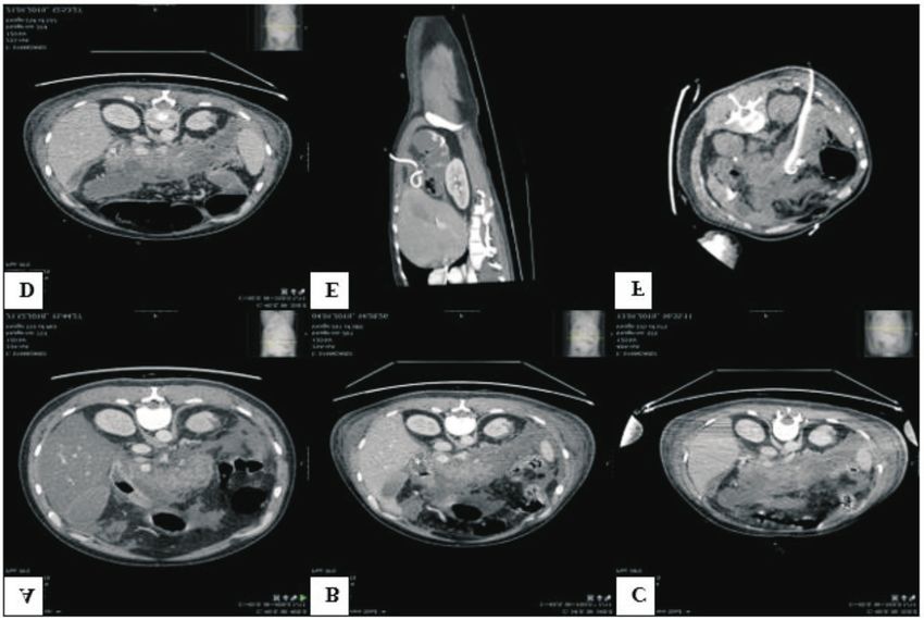
Proc. Latvian Acad. Sci., Section B, Vol. 75 (2021), No. 2. 137Fig. 1. Abdominal CT scan images. A, 48 hours after hospitalisation; B, 2 weeks after hospitalisation; C, 3 weeks after hospitalisation; D, 4 weeks after hos- pitalisation; E, F, 14 Fr pigtail drain inserted during US-guided puncture. Fig. 2. Abdominal CT scan images (cont.). G, progression of the necrotic changes on the 55th day; H, reduction of the necrotic tissue volume on the 69th day; I, new collection in pelvic retroperitoneal region on the 69th day; J, transhepatic puncture on the 110th day; K, abdomen on the 112th day; L, dynamics of the local status on the 185th day. 138 Proc. Latvian Acad. Sci., Section B, Vol. 75 (2021), No. 2.
talisation — 350 ml of necrotic tissues were evacuated and        lution 5l/24 h for the next 14 days. Irrigation of the retro-
pelvic collection was drained. Follow-up CT of the abdo-          peritoneal space resulted in almost complete clean-up of the
men was performed seven days later; improvement of local          necrotic collections and liver abscess formation was re-
status and reduction of necrotic tissues were found.              solved.

The third VARD procedure was done on the 78th day after           However, progressive accumulation of ascites in abdominal
hospitalisation, 200 ml of necrotic tissues were evacuated.       cavity developed during the following weeks, and therefore
Pseudomonas aeruginosa and Klebsiella pneumonia were              laparocentesis was performed; 3000 ml of ascitic fluid with-
revealed in microbiologic analysis of the acquired material.      out signs of inflammation was obtained.
Thus, antibacterial therapy was switched to Fosfomycin 4 g
q.i.d. and Colistin 3000 IU t.i.d. intravenously. The general     Gradual progression of an ileus was observed, and an addi-
condition of the patient improved, and renal and respiratory      tional CT scan of the abdomen was carried out. Dilatation
failure was resolved as well. Ten days after the last VARD        of intestines and colon, and thickening of the proximal
procedure, the retroperitoneal drains were removed and the        loops of the intestines (up to 6 mm) with signs of inflamma-
patient was moved to the regular surgery department.              tion were observed. For approximately five days, the patient
                                                                  had lack of stool — bowel stimulation with laxative drugs
On the 97th day after hospitalisation, an episode of clinical     and enemas did not give any positive result. An upper endo-
death and successful cardiopulmonary resuscitation oc-            scopy was performed on the 137th day after hospitalisation
curred. The patient was moved back to the intensive care          and diagnosis of ulcerous-necrotic enteritis was confirmed.
unit (ICU). Magnetic resonance imaging (MRI) of the head
                                                                  Although the patient still had signs of sepsis, intravenous
was performed; no signs of brain damage was detected. On
                                                                  steroid therapy was started (Medrol 40 mg/d). Additionally,
the 105th day after hospitalisation, the patient was moved
                                                                  peroral Metronidazol 0.5 g t.i.d. and activated carbon was
back to the regular surgery department.
                                                                  started with maximum dosage 50 mg/1kg/day. Ileus signs
On the 109th day, massive new collection in the retroperito-      were resolved, and bowel passage was renewed within 48
neal space and in the left subdiaphragmatic region was de-        hours.
tected on the follow-up CT of the abdomen. There were no          On the 144th day after hospitalisation, a VAC (Vacuum As-
possibilities to obtain the collection via regular US-guided      sisted Closure) system was attached to the retroperitoneal
puncture. CT-guided transhepatic puncture and drainage            wound gate. All irrigation drains were gradually removed
procedures were performed on the 110th day after hospitali-       during the following week. Local status improvement of the
sation. A 16 Fr pigtail drain was placed into the collection      retroperitoneal space, no necrotic changes, and no signs of
and 500 ml of pus was obtained for the first 48 hours (Fig.       the liver abscess were seen on the follow-up CT. The pa-
2J). Forty-eight hours after the procedure, significant sinus     tient was moved to the regular surgery department and ac-
tachycardia with febrile temperature up to 39.9 °C was ob-        tive physiotherapy was started.
served. The procalcitonin level increased up to 900.1 ng/ml.
Signs of the sepsis and portal vein pylephlebitis were            The VAC system was removed two weeks after insertion
shown, and patient was moved to the ICU. Antibacterial            and a mild amount of serous discharge from the etroperito-
treatment was initiated according to the hospital’s internal      neal space was excreted, requiring wound dressing twice a
guidelines.                                                       day. On the 175th day after hospitalisation, antibacterial
                                                                  therapy was stopped. For the next ten days, the CRP was
On the 112th day, the pigtail drain was removed, and addi-        normalised; no body temperature alteration occurred. On
tional CT of abdomen was performed (Fig. 2K), showing             the 185th day, the last follow-up CT was performed; no
reduction of the subdiaphragmatic collection and formation        clinically significant changes were found (Fig. 2L). The pa-
of liver abscess in the pigtail drain insertion area. As the      tient was discharged 187 days after admission for further
signs of sepsis were not resolved, on the 114th day after         ambulatory rehabilitation.
hospitalisation, a fourth VARD procedure was performed
and the splenic artery was ligated because of massive in-         During the last year, additional follow-up visits occurred
traoperative bleeding (blood loss in total ~1500 ml). The         monthly. The postoperative wound completely healed, no
retroperitoneal space was packed, and drains were not in-         specific complaints were reported and the general physical
serted at this time.                                              condition was satistactory as well. As a result of the disease,
                                                                  pancreatogenic diabetes developed, and therefore monitor-
Four days later, the fifth VARD procedure was carried out,        ing under the supervision of an endocrinologist and
pledgets were evacuated from the retroperitoneal space and        gastroenterologist team was started. Underlying Chron’s
no significant bleeding was observed. Two 30 Fr silicone          disease was monitored by a gastroenterologist, and addi-
drains for drainage and one 24 Fr silicone drain for irriga-      tional endoscopic investigations were performed.
tion were placed into the retroperitoneal space. An addi-
tional drain was inserted under US guidance in the previous       DISCUSSION
pigtail insertion place and an irrigation system to this region
was attached, which provided circulation of the rinsing flu-      Acute necrotising pancreatitis accounts for 10% of acute
ids. The retroperitoneal cavity was irrigated with saline so-     pancreatitis (AP) cases, associated with significant mortality
Proc. Latvian Acad. Sci., Section B, Vol. 75 (2021), No. 2.                                                                   139
and morbidity. The acute necrotic collections most com- methods. Open surgery is only indicated when a minimally
monly involve both pancreatic and peripancreatic fat tissue invasive approach fails (Navadgi et al., 2021). The choice
(up to 75–80%). In some instances, the necrotic collections of procedures usually has been adapted to the individual pa-
can be limited to the pancreas alone (5%) or to the tient with respect to timing, degree, and anatomic location
peripancreatic fat alone (20%). There are some predisposing of the necrosis (Dua et al., 2015).
factors for acute necrotising pancreatitis: gallstones, alcohol
abuse, drug intake, invasive procedures, trauma and A VARD step-up approach was successfully performed in
hypertriglyceridemia (Yadav and Lowenfels, 2013). Auto- our case as well. One US-guided and two CT-guided punc-
immune diseases also can trigger onset of the pancreatitis. tures were applied and five VARD procedures were made,
Acute pancreatitis is uncommon in patients suffering from strictly adapted to CT findings and clinical data. Thus, the
Crohn’s disease (Yadav et al., 2013). In some cases, mani- application of a minimally invasive approach slowly im-
festation of pancreatitis occurred before the onset of the in- proved the patient’s general condition.
testinal symptoms (Yadav and Lowenfels, 2013; Fotios et
al., 2018). In our case, Crohn's disease was discovered ac- Pylephlebitis is a rare complication of intraabdominal infec-
cidentally after a bleeding episode from the intestinal tract tion increasing the mortality rate (Choudhry et al., 2016).
43 days after admission. Clinical signs of pancreatitis potentially overlap the symp-
toms of pylephlebitis, which complicates the early diagnosis
Up to 70% of patients with necrotising pancreatitis develop of pylephlebitis (Aggarwal et al., 2016). In our case, 48
infected pancreatic necrosis later in the course of their dis- hours after the transhepatic CT-guided puncture, the clinical
ease. Infection of the necrosis is the predominant risk factor condition worsened. Following CT revealed pylephlebitis of
for multi-organ dysfunction and death (Dua et al., 2015). the portal vein. Fast recognition of portal vein inflammation
Mortality rates at the event of infected necrotising pancrea- allowed treating it successfully in an early stage. Iatrogenic
titis increases up to 30% with surgical intervention and pylephlebitis was not anticipated and not assessed due to
nearly 100% in the absence of any intervention (Kokosis et lack of objective data supporting risk of development of
al., 2021). Failure to achieve source control in patients with complications. In general, such cases are very rare and the
clearly infected necrosis has been associated with nearly authors did not have experience with such cases.
100% mortality (Dua et al., 2015). A contemporary ap-
proach to patients with necrotising pancreatitis and/or infec-
tious pancreatitis is summarised in the 3Ds: Delay, Drain CONCLUSIONS
and Debride (Kokosis et al., 2021). Acute pancreatitis is one of the most unpredictable and
challenging medical conditions in a surgeon’s practice,
There is the general consensus to delay intervention for at
which demands high consumption of financial, time and hu-
least 3–4 weeks after onset of disease and preferably as late
man resources and involvement of a multi-disciplinary
as it is feasible. Over the last decade, a variety of minimally
team. There are a variety of complications of acute pancrea-
invasive interventions for the treatment of acute necrotising
titis and associated co-morbidities that overlap with the
pancreatitis have been introduced as alternatives to the tra-
symptoms of acute pancreatitis, which worsen the course of
ditional open necrosectomy (Dua et al., 2015). The main
the disease. Recognition of pancreatitis complications at an
principles of invasive treatment have shifted away from
early stage can significantly affect the outcome of the dis-
open surgical interventions to laparoscopic access, radiol-
ease.
ogy, and endoscopy with an emphasis on less invasive ap-
proaches (Boumitri et al., 2021). Mini-invasive procedures A step-up approach using minimally invasive procedures
include: retroperitoneal pancreatic necrosectomy, referred (CT guided punctures and VARD procedures) has many
to as MIRP (minimally invasive retroperitoneal pancreatic benefits in comparison with open surgery. One of the essen-
necrosectomy) or VARD (Video-Assisted Retroperitoneal tial aspects in the clinical course of the disease is adequate
Debridement); laparoscopic or laparoscopic-assisted necro- timing for invasive interventions. Nowadays, mini-invasive
sectomy; endoscopic necrosectomy and various percutane- techniques have a key role in sparing and non-traumatic sur-
ous approaches, used alone or in combination with other gical solutions in treatment of acute necrotising pancreatitis.
techniques (US or CT-guided) (Dua et al., 2015). Interven-
tion is usually performed four weeks after onset of pancre-
atitis (Brunshot et al., 2013). Percutaneous approach alone ACKNOWLEDGEMENTS
without subsequent necrosectomy is effective in less than a
Gratitude to the all involved medical staff.
half of cases (Boumitri et al., 2021). One approach is not
considered to be the optimal method of choice when used as
a stand-alone procedure. REFERENCES
Aggarwal, H. K., Jain, D., Rao, A. (2016). Venous thrombosis complicating
The key to success is combination of correctly selected acute pancreatitis. JK Science, 18 (2), 120–122.
methods by using the step-up approach. Drainage proce- Al-Hamid, H., Manatsathit, W., Johnson, L., Barawi, M. (2015). Amer. J.
dures (either endoscopic or percutaneous) should be fol- Gastroenterol., 110, S101.
lowed by delay for maturing of walled-off necrosis and then Algahtani, F. H., Farag, Y. M. K., Aljebreen, A. M., Alazzam, N. A., Aleem,
debridement by endoscopic or minimally invasive surgical A. S., Jabri, F. F., Rajab, M. H., Shoukri, M. M. (2016). Thromboembolic
140 Proc. Latvian Acad. Sci., Section B, Vol. 75 (2021), No. 2.events in patients with inflammatory bowel disease. Saudi J. Gastro- Fousekis, F. S., Theopistos, V. I., Katsanos, K. H., Christodoulou, D. K.
enterol., 22 (6), 423–427. (2018). Pancreatic involvement in inflammatory bowel disease. J. Clin.
Med. Res., 10 (10), 743–751.
Boumitri, C., Brown, B., Kahaleh, M. (2017). Necrotizing pancreatitis: Cur-
Jasdanwala, S., Babyatsky, M. (2015). Crohn’s disease and acute pancreati-
rent management and therapies. Clin. Endosc., 50 (4), 357–365.
tis. A review of literature. J. Pancreas, 16 (2), 136–142.
Brunschot, S.van, Grinsven, J. van, Voermans, V. P., Bakker, O. J., Karakayali, F. Y. (2014). Surgical and interventional management of com-
Besserlink, M. G. H., Boermeester, M. A., Bollen, T. L., Bosscha, K., plications caused by acute pancreatitis. World J. Gastroenterol., 20 (37),
Bouwense, S. A., Bruno, M. J., et al. (2013). Transluminal endoscopic 13412–13423.
step-up approach versus minimally invasive surgical step-up approach in
Kokosis, G., Perez, A., Pappas, T. N. (2014). Surgical management of
patients with infected necrotising pancreatitis (TENSION trial): Design
necrotizing pancreatitis: An overview. World J. Gastroenterol , 20 (43),
and rationale of a randomised controlled multicenter trial
16106–16112.
[ISRCTN09186711]. BMC Gastroenterology, 13, 161.
Navadgi, S., Pandanaboyana, S., Windsor, J. A. (2015). Surgery for acute
Choudhry, A. J., Baghdadi, Y. M. K., Amr, M. A., Alzghari, M. J., Jenkins, pancreatitis. Indian J. Surg., 77 (5), 446–452.
D. H., Zielinski, M. D. (2016). Pylephlebitis: A review of 95 cases. J.
Tandon, R., Davidoff, A., Worthington, M. G., Ross, J. J. (2005). Pyleph-
Gastrointest. Surg., 20 (3), 656–661.
lebitis after CT-guided percutaneous liver biopsy. Amer. J. Roentgenol.,
184 (3 Suppl), S70–72.
Dua, M. M., Worhunsky, D. J., Tran, T. B., Friedland, S., Park, W. G.,
Visser, B. C. (2015). Surgical strategies for the management of necrotizing Yadav, D., Lowenfels, A. B. (2013). The epidemiology of pancreatitis and
pancreatitis. J. Pancreas, 16 (6), 547–558. pancreas cancer. Gastroenterology, 144 (6), 1252–1261.
Received 3 June 2020
Accepted in the final form 31 March 2021
AKÛTS KOMPLICÇTS NEKROTIZÇJOÐS PANKREATÎTS AR VIDEO ASISTÇTU NEKROTOMIJU PIELIETOJUMU ÂRSTÇÐANÂ
Akûts nekrotizçjoðs pankreatîts ir kompleksa slimîba ar augstiem saslimstîbas un mirstîbas râdîtâjiem. Inficçtas nekrozes gadîjumâ
ârstçðana ietver step-up pieeju, kas ietver endoskopisku vai minimâli invazîvu íirurìisku metodi. Daþos gadîjumos nâkas sastapties ar
ârkârtîgi retâm komplikâcijâm. Papildus tam arî blakusslimîbas pasliktina slimîbas klînisko gaitu. Ðajâ darbâ tiek aprakstîts 32 gadus vecs
pacients ar akûtu nekrotizçjoðu pankreatîtu, kas komplicçjâs ar rekurentiem retroperitoneâliem abscesiem, sepsi, jatrogçno pieloflebîtu un
esoðâs Krona slimîbas saasinâðanos. Terapijas iznâkums bija veiksmîgs. Hospitalizâcijas laikâ tika veikta viena ultrasskaòas vadîta
perkutâna drenâþa, divas datortomogrâfijas vadîtas retroperitonealâs telpas punkcijas (perkutâna un transhepâtiska) un piecas video vadîtas
retroperitoneâlas nekrotomijas. Pacients tika izrakstîts pçc 185 dienâm.
Proc. Latvian Acad. Sci., Section B, Vol. 75 (2021), No. 2. 141You can also read


