The impact of damage associated molecules released from canine tumor cells on gene expression in macrophages - Nature
←
→
Page content transcription
If your browser does not render page correctly, please read the page content below
www.nature.com/scientificreports
OPEN The impact of damage‑associated
molecules released from canine
tumor cells on gene expression
in macrophages
Shotaro Eto1,2, Hideyuki Yanai2, Sho Hangai2, Daiki Kato1, Ryohei Nishimura1 &
Takayuki Nakagawa1*
Dying or damaged cells that are not completely eradicated by the immune system release their
intracellular components in the extracellular space. Aberrant exposure of the damage-associated
molecules to the immune system is often associated with inflammation and cancer pathogenesis.
Thus, elucidating the role of damage-associated molecules in inducing sterile immune responses is
crucial. In this study, we show that prostaglandin E2 (PGE2) is produced in the supernatants from
several types of canine necrotic tumor cell lines. Inhibition of PGE2 production by indomethacin,
a potent inhibitor of cyclooxygenase (COX) enzymes, induces the expression of tumor necrosis
factor (Tnf) mRNA in the necrotic tumor cell supernatants. These results comply with the previous
observations reported in mouse cell lines. Furthermore, comprehensive ribonucleic acid-sequencing
(RNA-seq) analysis revealed that three categories of genes were induced by the damage-associated
molecules: (i) a group of PGE2-inducible genes, (ii) genes that promote inflammation and are
suppressed by PGE2, and (iii) a group of genes not suppressed by PGE2. Collectively, our findings
reveal the hitherto unknown immune regulatory system by PGE2 and damage-associated molecules,
which may have clinical implications in inflammation and cancer.
In the human body, ~ 105 cells die every second by programmed cell d eath1. These dead cells are quickly sensed
and eradicated by the macrophages and other phagocytes2. Despite the protective mechanisms intended for the
removal of dead cells, several recent studies have shown that excessive cell death beyond the phagocytic elimina-
tion limits can result in trauma and sterile inflammation, which are closely associated with the pathogenesis of
cancer and autoimmune diseases3. Therefore, understanding the precise mechanisms underlying the induction
of sterile inflammation by the self-derived, damage-associated molecules is crucial.
Dying and damaged cells release their intracellular components into the extracellular space. The self-derived,
damage-associated molecules (also known as damage-associated molecular patterns; D AMPs3–6), induce acti-
vation of immune responses through innate immune pattern recognition receptors (PRRs) such as the Toll-
like receptors (TLRs)3–6. To date, various types of immune-activating damage-associated molecules have been
identified, including high-mobility group box protein 1 (HMGB1), heat shock proteins (HSPs), S100 proteins
(calcium-binding cytosolic proteins), interleukin (IL)-1A, IL-33, nucleic acids (NAs), adenosine triphosphate
(ATP), and uric acids3–6. Some of these are recognized by the TLRs and reportedly induce sterile inflammation4.
Although several damage-associated molecules have been identified, we unexpectedly found that the super-
natants from necrotic dead cells did not induce the tumor necrosis factor-α (TNF-α), a potent pro-inflammatory
cytokine induced by the TLR activation in peritoneal m acrophages7. We also found that the supernatant con-
tains a substantial amount of prostaglandin E2 (PGE2), which is synthesized by the cyclooxygenase (COX)
enzymes, COX-1 and COX-2. PGE2 released from the dying or dead cells exerts immunosuppressive effects on
macrophages via its receptors, EP2/EP48,9. Indeed, Tnf mRNA induction by lipopolysaccharide (LPS), a TLR4
agonist, was suppressed by treatment with the necrotic supernatant. Furthermore, we also found that treatment
of supernatants from necrotic tumor cells with indomethacin, an inhibitor of the COX enzymes, reduced the
production of PGE2 and enhanced the expression of Tnf mRNA in m acrophages7. Therefore, both immunoactive
1
Laboratory of Veterinary Surgery, Graduate School of Agricultural and Life Sciences, The University of Tokyo,
1‑1‑1 Yayoi, Bunkyo‑ku, Tokyo 113‑8657, Japan. 2Department of Inflammology, Research Center for Advanced
Science and Technology, The University of Tokyo, Komaba 4‑6‑1, Meguro‑ku, Tokyo 153‑8505, Japan. *email:
anakaga@g.ecc.u-tokyo.ac.jp
Scientific Reports | (2021) 11:8525 | https://doi.org/10.1038/s41598-021-87979-1 1
Vol.:(0123456789)
www.nature.com/scientificreports/
Tissue of origin Cell lines References
33
CHMm
Mammary gland carcinoma 33
CTBm
34
Sora
34
Urothelial cell carcinoma Love
Nene Originally established
35
KMeC
36
Malignant Melanoma Pu
35
LMeC
37
HOS
37
Osteosarcoma OOS
38,39
HMPOS
Table 1. Summary of 11 canine tumor cell lines used in this study.
and immunosuppressive molecules released from dying or dead necrotic tumor cells, tune the resulting sterile
inflammation.
Although immune responses induced by self-derived damage-associated molecules have been studied, the
detail mechanisms underlying the regulation of sterile inflammation by the self-derived damage-associated
molecules remain unclear. In addition, most studies have focused on human and mouse cells, while cells from
other species have not been studied. Recently, dogs have been highlighted as a promising model for human
diseases such as cancer and genetic disease, as many naturally occurring diseases in dogs closely resemble those
in humans10–12. Dogs as a model organism provide an ideal solution to the gap between laboratory rodents and
humans for research on translational medicine, but research on damage-associated molecules using canine cells
is limited.
In this study, by employing a series of canine tumor cell lines, we showed that necrotic supernatants from
all the cell lines did not induce Tnf mRNA expression in either canine or mouse macrophage cell lines. We also
confirmed that necrotic supernatants from the canine tumor cells containing PGE2 and supernatants from
indomethacin-treated cells induced Tnf mRNA expression in mouse macrophages. Furthermore, we performed
comprehensive ribonucleic acid-sequencing (RNA-seq) to analyze the gene expression profiles in cells treated
with necrotic tumor cell supernatants with or without indomethacin treatment. These data are believed to reveal
information on the previously unknown regulatory mechanisms of innate immune responses by self-derived
damage-associated molecules.
Results
Necrotic supernatants from most canine tumor cells did not induce Tnf mRNA expression in
macrophages. We first analyzed the mRNA expression levels of damage-associated molecules (HMGB1,
HSP60, HSP70, S100A8, IL-1A, IL-33) in 11 canine tumor cell lines (Table 1). The 11 canine tumor cell lines
comprised of two breast cancer (CHMm and CTBm), three urothelial cell carcinoma (Sora, Love, Nene), three
malignant melanoma (KMeC, Pu, LMeC), and three osteosarcoma (HOS, OOS, HMPOS) cell lines. These four
different types of canine tumors reportedly possess similarities with human tumors in terms of clinical behavior
and molecular mechanisms13–16. Reverse transcription-quantitative polymerase chain reaction (RT-qPCR) anal-
ysis revealed that all canine tumor cells express several damage-associated molecules at various levels (Fig. 1).
Il1a mRNA expression was not detected in these cancer cell lines. We also analyzed the mRNA expression of
damaged-associated molecules in canine urothelial carcinoma tissues and adjacent normal tissues. The expres-
sion levels of damage-associated molecules in cancer tissues are not significantly different from those in normal
tissues (Supplementary Fig.S1), indicating that damage-associated molecules are not highly expressed in cancer
tissues but also in normal tissues.
We next prepared necrotic supernatants from 11 canine tumor cell lines by freeze–thaw cycles to examine
whether the necrotic canine tumor cell-derived damage-associated molecules induce an inflammatory response.
A murine macrophage cell line (RAW264.7) and a canine histiocytic sarcoma cell line (DH82) with macrophage-
like phenotypes17 were treated with these necrotic canine tumor cell supernatants and then analyzed for Tnf
mRNA expression (Fig. 2A). Although the representative damage-associated molecules were expressed in all
canine cancer cell lines (Fig. 1), we did not observe the expression of Tnf mRNA expression in both RAW264.7
and DH82 cells, when treated with the necrotic tumor cell supernatants of all canine tumor cell lines (Fig. 2B,C).
These data are consistent with our previous results showing that supernatants derived from necrotic murine cell
lines do not induce Tnf mRNA e xpression7.
PGE2 produced by dying and dead canine tumor cells suppresses innate immune activa‑
tion. We next sought to examine PGE2 production in 11 necrotic canine tumor cell supernatants, as PGE2
produced by necrotic murine tumor cells functions as a suppressor for the activation of innate immune responses
by dead tumor cells7. Consistent with our previous report, PGE2 was significantly detected in 9 of 11 necrotic
canine tumor cell supernatants (Fig. 3A). Based on these data, we next examined whether PGE2-containing
Scientific Reports | (2021) 11:8525 | https://doi.org/10.1038/s41598-021-87979-1 2
Vol:.(1234567890)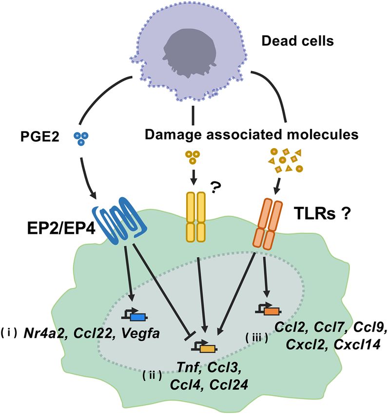
www.nature.com/scientificreports/
A B
HMGB1 mRNA HSP60 mRNA
Relative expression
Relative expression
3.0
1.2
2.0
0.8
1.0
0.4
0.0 0.0
Pu
Pu
HOS
HOS
KMeC
KMeC
OOS
OOS
Sora
Nene
Sora
Nene
LMeC
LMeC
CTBm
CTBm
CHMm
CHMm
Love
Love
HMPOS
HMPOS
C HSP70 mRNA D IL-33 mRNA
1.2
12
Relative expression
Relative expression
0.8
8
0.4
4
0.0
0
Pu
HOS
KMeC
OOS
Sora
Nene
LMeC
CTBm
Pu
CHMm
Love
HMPOS
KMeC
OOS
Sora
Nene
LMeC
CTBm
CHMm
Love
HOS
HMPOS
E S100A8 mRNA
Relative expression
1.2
0.8
0.4
0.0
OOS
Sora
Nene
LMeC
CTBm
CHMm
Love
Pu
HOS
HMPOS
KMeC
Figure 1. Gene expression of damage-associated molecules (HMGB1, HSP60, HSP70, IL33, S100A8) in 11
canine tumor cell lines. (A–E) Total RNA was extracted from cell lines in the logarithmic growth phase. HMGB1
(A), HSP60 (B), HSP70 (C), IL33 (D), and S100A8 (E) mRNA expression levels were examined by RT-qPCR
analysis. Data are presented as mean ± SD.
Scientific Reports | (2021) 11:8525 | https://doi.org/10.1038/s41598-021-87979-1 3
Vol.:(0123456789)www.nature.com/scientificreports/
Figure 2. Lack of immunostimulatory activities of canine tumor cell-derived necrotic supernatants. (A)
Schematic illustration of the preparation of necrotic tumor cell supernatants and RT-qPCR analysis for Tnf
mRNA expression in macrophage cell lines. In each tumor cell line, necrosis was induced by freeze–thaw cycles.
(B,C) Murine macrophages (RAW264.7) (B) and canine macrophages (DH82) (C) were exposed to the necrotic
supernatant (the volume of the supernatant is equivalent to 5 × 10⁶ cells) for 2 h. Tnf mRNA expression was
examined by RT-qPCR analysis. Data are presented as mean ± SD. Samples that showed more than two-fold
induction compared to PBS samples are indicated in red bars.
Scientific Reports | (2021) 11:8525 | https://doi.org/10.1038/s41598-021-87979-1 4
Vol:.(1234567890)www.nature.com/scientificreports/
Figure 3. Suppression of LPS-induced Tnf mRNA expression by dying cells-derived necrotic supernatant or PGE2 treatment. (A)
Production of PGE2 in necrotic supernatants derived from canine tumor cell lines. PGE2 in necrotic tumor cell supernatants was
measured by ELISA and normalized to cell number. (B) RAW264.7 cells (left panel) and DH82 cells (right panel) were left untreated
or stimulated with LPS (10 ng/ml) for 2 h in the presence of PGE2 (50 nM or 500 nM) or absence (PBS). Tnf mRNA expression levels
were determined by RT-qPCR analysis. (C,D) RAW264.7 cells (C) and DH82 cells (D) were stimulated with LPS (10 ng/ml) for 2 h
and co-cultured with an increasing volume of necrotic supernatants from Sora cells (left panels) and HMPOS (right panels). The
volume of the supernatant is equivalent to 5 × 10⁵ or 5 × 10⁶ cells. Expression of Tnf mRNA was then measured by RT-qPCR analysis.
(E,F) RAW264.7 cells were stimulated with LPS for 2 h in combination with or without the same volumes of necrotic supernatants
from LMeC cells (E) and HOS cells (F). The induction levels for Tnf mRNA were determined by RT-qPCR. Data are presented as
mean ± SD.*, p < 0.05 compared with the indicated samples.
Scientific Reports | (2021) 11:8525 | https://doi.org/10.1038/s41598-021-87979-1 5
Vol.:(0123456789)www.nature.com/scientificreports/
necrotic canine tumor cell supernatants impede the activation of innate immune responses in RAW264.7 and
DH82 cells. PGE2 treatment of RAW264.7 and DH82 cells suppressed Tnf mRNA expression induction by LPS
stimulation in the macrophages (Fig. 3B), suggesting that PGE2 from canine tumor dead cells functions as
an immunosuppressor. To examine the immunosuppressive activity of PGE2 in the necrotic canine tumor cell
supernatants, we selected Sora and HMPOS cells because they showed relatively high PGE2 production in the
supernatants (Fig. 3A). We stimulated the macrophages with LPS in combination with the necrotic canine tumor
cell supernatants, and then examined Tnf mRNA expression. As shown in Fig. 3C,D, the PGE2-containing
supernatants massively suppressed LPS-induced Tnf mRNA expression in both RAW264.7 and DH82 cells. The
inhibition of LPS-induced TNF-α production by PGE2 was also observed at the protein levels by ELISA (Sup-
plementary Fig. S2). In contrast, there was little, if any, suppressive activity in LMeC- or HOS-derived necrotic
canine tumor cell supernatant (Fig. 3E,F), which does not contain PGE2, suggesting that PGE2 in the necrotic
canine tumor cell supernatants plays an important role in Tnf mRNA suppression.
Next, we examined whether necrotic canine tumor cell self-derived damage-associated molecules activate
the innate immune responses in the absence of PGE2. We pretreated Sora and HMPOS cells with indometha-
cin, a potent inhibitor of the COX enzymes, and then prepared a necrotic supernatant of the cells. As expected,
PGE2 production in the supernatant was markedly reduced by treatment with indomethacin (Fig. 4A). Notably,
indomethacin-treated necrotic supernatants from canine tumor cell did not show any suppressive effect on LPS-
induced Tnf expression in RAW264.7 and DH82 cells (Fig. 4B,C). We then treated LPS-unstimulated RAW264.7
cells with PGE2-reduced supernatant, which interestingly upregulated Tnf mRNA expression (Fig. 4D). TNF-α
production in RAW264.7 cells was also observed (Supplementary Fig. S3). Collectively, these data indicate that
dying and dead canine tumor cells release PGE2 and inhibit innate immune activation by damage-associated
molecules.
We further examined how PGE2 released from necrotic cells suppresses LPS-induced Tnf mRNA expression
in macrophage cell lines. We first used specific inhibitors targeting four different PGE2 receptors (EP1–EP4)8,9.
The suppression of LPS-induced Tnf mRNA expression by necrotic cell supernatants was attenuated by EP2
and EP4 inhibitors, and the suppression was further significantly reduced by the combination of both inhibi-
tors (Supplementary Fig. S4). These results indicated that PGE2 released from dying cells exerts its suppressive
effect through the EP2 and EP4 receptors signaling. To investigate further the mechanism of immunosuppressive
effect by PGE2-EP2/EP4 axis, we examined the activation of NF-κB, a critical transcription factor for TNF-α,
by NF-κB luciferase reporter assay. Notably, the addition of necrotic cell supernatants did not inhibit the activa-
tion of NF-κB by LPS stimulation (Supplementary Fig. S5), suggesting that PGE2 in the supernatant suppresses
induction of Tnf mRNA in an NF-kB-independent manner (See also “Discussion”).
Gene expression profiles in macrophages treated with necrotic canine tumor cell superna‑
tants in the presence and absence of PGE2. The induction of Tnf mRNA expression and TNF-α pro-
duction in RAW264.7 cells by PGE2-reduced supernatants (Fig. 4D and Supplementary Fig. S3) prompted us
to investigate which genes are induced by the damage-associated molecules by comprehensive gene expression
analysis. We performed RNA-seq analysis with the total RNA extracted from the RAW264.7 cells treated with
the necrotic supernatant from canine tumor cells subjected to indomethacin treatment. As shown in Fig. 5A,B,
differential expression analysis identified 73 upregulated differentially expressed genes (DEGs) in the RAW264.7
cells treated with necrotic supernatant prepared from the Sora cells in the presence of indomethacin (Indo),
compared to untreated cells (PBS). We also found 133 DEGs in the RAW264.7 cells treated with the necrotic
supernatant derived from Sora cells in the absence of indomethacin (Mock). All DEGs and their fold changes
are listed in Supplementary Table S1.
To gain further insights into the upregulated DEGs, we performed Gene Ontology (GO) analysis to exam-
ine the functional changes between PBS vs. Mock and between PBS vs. Indo. The analysis identified 164 (PBS
vs. Mock) and 107 (PBS vs. Indo) GO terms with p-value < 0.01 and false discovery rate (FDR) < 0.01. The top
10 terms in each condition were dominated by terms involved in the activation of immune responses, such as
response to external stimulus, inflammatory response, and response to cytokines (Fig. 5C,D). We also performed
a Kyoto encyclopedia of genes and genomes (KEGG) pathway enrichment analysis and found 8 (PBS vs. Mock)
and 9 (PBS vs. Indo) signal pathways with p-values < 0.05. Notably, the nuclear factor-kB (NF-kB) signaling
was the most upregulated pathway under the stimulation by each necrotic supernatant from canine tumor cells
(Fig. 5C,D).
The Venn diagram shows that 76 DEGs (group i) were selectively upregulated by the necrotic supernatant
from Mock (Fig. 5B), indicating that PGE2 affects expression of several DEGs. These DEGs include Ccl22 and
Nr4a2 (Supplementary Table S1), which are reported to be induced by P GE218,19. Sixteen DEGs (group ii) were
specifically upregulated by PGE2-reduced necrotic supernatant (Fig. 5B). These DEGs included Tnf and pro-
inflammatory chemokines such as Ccl3, Ccl4, and Ccl24 (Supplementary Table S1). Fifty-seven DEGs (group
iii) including Ccl2, Ccl7, Ccl9, Cxcl2, and Cxcl14 chemokines were upregulated in both Indo and PBS (Fig. 5B),
indicating that the damage-associated molecules induce these DEGs in a PGE2-independent manner. These
DEGs Expression profiles of the genes were confirmed by measuring the relative mRNA levels through RT-qPCR
analysis (Fig. 6). Taken together, these data revealed hitherto unknown complex immune regulatory mechanisms
by immune-activating damage-associated molecules and PGE2 (Fig. 7).
Discussion
In the recent years, numerous studies have reported that innate immune activation by the self-derived damage-
associated molecules obtained from dead cells promotes inflammation, which exacerbates pathogenesis of can-
cer, autoimmune diseases, and chronic inflammation3–6. Although damage-associated molecules have gained
Scientific Reports | (2021) 11:8525 | https://doi.org/10.1038/s41598-021-87979-1 6
Vol:.(1234567890)www.nature.com/scientificreports/
Figure 4. Suppression of PGE2 production and enhancement of immunostimulatory activity of necrotic
canine tumor cell supernatants. (A) Production of PGE2 in necrotic supernatant from DMSO (Mock)- and
indomethacin (Indo)-treated tumor cells. PGE2 levels were determined by ELISA and normalized to cell
number. (B,C) RAW264.7 cells (B) and DH82 cells (C) were stimulated with LPS (10 ng/ml) for 2 h in
combination with necrotic supernatants (5 × 10⁶ cells) from Sora cells (left panels) and HMPOS cells (right
panels). The induction levels for Tnf mRNA were then determined by RT-qPCR analysis. (D) RAW264.7 cells
were treated for 2 h with necrotic supernatants from Mock-or Indo-treated Sora (left panel) and HMPOS (right
panel) cells (5 × 10⁶ cells). Tnf mRNA expression levels were examined by RT-qPCR analysis. Data are presented
as mean ± SD. *p < 0.05 compared with the indicated samples.
Scientific Reports | (2021) 11:8525 | https://doi.org/10.1038/s41598-021-87979-1 7
Vol.:(0123456789)www.nature.com/scientificreports/
Figure 5. Gene expression analysis of RAW264.7 cells treated with necrotic supernatants by comprehensive RNA-seq analysis.
RAW264.7 cells were treated for 4 h with necrotic supernatant from Sora cells pretreated with indomethacin (Indo) or DMSO (Mock).
Total RNA was extracted and subjected to RNA-seq analysis. Differentially expressed genes (DEGs) were identified using FDR < 0.05 as
the statistical criterion for filtering. (A) Heat map of the different gene expression patterns in each sample. (B) Venn diagrams showing
commonly and differentially regulated genes by each necrotic supernatant. Group (i) contains 76 genes induced only by necrotic
supernatant from Mock. Group (ii) represents 16 genes induced only by necrotic supernatant from Indo. Group (iii) includes 57 genes
commonly induced by necrotic supernatants from Mock and Indo. (C,D) Biological processes and signaling pathways affected by
necrotic supernatants from Mock (C) and Indo (D) Sora cells. GO analysis (upper panels) and KEGG pathway enrichment analysis
(lower panels) were employed.
Scientific Reports | (2021) 11:8525 | https://doi.org/10.1038/s41598-021-87979-1 8
Vol:.(1234567890)www.nature.com/scientificreports/
Figure 6. Validation of representative DEGs derived from RNA-seq analysis by RT-qPCR. (A–K) RAW264.7
cells were treated for 4 h with the necrotic supernatant from PBS or Indo. Gene expression of group (i) (Nr4a2
(A), Ccl22 (B), and Vegfa (C)), group (ii) (Ccl3 (D), Ccl4 (E), and Ccl24 (F)), and group (iii) (Ccl2 (G), Ccl7
(H), Ccl9 (I), Cxcl2 (J), and Cxcl14 (K)) was examined by RT-qPCR analysis. Data are presented as mean ± SD.
*p < 0.05 compared with the indicated samples.
Scientific Reports | (2021) 11:8525 | https://doi.org/10.1038/s41598-021-87979-1 9
Vol.:(0123456789)www.nature.com/scientificreports/
Figure 7. Schematic diagram of the interaction of PGE2 and damage-related molecules in the induction
of genes regulating the immune response in macrophages. PGE2 released from dead cells induces
immunosuppressive molecules such as Nr4a2, Ccl22, and Vegfa via EP1-4, PGE2 receptors (i). Damage-
associated molecules induce a variety of pro-inflammatory chemokines, probably via TLRs. Some of these genes
(Tnf, Ccl3, Ccl4 and Ccl24) are suppressed by PGE2 (ii) and others (Ccl2, Ccl7, Ccl9. Cxcl2 and Cxcl14) are not
(iii). Since there are no reports on the relationship between Ccl24 mRNA and TLR signaling, new damage-
related molecules may be involved in the induction of Ccl24 mRNA.
attention for their pro-inflammatory a ctivity3–6, it remains unclear whether and how the damage-associated
molecules regulate the inflammatory innate immune responses. To further characterize the activation of innate
immune responses by the damage-associated molecules, in this study, we examined innate immune activation
by several types of damage-associated molecules obtained from the canine tumor cell lines.
Consistent with our previous findings7, PGE2 was indeed produced in the necrotic supernatants from major-
ity of the canine tumor cell lines (9/11) (Fig. 3A). In particular, the necrotic supernatants from 3 cell lines of
urothelial carcinoma (Sora, Love, and Nene) contained large amounts of PGE2 (Fig. 3A). This observation may
reflect our previous result that the COX genes are highly expressed in these cell lines20,21.
Furthermore, despite the fact that LMeC-derived supernatants without PGE2 production did not suppress
LPS-induced Tnf mRNA expression, the HOS-derived supernatants also without PGE2 production, showed
relatively weak innate immune suppression at high concentrations (Fig. 3E,F). These results suggest the pres-
ence of previously unidentified new immunosuppressive damage-associated molecule(s) in the supernatant.
Identification and characterization of the immunosuppressive molecule(s) released from dying cells need further
clarification.
Based on our data and previous reports8,9, PGE2 released from dying cells exerted its immunosuppressive
effects mainly through EP2 and EP4 receptors (Supplementary Fig.S4). Although the mechanism of suppression
of inflammation induction by the PGE2-EP2/4 axis is not well understood, we observed PGE2 treatment did not
affect the transcriptional activation of NF-κB (Supplementary Fig. S5). In this context, we found that 76 DEGs
(group i) including Nr4a2, Ccl22 and Vegfa were induced specifically by PGE2-containing necrotic supernatant
(Figs. 5B, 6A–C, Supplementary Table S1). It is plausible that PGE2 in the supernatant induces the expression
of the g enes18,19,22,23. Since Nr4a2 is known to suppress induction of Tnf mRNA expression by LPS stimulation
in monocytes/macrophages in an NF-κB-independent m anner24, the suppression of Tnf mRNA induction may
be mediated by Nr4a2. In addition, CCL22 promotes the migration of regulatory T cells (Tregs) through the
CCR4 receptor25,26, and VEGF-A in addition to its angiogenic effects exerts immunosuppressive effects leading
to the accumulation of Tregs and myeloid-derived suppressor cells in the tumor m icroenvironment27. Therefore,
PGE2 released from the dying and dead cells may suppress inflammation and promote wound healing through
the induction of these immunosuppressive mediators.
RNA-seq analysis also revealed that there are two groups of DEGs induced by damage-associated molecules
(Fig. 5B). One group consisted of 16 DEGs (group ii), including Tnf, Ccl3, Ccl4, and Ccl24, whose expressions are
suppressed by PGE2 (Figs. 5B, 6D–F, Supplementary Table S1). Since PGE2 is known to suppress the expression
Scientific Reports | (2021) 11:8525 | https://doi.org/10.1038/s41598-021-87979-1 10
Vol:.(1234567890)www.nature.com/scientificreports/
of Ccl3 and Ccl4 induced by TLR2/TLR428, damage-associated molecules may activate the TLR signaling pathway.
In contrast, Ccl24 mRNA expression is mainly induced by the T helper cell type 2 (Th2) cytokines, such as IL-4
and IL-1329, but its association with TLR signaling has not been reported. Therefore, it is expected that a new
damage-associated molecule may be involved in the induction of Ccl24 mRNA. Another group contained 57
DEGs (group iii), including Ccl2, Ccl7, Ccl9, Cxcl2, and Cxcl14. The expression of these DEGs was unaffected by
indomethacin treatment (Figs. 5B, 6G–K). This indicates that the induction of these mRNA was not repressed
by PGE2. Furthermore, KEGG pathway analysis suggested that the NF-kB signaling pathway was activated by
damage-associated molecules (Fig. 5C,D). Activation of the NF-kB signaling pathway has been observed in a
variety of inflammatory diseases and may contribute to the exacerbation of disease p athology30. It remains to be
clarified which damage-associated molecules activate the NF-kB signaling pathway.
In the GO analysis, several terms related to cell death were included in the top of the list in addition to those
related to immune response (Fig. 5C,D). Originally, TLR signaling, extracellular HMGB1 and histones induce
apoptosis in macrophages31; however, in the present study, necrosis supernatants enhanced the expression of the
genes involved in anti-apoptosis, such as Bcl2a1d, Bcl2a1b, and Bcl2l1. This may be involved in the activation of
NF-kB signaling, reported to suppress apoptosis downstream of T LRs32, but it is unknown how damage-related
molecules promote cell survival in macrophages.
In summary, this study reveals a hitherto unknown mechanism for the regulation of innate immune responses
by the damage-associated molecules. The identification of the damage-associated molecules involved in the
regulation of innate immune responses may be critical for understanding their role in the immune regulatory
processes and clinical pathogenesis of inflammatory diseases.
Methods
Reagents and cell culture. LPS (O55:B4), SC51089 (EP1 inhibitor), TG4-155(EP2 inhibitor), L-798106
(EP3 inhibitor) and ONO-AE3-208 (EP4 inhibitor) was purchased from Sigma-Aldrich. PGE2 and indometha-
cin were purchased from Cayman Chemical. These reagents were reconstituted in dimethyl sulfoxide (DMSO)
or phosphate buffered saline (PBS) and stored at − 20 °C or − 80 °C. Two canine mammary gland tumor cell
lines (CHMm and CTBm), 3 canine urothelial cell carcinoma cell lines (Sora, Love, Nene), 3 canine malignant
melanoma cell lines (KMeC, Pu, LMeC), and 3 canine osteosarcoma cell lines (HOS, OOS, HMPOS) used in this
study were established in our laboratory33–39. We confirmed no mycoplasma contamination in any of these cell
lines using the TaKaRa PCR Mycoplasma Detection Set (TaKaRa Bio). Each canine tumor cell line was main-
tained in Roswell Park Memorial Institute (RPMI) 1640 Medium (RPMI-1640) supplemented with 10% heat-
inactivated fetal bovine serum (FBS) and 5 mg/L gentamicin (Sigma-Aldrich). RAW264.7 cells were maintained
as described previously40. DH82 cells were kindly provided by Dr. Uchida (Laboratory of Veterinary Pathology,
The University of Tokyo) and was cultured in Dulbecco’s Modified Eagle’s medium (DMEM) supplemented with
10% heat-inactivated FBS and 5 mg/L gentamicin. All cell lines were cultured at 37 °C in a humidified atmos-
phere with 5% carbon dioxide (CO2).
Clinical canine samples. Surgically resected canine urothelial carcinoma tissue samples and adjacent nor-
mal tissues from the same case (n = 4) were obtained from the archival collection of the University of Tokyo Vet-
erinary Medical Center (samples collected in 2017–2018). The client of each hospital provided informed consent
for the use of these samples for this study.
Preparation of necrotic tumor cell supernatants. Induction of necrosis was performed by a freeze–
thaw method as described previously41,42. For the preparation of a PGE2-depleted necrotic supernatant, cells
were treated with indomethacin (10 μM) for 24 h and subjected to freeze–thaw cycles in the presence of indo-
methacin (10 μM).
Enzyme‑linked immunosorbent assay (ELISA). PGE2 concentration in the necrotic cell supernatant
was determined using PGE2 ELISA Kit (Cayman Chemical). TNF-α production levels were examined using
mouse TNF-α DuoSet ELISA (R&D Systems) according to the manufacturer’s instructions.
Reporter assay. RAW 264.7 cells (5 × 10⁶) were seeded on 10 cm Petri dishes and transiently co-transfected
with X-tremeGENE 9 DNA transfection reagent (Roche Life Science) and 5 μg firefly luciferase reporter plasmid
for NF-κB43 (Stratagene) on the following day. After 24 h, cells were re-seeded on 24-well plates and stimulated
with LPS (10 ng/mL) in the presence or absence of necrotic cell supernatants. After 2 h of incubation, cells were
harvested with lysis buffer and luciferase activity was measured using PicaGene Luminescence Kit (Toyo BeNet
Co., Ltd.).
RT‑qPCR analysis. Total RNA from tissues or cells was extracted using RNAiso (TaKaRa Bio) or NucleoSpin
RNA II (Macherey–Nagel) and reverse-transcribed with PrimeScript RT Master Mix (TaKaRa Bio). RT-qPCR
was performed on a LightCycler 480 using the SYBR Green PCR Master Mix (Roche Life Science), and values
were normalized to the expression of Gapdh mRNA. Primer sequences are listed in Supplementary Table S2.
RNA‑sequencing (RNA‑seq) analysis. Total RNA was extracted from RAW264.7 cells treated with PBS
or treated with necrotic supernatants obtained from tumor cell lines with (Indo) or without (Mock) indometha-
cin treatment using NucleoSpin RNA II (Macherey–Nagel). RNA-seq (150 bp paired-end) analysis was per-
formed by GENEWIZ using DNBSEQ-G400 sequencer, generating a minimum of 4.5 million read pairs for each
Scientific Reports | (2021) 11:8525 | https://doi.org/10.1038/s41598-021-87979-1 11
Vol.:(0123456789)www.nature.com/scientificreports/
sample. In order to remove technical sequences, including adapters, polymerase chain reaction (PCR) primers,
or fragments thereof, and quality of bases lower than 20, pass filter data of fastq format were processed by Cuta-
dapt (V1.9.1). Data were aligned to reference genome via software Hisat2 (v2.0.1). For gene expression analysis,
FPKM (Fragments per kilo bases per million reads) was calculated based on the number of reads in HTSeq
(v0.6.1). Differential gene expression analysis was performed using EdgeR44,45 using an FDR < 0.05 as the cutoff
criterion. GO and KEGG pathway enrichment analysis were performed using the Database for Annotation,
Visualization, and Integrated Discovery (DAVID, https://david.ncifcrf.gov/, version 6.8)46,47. For GO analysis,
p-value < 0.01 and FDR < 0.01 were used as cutoff criteria. A p-value < 0.05 was used for the KEGG functional
analysis.
Data availability. The data obtained during the current study will be available from the corresponding
author upon reasonable request. RNA-seq data were deposited in the E-GEAD-394.
Statistical analysis. Statistical analysis was performed using R software (https://www.R-project.org/). Data
are expressed as mean ± SD. The p-values were determined by Tukey–Kramer analysis and paired T test, and the
difference was considered to be statistically significant at p < 0.05.
Received: 30 September 2020; Accepted: 6 April 2021
References
1. Fuchs, Y. & Steller, H. Programmed cell death in animal development and disease. Cell 147, 742–758 (2011).
2. Nagata, S., Hanayama, R. & Kawane, K. Autoimmunity and the clearance of dead cells. Cell 140, 619–630 (2010).
3. Chen, G. Y. & Nuñez, G. Sterile inflammation: Sensing and reacting to damage. Nat. Rev. Immunol. 10, 826–837 (2010).
4. Gong, T., Liu, L., Jiang, W. & Zhou, R. DAMP-sensing receptors in sterile inflammation and inflammatory diseases. Nat. Rev.
Immunol. 20, 95–112 (2020).
5. Bianchi, M. E. DAMPs, PAMPs and alarmins: All we need to know about danger. J. Leukoc. Biol. 81, 1–5 (2007).
6. Krysko, D. V. et al. Immunogenic cell death and DAMPs in cancer therapy. Nat. Rev. Cancer 12, 860–875 (2012).
7. Hangai, S. et al. PGE2 induced in and released by dying cells functions as an inhibitory DAMP. Proc. Natl. Acad. Sci. U. S. A. 113,
3844–3849 (2016).
8. Sugimoto, Y. & Narumiya, S. Prostaglandin E receptors. J. Biol. Chem. 282, 11613–11617 (2007).
9. Rodríguez, M. et al. Polarization of the innate immune response by prostaglandin E2: A puzzle of receptors and signals. Mol.
Pharmacol. 85, 187–197 (2014).
10. Paoloni, M. & Khanna, C. Translation of new cancer treatments from pet dogs to humans. Nat. Rev. Cancer 8, 147–156 (2008).
11. Starkey, M. P., Scase, T. J., Mellersh, C. S. & Murphy, S. Dogs really are man’s best friend—Canine genomics has applications in
veterinary and human medicine!. Brief. Funct. Genom. 4, 112–128 (2005).
12. Momozawa, Y. The potential of translational research in dogs in human medicine. Translat. Regulat. Sci. 1, 31–36 (2019).
13. Abdelmegeed, S. M. & Mohammed, S. Canine mammary tumors as a model for human disease. Oncol. Lett. 15, 8195–8205 (2018).
14. Knapp, D. W. et al. Naturally-occurring canine transitional cell carcinoma of the urinary bladder A relevant model of human
invasive bladder cancer. Urol. Oncol. 5, 47–59 (2000).
15. Hernandez, B. et al. Naturally occurring canine melanoma as a predictive comparative oncology model for human mucosal and
other triple wild-type melanomas. Int. J. Mol. Sci. 19, 394 (2018).
16. Mueller, F., Fuchs, B. & Kaser-Hotz, B. Comparative biology of human and canine osteosarcoma. Anticancer Res. 27, 155–164
(2007).
17. Wellman, M. L., Krakowka, S., Jacobs, R. M. & Kociba, G. J. A macrophage-monocyte cell line from a dog with malignant histio-
cytosis. In Vitro Cell. Dev. Biol. 24, 223–229 (1988).
18. Hibino, S. et al. Inhibition of Nr4a receptors enhances antitumor immunity by breaking Treg-mediated immune tolerance. Cancer
Res. 78, 3027–3040 (2018).
19. McIlroy, A. et al. Histamine and prostaglandin E up-regulate the production of Th2-attracting chemokines (CCL17 and CCL22)
and down-regulate IFN-gamma-induced CXCL10 production by immature human dendritic cells. Immunology 117, 507–516
(2006).
20. Yoshitake, R. et al. Aberrant expression of the COX2/PGE2 axis is induced by activation of the RAF/MEK/ERK pathway in
BRAFV595E canine urothelial carcinoma. Sci. Rep. 10, 7826 (2020).
21. Yoshitake, R. et al. Molecular investigation of the direct anti-tumour effects of nonsteroidal anti-inflammatory drugs in a panel of
canine cancer cell lines. Vet. J. 221, 38–47 (2017).
22. Wang, D. & Dubois, R. N. Eicosanoids and cancer. Nat. Rev. Cancer 10, 181–193 (2010).
23. Wu, W.-K., Llewellyn, O. P. C., Bates, D. O., Nicholson, L. B. & Dick, A. D. IL-10 regulation of macrophage VEGF production is
dependent on macrophage polarisation and hypoxia. Immunobiology 215, 796–803 (2010).
24. Saijo, K. et al. A Nurr1/CoREST pathway in microglia and astrocytes protects dopaminergic neurons from inflammation-induced
death. Cell 137, 47–59 (2009).
25. Shevach, E. M. Fatal attraction: Tumors beckon regulatory T cells. Nat. Med. 10, 900–901 (2004).
26. Rapp, M. et al. CCL22 controls immunity by promoting regulatory T cell communication with dendritic cells in lymph nodes. J.
Exp. Med. 216, 1170–1181 (2019).
27. Yang, J., Yan, J. & Liu, B. Targeting VEGF/VEGFR to modulate antitumor immunity. Front. Immunol. 9, 978 (2018).
28. Parker, L. C., Whyte, M. K. B., Vogel, S. N., Dower, S. K. & Sabroe, I. Toll-like receptor (TLR)2 and TLR4 agonists regulate CCR
expression in human monocytic cells. J. Immunol. 172, 4977–4986 (2004).
29. Makita, N., Hizukuri, Y., Yamashiro, K., Murakawa, M. & Hayashi, Y. IL-10 enhances the phenotype of M2 macrophages induced
by IL-4 and confers the ability to increase eosinophil migration. Int. Immunol. 27, 131–141 (2015).
30. Taniguchi, K. & Karin, M. NF-κB, inflammation, immunity and cancer: Coming of age. Nat. Rev. Immunol. 18, 309–324 (2018).
31. Chen, R. et al. Nuclear DAMP complex-mediated RAGE-dependent macrophage cell death. Biochem. Biophys. Res. Commun. 458,
650–655 (2015).
32. Aliprantis, A. O., Yang, R. B., Weiss, D. S., Godowski, P. & Zychlinsky, A. The apoptotic signaling pathway activated by Toll-like
receptor-2. EMBO J. 19, 3325–3336 (2000).
Scientific Reports | (2021) 11:8525 | https://doi.org/10.1038/s41598-021-87979-1 12
Vol:.(1234567890)www.nature.com/scientificreports/
33. Uyama, R. et al. Establishment of four pairs of canine mammary tumour cell lines derived from primary and metastatic origin
and their E-cadherin expression. Vet. Comp. Oncol. 4, 104–113 (2006).
34. Eto, S. et al. Anti-tumor effects of the histone deacetylase inhibitor vorinostat on canine urothelial carcinoma cells. PLoS ONE 14,
e0218382 (2019).
35. Inoue, K. et al. Establishment and characterization of four canine melanoma cell lines. J. Vet. Med. Sci. 66, 1437–1440 (2004).
36. Ohashi, E. et al. Effect of retinoids on growth inhibition of two canine melanoma cell lines. J. Vet. Med. Sci. 63, 83–86 (2001).
37. Meyer, F. R. L. & Walter, I. Establishment and characterization of new canine and feline osteosarcoma primary cell lines. Vet. Sci.
China 3, 9 (2016).
38. Kadosawa, T., Nozaki, K., Sasaki, N. & Takeuchi, A. Establishment and characterization of a new cell line from a canine osteosar-
coma. J. Vet. Med. Sci. 56, 1167–1169 (1994).
39. Barroga, E. F., Kadosawa, T., Okumura, M. & Fujinaga, T. Establishment and characterization of the growth and pulmonary
metastasis of a highly lung metastasizing cell line from canine osteosarcoma in nude mice. J. Vet. Med. Sci. 61, 361–367 (1999).
40. Takaoka, A. et al. Integral role of IRF-5 in the gene induction programme activated by Toll-like receptors. Nature 434, 243–249
(2005).
41. Eigenbrod, T., Park, J.-H., Harder, J., Iwakura, Y. & Núñez, G. Cutting edge: Critical role for mesothelial cells in necrosis-induced
inflammation through the recognition of IL-1 alpha released from dying cells. J. Immunol. 181, 8194–8198 (2008).
42. Scaffidi, P., Misteli, T. & Bianchi, M. E. Release of chromatin protein HMGB1 by necrotic cells triggers inflammation. Nature 418,
191–195 (2002).
43. Honda, K. et al. Role of a transductional-transcriptional processor complex involving MyD88 and IRF-7 in Toll-like receptor
signaling. Proc. Natl. Acad. Sci. U. S. A. 101, 15416–15421 (2004).
44. Zhou, X., Lindsay, H. & Robinson, M. D. Robustly detecting differential expression in RNA-sequencing data using observation
weights. Nucleic Acids Res. 42, e91 (2014).
45. Robinson, M. D., McCarthy, D. J. & Smyth, G. K. edgeR: A bioconductor package for differential expression analysis of digital gene
expression data. Bioinformatics 26, 139–140 (2010).
46. Huang, D. W., Sherman, B. T. & Lempicki, R. A. Bioinformatics enrichment tools: Paths toward the comprehensive functional
analysis of large gene lists. Nucleic Acids Res. 37, 1–13 (2009).
47. Huang, D. W., Sherman, B. T. & Lempicki, R. A. Systematic and integrative analysis of large gene lists using DAVID bioinformatics
resources. Nat. Protoc. 4, 44–57 (2009).
Acknowledgements
We are grateful to Kazuyuki Uchida (The University of Tokyo) for providing the cell lines (DH82). This work
was supported in part by Grant-In-Aid for Scientific Research18K07167, 19K22362, 20H03141, 20H00504, and
20H03144 from the Ministry of Education, Culture, Sports, Science, and Technology (MEXT), AMED-CREST/
PRIME (20gm6110008h) from the Japan Agency for Medical Research and Development, and the Naito Founda-
tion. S.E. is a research fellow of the Japan Society for the Promotion of Science.
Author contributions
H.Y. and S.H. designed the study. S.E. performed the experiments and contributed to the data analysis. S.E.
and H.Y. prepared the figures and wrote the manuscript. H.Y., S.H., D.K., R.N., and T.N. supervised this study.
Competing interests
The authors declare no competing interests.
Additional information
Supplementary Information The online version contains supplementary material available at https://doi.org/
10.1038/s41598-021-87979-1.
Correspondence and requests for materials should be addressed to T.N.
Reprints and permissions information is available at www.nature.com/reprints.
Publisher’s note Springer Nature remains neutral with regard to jurisdictional claims in published maps and
institutional affiliations.
Open Access This article is licensed under a Creative Commons Attribution 4.0 International
License, which permits use, sharing, adaptation, distribution and reproduction in any medium or
format, as long as you give appropriate credit to the original author(s) and the source, provide a link to the
Creative Commons licence, and indicate if changes were made. The images or other third party material in this
article are included in the article’s Creative Commons licence, unless indicated otherwise in a credit line to the
material. If material is not included in the article’s Creative Commons licence and your intended use is not
permitted by statutory regulation or exceeds the permitted use, you will need to obtain permission directly from
the copyright holder. To view a copy of this licence, visit http://creativecommons.org/licenses/by/4.0/.
© The Author(s) 2021
Scientific Reports | (2021) 11:8525 | https://doi.org/10.1038/s41598-021-87979-1 13
Vol.:(0123456789)You can also read