Air-Liquid Interface Culture System for Standardized Respiratory Research
←
→
Page content transcription
If your browser does not render page correctly, please read the page content below
Air-Liquid Interface Culture
System for Standardized
Respiratory Research
Application Note

                            Generation of a stable and functional 3D bronchial epithelial cells using the PromoCell
                            human airway model with primary human Air-Liquid Interface Culture System.

             Day 0:                                           Thaw and plate ALI
                                                              pre-screened HBEpC

                                                                                                                       2D phase:
                                                                                                                       Expansion in Airway
                                                                                                                       Epithelial Cell Growth
                                                                                                                       Medium
                                                                    Medium change
                                                                                                                       See part A of protocol
             Cultivate for 5 – 7 days                     Detach cells

                                                                         2
                                                                     Seed single cell
                                                                     suspension on inserts
                               1
                    Coat porous                                                                                        3D phase (submerged):
                  membrane with                                                                                        Proliferation in Airway
                       collagen
                                                                                                                       Epithelial Cell Growth
                                                                               Submerged culture                       Medium on collagen coated
                                                                               Collagen                                Transwell®
                                                                               Porous membrane
                                                                                                                       See part B of protocol
                                                                  Medium change
             Cultivate for 3 – 4 days                                                                                See part B of protocol

             Airlift:

                                                                               Air exposure

                                                                               100% confluent cell layer

                                                                                                                       3D phase (airlift):
                                                                    Medium change/wash insert                          Differentiation in
                                                                                                                       ALI-Airway Medium
                                                                                                                       on Transwell® during
             Cultivate for 28 days                                                                                     air exposure
             after airlift
                                                                               Air exposure                            See part C of protocol

                                                                               Polarized epithelium
                                                                               with barrier function

Fig. 1: Schematic overview of culture phases for differentiation of HBEpC in the PromoCell ALI culture media system. The workflow can be divided into various
phases: A, Expansion of human primary cells in 2D culture on plastic. B, Re-seeding and expansion of the cells in 3D culture on collagen type I coated porous
membranes in submerged culture. C, Induction of the differentiation phase by airlift in ALI-Airway Medium. The formation of a tight epithelial barrier occurs in
the differentiation phase lasting at least 28 days post-airlift. For the detailed steps, please see the protocol below post-airlift. For the detailed steps, see the
following protocol.

Application Note | Air-Liquid Interface Culture System                                                                                                            1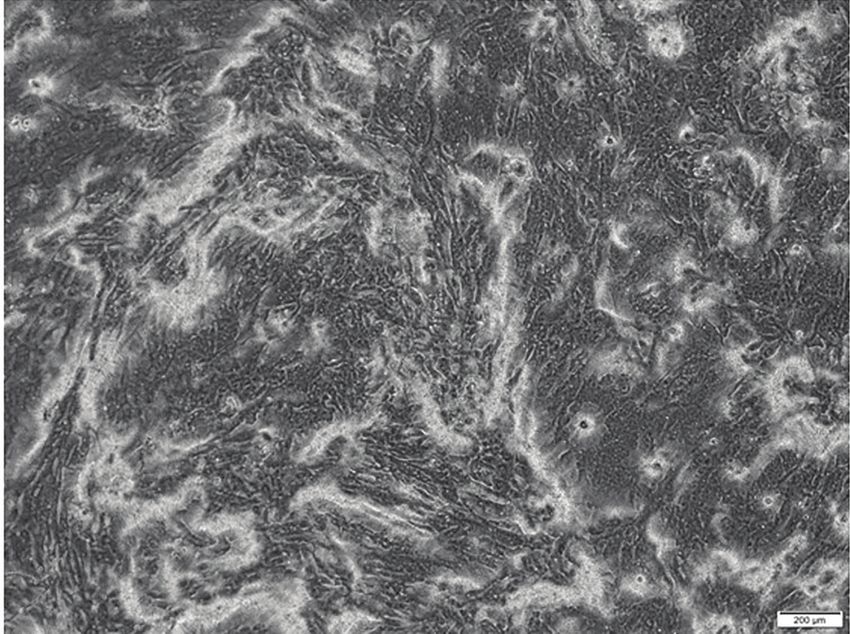
Background
The transcriptional profile of an in vivo air-       Well-differentiated in vitro airway epithelial   membrane. The barrier function can be
way epithelium very closely resembles the         cultures are characterized by the formation of      quantified by measuring TEER (> 500 Ω*cm2
differentiated primary cells grown at the air-    a pseudostratified epithelium and a barrier         for 4 weeks in culture).
liquid interface (ALI), suggesting that the use   function between adjacent environments.                 The PromoCell Air-Liquid Interface Culture
of primary cultures and the presence of an air-   Although airway epithelial cells do not dif-        System workflow is divided into a 2D expan-
liquid interface are important to recapitulate    ferentiate in 2D culture on plastic, they can       sion phase and a 3D differentiation phase (see
airway epithelial biology. In addition, the       undergo mucociliary differentiation when            Figure 1). After 2D expansion of ALI pre-scree-
close similarity between cells of tracheal and    grown on porous membranes at an air-liquid          ned HBEpCs in Airway Epithelial Cell Growth
bronchial origin within and between different     interface (ALI). The air-liquid interface permits   Medium (C-21060), the cells have to be seeded
human donors suggests a robust expression         polarization of epithelial cells by supporting      into Transwell® inserts as submerged cultures
profile that is specific to airway cells [1].     their differentiation [3].                          and allowed to grow until they reach conflu-
    In vitro ALI models have therefore been          An epithelial barrier is generated by            ence. Quality control evaluated ALI pre-scree-
recommended for studying the physiological        a high-integrity apico-lateral junctional           ned HBEpC lots are tested for proper barrier
and pathophysiological responses of the           complex composed of tight and adherens              function.
respiratory tract, molecular events, and          junctions. The integrity of these tight junction        The ALI-Airway medium lacks attach-
modes of action and interaction of different      dynamics in cell culture models of epithelial       ment factors, and collagen type I is therefo-
cell types [2]. Well-differentiated in vitro      monolayers can be quantified by measuring           re a prerequisite for optimal cell attachment.
airway epithelial cultures are characterized by   the transepithelial electrical resistance           Differentiation of ALI pre-screened HBEpC
the generation of a stable and functional 3D      (TEER). TEER values are widely accepted             is stimulated by changing to the Air-Liquid
human airway cell model with primary human        as strong indicators of the integrity of the        Interface Medium (ALI-Airway, C-21080) and
bronchial epithelial cells using the PromoCell    cellular barriers. TEER measurements can be         exposure to air. The ALI medium consists of
Air-Liquid Interface Culture System.              performed in real time without cell damage          a Basal Medium and a BPE- and serum-free
    The transcriptional profile of an in vivo     and are generally based on measuring ohmic          SupplementMix that enhances the barrier-for-
airway epithelia very closely resembles the       resistance or impedance [4], [5], [6], [7], [8].    ming function of HBEpCs (see Figure 1). Our
differentiated primary cultures grown at the         ALI cultures can also be used for multiple       ALI pre-screened HBEpC are available on re-
air-liquid interface (ALI), suggesting that the   readouts, e.g. viability assays, analysis of        quest by contacting our Scientific Support.
use of primary cultures and the presence          virus infection and respiratory disease                 The following application note describes
of an air-liquid interface are important to       mechanisms, cytokine release, and expression        the ALI cell culture procedure in detail and re-
recapitulate airway epithelia biology. In         of messenger RNA.                                   commended TEER measurement technology.
addition, the close similarity between cells         We have developed a standardized ALI
of tracheal and bronchial origin within and       culture system that ensures epithelial barriers
between different human donors suggests           of highly viable primary human bronchial
a robust expression profile that is specific to   epithelial cells (HBEpC) over a period of
airway cells [1].                                 28 days when cultured in 3D on a porous

           Air-Liquid Interface Culture System Protocol Part A
                                    2D expansion of ALI pre-screened Human Bronchial Epithelial Cells

                        The protocol in section A describes the pro-        required amount of ALI pre-screened HBEpC
                        cedure for thawing and expanding of the             in 2D culture on plastic.

I. Materials                                      ■

                                                  ■
                                                         ALI pre-screened Human Bronchial Epithelial Cells (HBEpC) C-12640
                                                         Airway Epithelial Cell Growth Medium, containing the Basal Medium and either
                                                         SupplementMix (Ready-to-use; C-21060) or SupplementPack (Kit; C-21160).
                                                  ■      Cell culture vessel (e.g. Falcon®; Corning® Inc.)
                                                  ■      6.5 mm of Transwell® inserts, 0.4 µm pore size, tissue culture treated polyester mem-
                                                         brane polystyrene plates (we strongly recommend Costar® from Corning® Inc., product
                                                         number 3470-Clear)
                                                  ■      Water bath at 37°C
                                                  ■      Timer
                                                  ■      Cell counting equipment

                                                      Use aseptic techniques and a laminar flow bench.

Application Note | Air-Liquid Interface Culture System                                                                                              2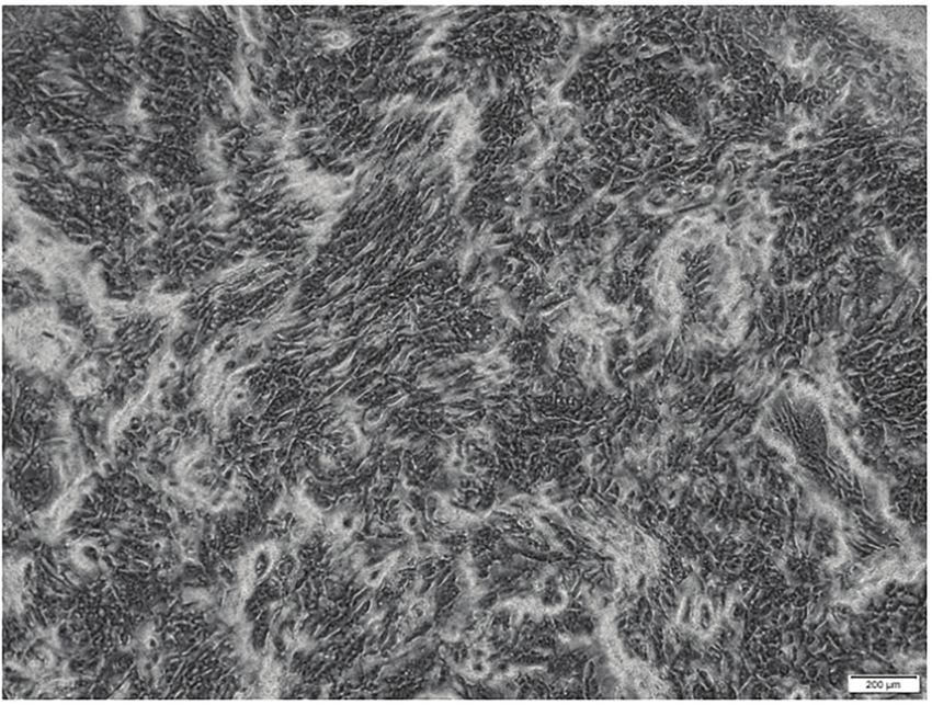
II. Protocol To prepare the medium, thaw the Supple-
mentMix or SupplementPack at 15 - 25°C.
homogenous mixture is formed. After additi-
on of the supplement(s) to the Basal Medium,
Aseptically mix the supplement by carefully its shelf life (complete growth medium) is 6
pipetting it up and down. Transfer all compo- weeks. Store the complete growth medium
nents to the 500 ml bottle of Basal Medium. at 2 - 8°C.
Close the bottle and swirl gently until a
1 2
Thaw the ALI pre-screened HBEpC Incubate the cells
Remove the cryovial from liquid nitrogen and transport the vial on dry Gently swirl the vessel containing the cell suspension and place it in an
ice. Under a laminar flow bench, release the pressure of the vial by incubator (37°C, 5% CO2). After 16 - 24 hours, check the cell adherence
briefly twisting the cap counterclockwise by a quarter turn and then under a microscop and replace the growth medium. There should be
retightening it. Allow the cell suspension to thaw in a water bath at only a few floating cells.
37°C for 2 minutes. Rinse the vial with 70% EtOH and place it under a
laminar flow bench. Aspirate the ethanol from the threads of the screw
cap. Carefully open the cryovial. Transfer the cell suspension to the cell
culture vessels containing the prewarmed medium from step 1.
Note: Our cryopreserved cells are frozen in Cryo-SFM C-29910, which
contains DMSO. Work quickly to prevent a longer incubation of the
cell suspension in Cryo-SFM, because the cells are very sensitive after
thawing.
3 4
Cultivate cells while regularly changing the medium Subcultivate the expanded cells
Change the medium every two to three days (e.g. Mon-Wed-Fri). Use Once the cells have reached 70 - 80% confluence, subculivate the
prewarmed complete Airway Epithelial Cell Growth Medium (180 µl cells as described in section B. The required confluence can typi-
medium/ cm2). Regularly check the confluence of the cells. Once they cally reached 5 - 7 days after thawing. The morphology of the cells
have reached 70 - 80% confluence, passage the cells. should be the typical cobblestone pattern of epithelial cells (see
Figure 2).
Note: Avoid confluence >90% for HBEpC. The cells can become con-
tact-inhibited, resulting in slower proliferation after passaging.
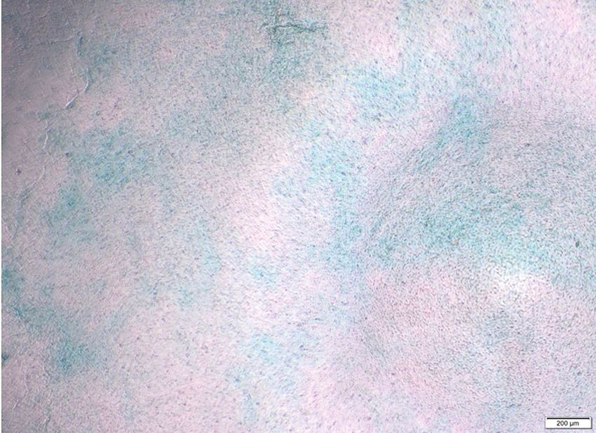
Fig. 2: Typical morphology of HBEpC in 2D culture. The cells were expanded
in PromoCell Airway Epithelial Cell Growth Medium until they reached 70 - 80%
confluence. The image was taken 7 days after seeding at 10x magnification
(scale bar 100 µm).
Application Note | Air-Liquid Interface Culture System 3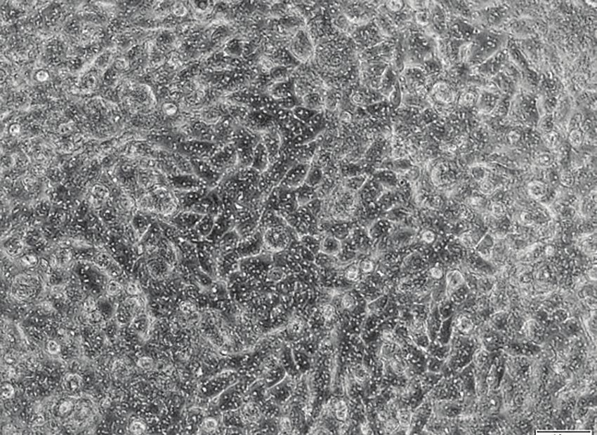
Air-Liquid Interface Culture System Protocol Part B
                               Subcultivation and re-plating of HBEpC on precoated Transwell® plates

                             This section describes detachment from 2D          on collagen type I precoated Transwell® in-
                             culture and seeding of HBEpC at high density       serts as a submerged 3D culture.

I. Materials                                  ■

                                              ■
                                                       Phosphate Buffered Saline w/o Ca++/ Mg++ (PBS, C-40232)
                                                       0.04 % Trypsin / 0.03% EDTA (Trypsin/ EDTA, C-41010)
                                              ■        0.05 % Trypsin Inhibitor, 0.1% BSA (TNS, C-41110)
                                              ■        Collagen Type I Solution (Rat Tail) (We strongly recommend Corning® Inc., product num-
                                                       ber 354236
                                              ■        6.5 mm of Transwell® inserts, 0.4 µm pore size, tissue culture treated polyester mem-
                                                       brane polystyrene plates (we strongly recommend Costar® from Corning® Inc., product
                                                       number 3470-Clear)

                                                   Use aseptic techniques and a laminar flow bench.

II. Protocol                                  On the day of use, coat the Transwells®
                                              with collagen type I and seed the HBEpC
                                                                                                   in early passages (P3) which results in high
                                                                                                   TEER-values. Passages >3 may result in a
                                              on the coated inserts. We strongly recom-            decrease of barrier formation indicated by
                                              mend the use of ALI pre-screened HBEpC               lower TEER-values

                                                  1

                                              Coating of Transwell® collagen type I solution and cell seeding

                                              Dilute collagen type I stock solution to a wor-      in an incubator (37°C, 5% CO2). Carefully
                                              king concentration of 0.03 mg/ ml. For 6.5 mm        aspirate collagen solution from the inserts*.
                                              Transwell® inserts use 100 µl of collagen type       Immediately wash the inserts with 150 µl of
                                              I solution per insert (area of insert = 0.33 cm2).   PBS. If you want to take a break, keep the PBS
                                              For each Transwell® a minimum collagen type I        on the inserts and store the plate at 37°C.
                                              working solution of 1.2 ml is needed.
                                                                                                   *Aspiration of inserts can be performed much more ea-
                                                                                                   sily if you stick a 1,000 μl pipette tip on top of the aspira-
                                              Example:
                                                                                                   tion pipette. This gives you better control and makes it
                                                                                                   easier to handle the tiny inserts. Be careful not to dama-
                                                                                                   ge the membrane with the pipette tip.
                                                      2 ml x 0.03 mg/ml
                                                                            = 0.015 ml of stock
                                                        3.9 mg/ml                                  Note: We cannot guarantee the barrier-for-
                                                  collagen stock solution                          ming function of HBEpC if another Transwell®
                                                                                                   or another coating is used. Collagen type I
                                                                                                   stock solution should be stored at 2 - 8°C.
                                              Add 0.015 ml of collagen type I stock solution       Let the stock solution acclimate to room tem-
                                              to 1.985 ml of PBS in a 15 ml conical tube and       perature (20 - 25°C) before diluting the wor-
                                              mix. Coat each Transwell® insert (the upper          king concentration. Cold collagen solution
                                              chamber only) with 100 µl of the collagen            is much more viscous and therefore more
                                              working concentration. For optimal colla-            difficult to pipette. Depending on the design
                                              gen distribution, gently rock the plate from         of your experiment, remember to include
                                              side to side and front to back. Do not swirl         one collagen-coated Transwell® insert as a
                                              the plate. Incubate the plate for 45 minutes         “blank” without cells.

Application Note | Air-Liquid Interface Culture System                                                                                                         4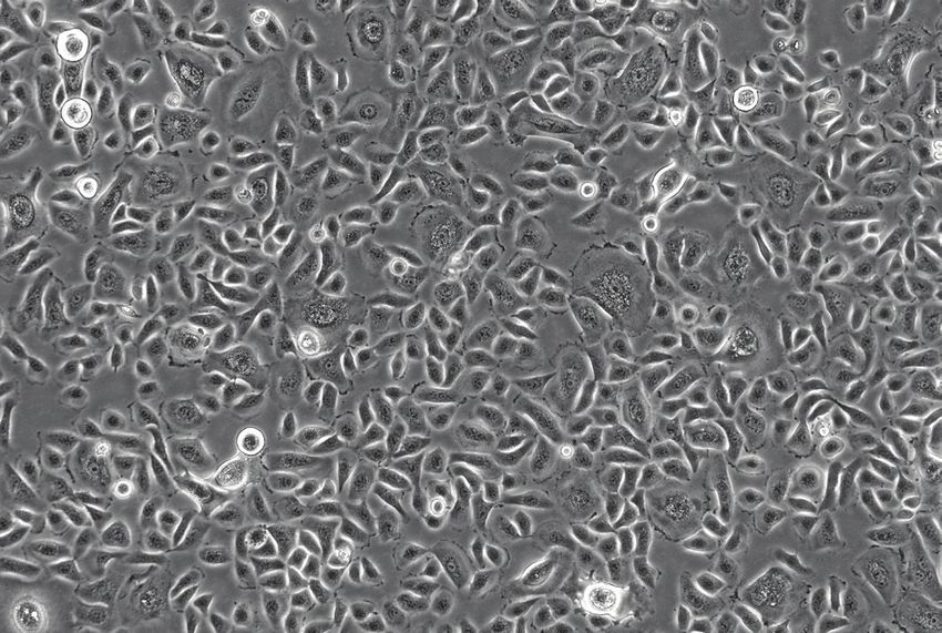
2                                                                           3

Wash the cells                                                             Detach the cells

Approximately 5 - 7 days after thawing, the HBEpC should reach 70 - Aspirate the PBS w/o Ca++/Mg++ from the vessel and add prewarmed
90% confluence. Aspirate the medium and wash the cells by adding Trypsin/ EDTA (100 µl/cm2) to the cells. Gently swirl the vessel to en-
an equal volume of PBS w/o Ca++/Mg++.                               sure that the cells are completely covered with Trypsin/ EDTA. Place
                                                                    the vessel in an incubator (37°C, 5% CO2) for 4 minutes. Check detach-
Note: Allow the PBS w/o Ca++/Mg++ to reach room temperature before ment under a microscope. The cells should be rounded. To encourage
adding to the cells.                                                detachment, you can gently tap the vessel horizontally against the tab-
                                                                    letop. Return the vessel to the laminar flow bench and add an equal
                                                                    amount of TNS to the cells. Gently swirl the vessel.

                                                                           Note: Epithelial cells stick tightly to plastic because of the large num-
                                                                           ber of adherens junctions. If the cells do not round after 4 minutes of
                                                                           incubation at 37°C, you can place the vessel in the incubator for 1 ad-
                                                                           ditional minute. Do not over-trypsinize them. If they are still sticking
                                                                           after 1 minute of incubation, use a 1,000 µl pipette to wash them down.

  4                                                                          5

Collect the cells and determine the cell number                            Plate the cells on collagen type I coated
and viability                                                              Transwell® plate (24-well plate)

Transfer the cell suspension to a 15 ml conical tube. To collect all re-   Make sure that the inserts of the Transwell® plate have been collagen-
maining cells, add complete Airway Epithelial Cell Growth Medium to        coated for at least 45 minutes at 37°C in an incubator, according to step
the vessel and transfer into the same 15 ml conical tube. Examine the      1. Aspirate the collagen solution from the inserts and wash each insert
vessel under a microscope to check if all cells have been collected.       with 150 µl of prewarmed PBS w/o Ca++/ Mg++. To avoid evaporation,
Use an appropriate volume of detached cell suspension for determin-        you can fill the outer wells of the plate with 200 µl PBS (optional). After
ing the cell number. Use your standard methods for cell counting and       removing the PBS w/o CA++/ Mg++ to seed the cells, work quickly
viability assessment. Spin down the cells (3 minutes at 300 x g) and as-   to avoid dryness of the semipermeable membrane. After cell coun-
pirate the supernatant. Transfer Airway Epithelial Cell Growth Medium      ting calculate the desired number of cells. For a 6.5 mm Transwell®
to the pellet and resuspend the cells by pipetting them up and down.       (24-well plate) use a seeding density of 150,000 cells/cm2. The volu-
Keep the cells under the laminar flow bench until you seed them.           me of the apical chamber is 100 µl (500,000 living cells/ml). Mix with
                                                                           an appropriate volume of Airway Epithelial Cell Growth Medium for
                                                                           a final concentration of 500,000 cells/ml in a conical tube. For one
                                                                           6.5 mm Transwell® plate, you need at least 600,000 living cells. If you
                                                                           plan to do TEER measurements, remember to keep one Transwell®
                                                                           insert as a blank without cells. Transfer 500 µl of Airway Epithelial Cell
                                                                           Growth Medium to the Transwell® in each basal chamber. Afterwards
                                                                           use a 1,000 µl pipette to transfer 100 µl cell suspension into each upper
                                                                           chamber. If you use a blank insert, use 500 µl of Airway Epithelial Cell
                                                                           Growth Medium in the lower chamber and 100 µl of Airway Epithelial
                                                                           Cell Growth Medium in the upper chamber (see Figure 3). For optimal
                                                                           distribution of the cells, gently rock the plate from side to side and
                                                                           front to back. Do not swirl the plate.

  6

Expansion of the cells in submerged culture

Change the medium 24 hours after seeding. Hold the plate at an angle contact of the pipette tip with the cell layer. Transfer 500 µl of Airway
and carefully collect the medium from the lower and upper chambers Epithelial Cell Growth Medium to the lower chamber and 100 µl of
using an aspiration pipette or 1,000 µl pipette. Be careful to avoid any Airway Epithelial Cell Growth Medium to the upper chamber.

Application Note | Air-Liquid Interface Culture System                                                                                              5
2
2
Submerged
Apical chamber: Fig. 3: Collagen type I coated Transwell®
100 µl Airway plate with submerged cultivated HBEpC. The
Epithelial Cell porous membrane was coated with collagen
Growth Medium type I solution. The basal chamber is only filled
Collagen with Airway Epithelial Cell Growth Medium,
Basal chamber: whereas the apical chamber is filled with cell
500 µl Airway Porous membrane suspension. Cells will attach to the collagen
Epithelial Cell coated membrane after a few hours.
Growth Medium
Proliferation phase
Air-Liquid Interface Culture System Protocol Part C
Differentiation of HBEpC at air-liquid interface
This section describes 3D culture of HBEpC medium to promote differentiation for 3 - 4
on Transwell® inserts cultivated in ALI-Airway weeks while exposed to air.
I. Materials ■
■
Air-Liquid Interface Medium (ALI-Airway; C-21080)
Phosphate Buffered Saline w/o Ca++/Mg++ (PBS, C-40232)
■ Gentamicin-Sulfate solution with a final concentration of 50 µg/ ml in the medium
Use aseptic techniques and a laminar flow bench.
II. Preparation The PromoCell Air-Liquid Interface Medium
(ALI-Airway) is designed for differentiating
mend the addition of 50 µg/ml of Gentamicin
(C-42060) for long-term cultivation, especi-
of ALI medium plated ALI pre-screened HBEpC on Transwell® ally if you want to perform TEER measure-
under airlift conditions. It does not contain ments with an electrode pair. After adding
adherence factors, so it is mandatory to use the SupplementMix, the shelf life of the ALI-
it with collagen coated Transwells®. To pre- Airway medium is about 4 weeks. Store the
pare the medium, thaw the SupplementMix complete growth medium at 2 - 8°C. Do not
at 15 - 25°C. Aseptically mix the supplement prewarm the bottle at 37°C. At the time of
by carefully pipetting up and down. Transfer use, allow the medium to warm up to room
all supplements to the 500 ml bottle of Basal temperature. ALI-Airway medium contains
Medium. Close the bottle and swirl gently until light-sensitive components and we therefore
a homogenous mixture is formed. We recom- recommend protecting it from light.
Application Note | Air-Liquid Interface Culture System 61
Initiate the differentiation and start the airlift culture
The cells should be 100% confluent 3 - 4 days after seeding on Note: Make a timetable for your ALI experiment to prevent problems
Transwell® (see Figure 4). Check the confluence under a microscope. over the weekend (e.g. thaw cells on Tuesday – passage cells on
Carefully aspirate the Airway Epithelial Cell Growth Medium in the low- Monday following week and seed on Transwell® – change medium
er and upper chambers. Transfer 500 µl of ALI-Airway medium to the on Tuesday – change to ALI-Airway medium on Thursday and airlift
lower chamber. Do not pipette any medium into upper chamber. The the cultures). If the cells do not reach 100% confluence on Transwell®,
upper chamber should remain empty – air exposure will stimulate the change medium and let them grow for another day. It is important for
differentiation process (see Figure 5). the cell layer to be completely closed when airlifting.
2
Cultivate the cells under airlift conditions
Replace the ALI-Airway medium in the lower chamber every 2 - 3 days diffuses to the insert it should be removed. Wash the upper chamber
(e.g. Mon-Wed-Fri). On Mondays, change the medium in the morning, once a week with 150 µl of prewarmed PBS w/o Ca++/ Mg++. Carefully
on Fridays in the evening. The intact cell layer will prevent diffusion aspirate the PBS without damaging the cell layer. Damaging the cell
of medium from the lower to the upper chamber. If some medium layer will disrupt the epithelial barrier.
Air exposure
Collagen
Porous
membrane
Basal chamber:
500 µl ALI medium
Fig. 4: HBEpC morphology in submerged culture on Transwell®. Image taken Fig. 5: IllustrationDifferentiation phase
of collagen type I coated Transwell® with air exposure of
on day 4 after seeding HBEpC on Transwell® in Airway Epithelial Cell Growth the epithelial layer. During the 3D culture differentiation phase, the ALI-Airway
Medium. Cells reached confluence in submerged culture, and differentiation medium is only filled in the basal chamber. Here, the medium is changed
can be initiated by ALI-Airway medium and starting the air-liquid culture (10x every 2 - 3 days whereas the apical chamber is only washed with PBS once
magnification). a week.
3
Differentiation until week 4 Epithelial barrier integrity
2.500
We guarantee TEER values >500 Ω*cm2 if you are using ALI pre-screened
HBEpC and follow these instructions. Differentiation will be completed 2.000
on day 28 after airlifting.
TEER [Ω*cm2]
1.500
Fig. 6: TEER values of barrier forming HBEpC over a culture period of 28 days 1.000
post-airlift. TEER measurement was performed using EVOM2® and STX2
Chopstick® Electrode Set (World Precision Instruments®). Two different HBEpC
500
pre-screened donors (Donor 1 and Donor 2) were compared. A competitor ALI
medium shows lower TEER values compared to the standardized PromoCell
0
ALI system. The barrier-forming function of PromoCell ALI-Airway medium re-
sults in an earlier rise of the TEER values (1st week). This quick rise of the TEER d3 d5 d7 d14 d21 d28
values makes it possible to analyze the epithelial barrier much earlier. Our ALI-
Donor 1 PromoCell ALI-Airway medium Donor 2 PromoCell ALI-Airway medium
Airway medium ensures stable TEER values over 500 Ω*cm2 for different end-
point measurements. Donor 1 competitor Medium Donor 2 competitor Medium
Application Note | Air-Liquid Interface Culture System 7A B
Fig. 7: Mucin production indicates differentiation of HBEpC cultured under air-liquid conditions. A, Microscopy of unfixed HBEpC after culturing in ALI-Airway
medium for 33 days. B, Alcian Blue staining of fixed cells after 33 days in culture. Alcian Blue stains sulfated and carboxylated and acid mucopolysaccharides
and sulfated and carboxylated sialomucins (glycoproteins), which are indicated in blue (4x magnification, scale bar 200 µm)
A B
Viability Profile
4
Live Dead
82.2% 17.8%
3
Nucleated Cells
2
1
0
0 1 2 3 4
Live Viability Dead
Fig. 8: After 4 weeks of airlift culture HBEpC show typical morphology and high viability. Cells were cultured in ALI-Airway medium while changing the medium
in the basal chamber on Mondays, Wednesdays and Fridays. Once a week, cells were washed with PBS. At this time TEER measurements should be >500 Ω*cm2
indicating a monolayer with high integrity and barrier function. A, Microscopy of Transwell® shows an intact cell layer of HBEpC (4x magnification, scale bar 200
µm). B, Cell viability was determined by MUSE Cell Analyzer (Merck Millipore®); and the viability of cells should be >70%.
Application Note | Air-Liquid Interface Culture System 8Air-Liquid Interface Culture System Protocol Part D
TEER measurement with an electrode pair
This section describes a method for studying transepithelial electrical resistance (TEER)
the formation of tight epithelial barriers by measurement.
I. Materials ■
■
EVOM2® voltohmmeter (World Precision Instruments®)
STX2® Electrode Set (World Precision Instruments®)
■ 1.000 Ω test resistor (World Precision Instruments®)
Use aseptic techniques and a laminar flow bench.
II. Equipment If you wish to measure TEER at different times,
always use aseptic techniques and a laminar
rinse with 70% EtOH. Let the electrode air dry
under the laminar flow bench. Afterwards
flow bench. Before you start using the volt- rinse the electrode 2x with pre-warmed PBS
ohmmeter, test it by connecting it to a 1,000 w/o Ca++/Mg++. The electrode can be stored
Ω test resistor. Place the STX2 Chopstick® for a short time in PBS w/o Ca++/Mg++.
Electrode Set under laminar flow bench and
1 2
Replace the medium in both chambers Measurement of epithelial resistance with EVOM2®
epithelial voltohmmeter
Aspirate ALI-Airway medium in the lower chamber and transfer 500
µl prewarmed ALI -Airway medium to the lower chamber and 100 µl Place the Transwell® plate under a laminar flow bench. Rinse the elec-
medium into upper chamber. Incubate the plate in an incubator (37°C, trode pair of the equibrilated EVOM2® twice with pre-warmed PBS
5% CO2) for 30 minutes. w/o Ca++/Mg++. Immerse the electrode pair in the „blank“ Transwell®
(without cells). It is very important for the shorter electrode to be in
the upper chamber (see Fig. 10). The electrical resistance of 6.5 mm
Transwell® without cells is typically between 120 and 180 Ω. Measure
the electrical resistance of your samples.
Note: It is not necessary to rinse the electrode between wells. Work
fast to avoid changes in the temperature of the culture medium.
3
Replace the medium in both chambers
The integrity of the epithelial barrier can be measured by the electrical The electrical resistance is inversely proportional to the area of the
resistance calculated based on Ohm’s law of the ratio of voltage and semipermeable membrane. The TEER values are typically reported
current. To avoid damage of the cells, when applying direct current as unit area resistance [Ω*cm2].
(DC) voltage an alternating current (AC) voltage signal with a square Resistance of a unit area = resistance [Ω] x effective membrane area
waveform is also applied (a square wave at a frequency of 12.5 Hz). [cm2]
The STX2 Chopstick® Electrode Set actually consists of an outer and Unit area = 1 cm2
an inner electrode. The outer electrode is the “current electrode” and Effective membrane area = see Transwell® product information from
the inner electrode is the “voltage electrode”. The current electrode manufacturer
passes current through the cell layer on the semipermeable mem-
brane, and the voltage electrode acts as a voltage sensor. The unit of Example: 6.5 mm Transwell® Corning® Inc., product number 3470
resistance is Ω. To calculate the tissue resistance, the blank resistance (membrane area = 0.33 cm2)
(membrane without cells) must be measured and subtracted from the RBlank= 140 Ω
total resistance: RTotal = 2.500 Ω
RTissue = 2.360 Ω
RTissue [Ω]= RTotal – RBlank RReported = 779 Ω*cm2
Application Note | Air-Liquid Interface Culture System 9Fig. 9: Measuring the transepithelial electri-
cal resistance with epithelial voltohmmeter
and STX2® electrode. Note that the shorter
electrode must be placed in the upper cham-
ber and the longer electrode in the lower
chamber. Variance in depth of the electrode Apical chamber:
100 µl ALI-Airway medium
position must be prevented by allowing the
longer electrode to touch the bottom of the
dish. Place the electrode pair right angles to Basal chamber:
the chamber. Do not touch the cell layer. 500 µl ALI-Airway medium
Trademark References Corning®, Costar® and Transwell® are regis-
tered trademarks of Corning® Incorporated.
are registered trademarks of World Precision
Instruments®.
EVOM2® and STX2 Chopstick® Electrode Set
Material
Product Size Catalog Number
500,000
ALI pre-screened Human Bronchial Epithelial Cells (HBEpC) C-12640
cryopreserved cells
Airway Epithelial Cell Growth Medium (Ready-to-use) 500 ml C-21060
Airway Epithelial Cell Growth Medium Kit 500 ml C-21160
Air-Liquid Interface Medium (ALI-Airway) 500 ml C-21080
Phosphate Buffered Saline w/o Ca++/ Mg++ 500 ml C-40232
Trypsin/ EDTA (0.04% (w/v) Trypsin/ 0.03% (w/v) EDTA) 125 ml C-41010
Trypsin Neutralizing Solution (0.05% (w/v) Trypsin Inhibitor, 0.1%
125 ml C-41110
(w/v) BSA)
Application Note | Air-Liquid Interface Culture System 10Additional products strongly recommended for ALI culture Product Size Catalog Number Collagen Type I (Rat Tail) (Corning®) 3 - 4 mg/ml 354236 Costar® Transwell® (24 well plate) (Corning®) - 3470 EVOM2® (World Precision Instruments®) - - STX2® Electrode Set (World Precision Instruments®) - - 1.000 Ω test resistor (World Precision Instruments®) - 91750 Related Products Product Size Catalog Number Colorimetric Cell Viability Kit I (WST-8) 500 assays PK-CA705-CK04 Colorimetric Cell Viability Kit III (XTT) 1000 assays PK-CA20-300-1000 Colorimetric Cell Viability Kit IV (MTT) 1000 assays PK-CA707-30006 Fluorometric Cell Viability Kit I (Resazurin) 500 assays PK-CA707-30025-0 Fluorometric Cell Viability Kit II (Calcein) 1000 assays PK-CA707-30026 Bioluminescent Cell Viability Kit I (ATP) 1000 assays PK-CA577-K254-1000 Senescence Detection Kit II (fluorometric) 100 assays PK-CA577-K991 Cell Proliferation Kit I (CFSE) 10 x 50 µg PK-CA707-30050 Cell Proliferation/Cytotoxicity Kit 1000 assays PK-CA577-K307 LDH Cytotoxicity Kits (colorimetric/fluorometric) 500 assays PK-CA577-K313/K314 Crystal Violet Cytotoxicity Assay Kit 1000 assays PK-CA577-K329 Neutral Red Cytotoxicity Assay Kit 1000 assays PK-CA577-K447 Apoptotic/Necrotic/Healthy Cells Detection Kit 50 assays PK-CA707-30018 Live/Dead Cell Staining Kits 500 assays PK-CA577-K315 PromoFectin-siRNA 1 ml PK-CT-2000-RNA-100 Cell Migration/Chemotaxis Assay Kits 12/96 assays PK-CA577-K906-K911 Phagocytosis Assay Kits (Zymosan; green or red-fluorescent ) 100 assays PK-CA577-K397/K398 Reactive Oxygen Species (ROS) Detection Assay Kit 250 assays PK-CA577-K936 Intracellular Hydrogen Peroxide Detection Kit 200 assays PK-CA577-K204 Intracellular Glutathione (GSH) Detection Kit 100 assays PK-CA577-K504 Cytokines & Growth Factors (e.g. Interleukins, MMP, Rantes, TNF-alpha) see our website ELISAs & Antibodies (e.g. Interleukins, MUC, Tubulin, Actin, E-Cadherin) see our website Intracellular Calcium Indicators (e.g. Fura-2, Fluo-3) see our website Application Note | Air-Liquid Interface Culture System 11
References
1.           Pezzulo, A.A., et al., The air-liquid interface and use of primary cell cultures are important to recapitulate the transcriptional profile of
             in vivo airway epithelia. American Journal of Physiology-Lung Cellular and Molecular Physiology, 2010. 300(1): p. L25-L31.
2.           Lacroix, G., et al., Air–liquid Interface in vitro models for respiratory toxicology research: consensus workshop and recommendations.
             Applied in vitro toxicology, 2018. 4(2): p. 91-106.
3.           Knowles, M.R., et al., Measurements of transepithelial electric potential differences in the trachea and bronchi of human subjects in
             vivo. American Review of Respiratory Disease, 1982. 126(1): p. 108-112.
4.           Fish, R.M. and L.A. Geddes, Conduction of electrical current to and through the human body: a review. Eplasty, 2009. 9.
5.           Bhat, M., et al., Regulation of tight junction permeability by calcium mediators and cell cytoskeleton in rabbit tracheal epithelium.
             Pharmaceutical research, 1993. 10(7): p. 991-997.
6.           Srinivasan, B., et al., TEER measurement techniques for in vitro barrier model systems. Journal of laboratory automation, 2015. 20(2):
             p. 107-126.
7.           Barar, J., A. Maleki, and Y. Omidi, Modulation of cellular transport characteristics of the human lung alveolar epithelia. Iranian Journal of
             Pharmaceutical Research, 2010: p. 163-171.
8.           Fulcher, M.L., et al., Well-differentiated human airway epithelial cell cultures, in Human cell culture protocols. 2005, Springer. p. 183-206.
9.           Spina, D., Epithelium smooth muscle regulation and interactions. American journal of respiratory and critical care medicine, 1998.
             158(supplement_2): p. S141-S145.
10.          Harkema, J., et al., Epithelial cells of the conducting airways: a species comparison. Lung biology in health and disease, 1991. 55:
             p. 3-39.
11.          Pilewski, J.M. and R.A. Frizzell, Role of CFTR in airway disease. Physiological reviews, 1999. 79(1): p. S215-S255.
12.          Inayama, Y., et al., In vitro and in vivo growth and differentiation of clones of tracheal basal cells. The American journal of pathology,
             1989. 134(3): p. 539.
13.          Ayers, M. and P. Jeffery, Proliferation and differentiation in mammalian airway epithelium. European Respiratory Journal, 1988. 1(1):
             p. 58-80.
14.          Ma, J., B.K. Rubin, and J.A. Voynow, Mucins, mucus, and goblet cells. Chest, 2018. 154(1): p. 169-176.
15.          Lumsden, A.B., A. McLean, and D. Lamb, Goblet and Clara cells of human distal airways: evidence for smoking induced changes in
             their numbers. Thorax, 1984. 39(11): p. 844-849.
16.          Hicks Jr, W., et al., Isolation and characterization of basal cells from human upper respiratory epithelium. Experimental cell research,
             1997. 237(2): p. 357-363.
17.          Hajj, R., et al., Basal cells of the human adult airway surface epithelium retain transit amplifying cell properties. Stem Cells, 2007. 25(1):
             p. 139-148.
18.          Brechbuhl, H.M., et al., β-catenin dosage is a critical determinant of tracheal basal cell fate determination. The American journal of
             pathology, 2011. 179(1): p. 367-379.
19.          Evans, M.J., et al., The role of basal cells in attachment of columnar cells to the basal lamina of the trachea. Am J Respir Cell Mol Biol,
             1989. 1(6): p. 463-469.
20.          Ganesan, S., A.T. Comstock, and U.S. Sajjan, Barrier function of airway tract epithelium. Tissue barriers, 2013. 1(4): p. e24997.
21.          Knight, D.A. and S.T. Holgate, The airway epithelium: structural and functional properties in health and disease. Respirology, 2003. 8(4):
             p. 432-446.
22.          Weiser, N., et al., Paracellular permeability of bronchial epithelium is controlled by CFTR. Cellular Physiology and Biochemistry, 2011.
             28(2): p. 289-296.
23.          Knowles, M., et al., Bioelectric properties and ion flow across excised human bronchi. Journal of Applied Physiology, 1984. 56(4):
             p. 868-877.
24.          Rezaee, F. and S.N. Georas, Breaking barriers. New insights into airway epithelial barrier function in health and disease. American
             journal of respiratory cell and molecular biology, 2014. 50(5): p. 857-869.

PromoCell GmbH                                        USA/Canada                                         United Kingdom
Sickingenstr. 63/65                                   Phone: 1 – 866 – 251 – 2860 (toll free)            Phone: 0800 96 03 33 (toll free)
69126 Heidelberg                                      Fax:   1 – 866 – 827 – 9219 (toll free)            Fax:    0800 169 85 54 (toll free)
Germany
                                                      Deutschland                                        Other Countries
                                                      Telefon: 0800 – 776 66 23 (gebührenfrei)           Phone: +49 6221 – 649 34 0
                                                      Fax:     0800 – 100 83 06 (gebührenfrei)           Fax:    +49 6221 – 649 34 40

                                                      France
info@promocell.com                                    Téléphone:    0800 – 90 93 32 (ligne verte)
                                                                                                                                                         2021

www.promocell.com                                     Téléfax: 		   0800 – 90 27 36 (ligne verte)        © PromoCell GmbH
Application Note | Air-Liquid Interface Culture System                                                                                                  12
You can also read