Extract of Dendropanax morbiferus H.Lév. Leaves Induces Apoptosis in Human Lung Carcinoma A549 Cells
←
→
Page content transcription
If your browser does not render page correctly, please read the page content below
Cancer Reports and Reviews

    Research Article                                                                                                                     ISSN: 2513-9290

Extract of Dendropanax morbiferus H.Lév. Leaves Induces
Apoptosis in Human Lung Carcinoma A549 Cells
Eun-Seon Yoo1, Sung-Hyun Kim1, Joong-Seok Woo1, Jae-Han Lee1, So-Hee Han1, Soo-Hyun Jung1, Sae Man Kim2, Eun Gee Kim2, Kwang
Joong Kim3 and Ji-Youn Jung1*
1
 Department of Companion and Laboratory Animal Science, Kongju National University, Daehak-ro, Yesan-eup, Yesan-gun, Chungcheongnam 32439, Republic
of Korea
2
 DPBioPharm, Wolmuncheon-ro, Wabu-eup, Namyangju-si, Gyeonggi-do 12213, Republic of Korea
3
 ELDO Healthcare Co. Ltd., Gwanpyeong-ro, Dongan-gu, Anyang-si, Gyeonggi-do 14066, Republic of Korea

Abstract
Dendropanax morbiferus H.Lév. has been used in traditional medicine for the treatment of skin diseases, migraine, dysmenorrhea and other maladies. Recent studies
have reported that extract of Dendropanax morbiferus H.Lév. has an anti-tumor effect on several types of cancer cells in vitro. In this study, we investigated the
anticancer effects and molecular mechanisms of extracts of Dendropanax morbiferus H. Lév. (DPL) in human lung carcinoma. In vitro studies were performed using
the MTT assay, DAPI staining, flow cytometry and western blot. In vivo studies were performed on four-week-old female BALB/c nude mice using xenografts
and oral administration, terminal deoxynucleotidyl transferase dUTP nick end labeling, immunohistochemistry, hematoxylin and eosin. The viability of A549
lung carcinoma cells assessed by MTT assay decreased in a concentration-dependent manner from 300 μg/mL. DAPI staining revealed that DNA fragmentation
significantly increased in a concentration-dependent manner, indicating apoptosis. Flow cytometry revealed that apoptosis was increased in a dose-dependent manner
following treatment with DPL. Western blotting revealed that the protein levels of Bax, cleaved-poly (ADP-ribose) polymerase, and phospho-p38 increased while
those of B-cell lymphoma 2 decreased. In addition, levels of phosphorylated ERK1/2 and p-c-Jun N-terminal kinases were significantly different from the control. We
also investigated the in vivo effects of DPL on tumor growth. Tumor size decreased in cells treated with 500 mg/kg DPL compared with the control group in vivo.
Apoptosis, assessed by terminal deoxynucleotidyl transferase dUTP nick end labeling, was significantly increased, and the inhibitory effect on tumors was confirmed.
Immunohistochemistry staining showed increased expression of phospho-p38 in the 500 mg/kg-treated group. The results indicate that DPL induced apoptosis
through the p38 mitogen-activated protein kinase signaling pathway in A549 cells.

Introduction                                                                        D. morbiferus confirmed the presence of 32 substances in the tree using
                                                                                    capture compound mass spectrometry, of which β-selinene is the most
     The incidence of cancer and the number of deaths from cancer                   abundant, followed by capnellane-8-one, a type of sesquiterpene with
are continually increasing, and cancer is the leading cause of death in             two annuli [8]. The extract of D. morbiferus has been reported to exhibit
South Korea [1]. Lung cancer is the most prevalent cancer following                 anti-tumor [9], antioxidant, whitening [10,11], and anti-inflammatory
stomach cancer, colorectal cancer, and thyroid cancer, comprising                   activities [12], and it is used to treat diabetes [13]. However, there is no
22.8% of total cancer deaths [2]. Lung cancer can be classified into                research on whether it exerts anti-tumor effects in vivo. Especially, it has
small cell lung carcinoma (SCLC) and non-small cell lung carcinoma                  not found to have antitumor effects in vivo in human lung carcinoma
(NSCLC); the latter represents 85-90% of the total cases of lung cancer             in vivo.
[3]. NSCLC is resistant to radiotherapy and chemotherapy, the most
commonly used treatments, and occurs at a later stage, which makes                      Apoptosis plays a role in homeostasis by maintaining cells during
surgical treatment difficult. For these reasons, the 5-year survival rate of        development and aging, and in tissues; it is initiated by a defensive
NSCLC is less than 15% [4]. There are many therapies for the treatment              mechanism following damage to the immune system caused by a
of lung cancer, including surgery, radiotherapy, chemotherapy, and                  disease or harmful substances. A variety of harmful irritations, such
targeted therapy, but there have been no studies indicating the optimal             as heat, radiation, or low oxygen, can induce apoptosis at a low level
therapy, suggesting the need for new strategies [5]. The study of                   and necrosis at a high level. Apoptosis occurs either exogenously or
natural materials is increasing as a source of future cancer treatments             endogenously. When the caspase cascade is initiated, the DNA is
because compounds extracted from natural sources have contributed
significantly to the development of new drugs; more than 60% of
approved drugs are derived from natural compounds, as shown by the                  *Correspondence to: Ji-Youn Jung, Department of Companion and Laboratory
investigation of chemotherapeutic agents and their sources [6].                     Animal Science, Kongju National University, Yesan-eup, Yesan-gun,
                                                                                    Chungcheongnam-do, 32439, Koera, Tel: +82-41-330-1526; Fax: +82-41-330-
    Dendropanax morbiferus H.Lév. is an evergreen broad-leaved                      1529; E-mail: wangza@kongju.ac.kr
tree in the Araliaceae family found in the Jeollanam-do coastal area
and Jeju island. Its yellow sap is used to paint and coat the surface of            Key words: apoptosis, A549 cells, Dendropanax morbiferus H.Lév., human lung
                                                                                    carcinoma, p38 MAPK pathway
wooden crafts. D. morbiferus extracts have been used to improve blood
circulation, boost immunity, and treat diabetes [7]. Recent reports on              Received: May 14, 2021; Accepted: May 24, 2021; Published: May 31, 2021

Cancer Rep Rev, 2021       doi: 10.15761/CRR.1000227                                                                                               Volume 5: 1-8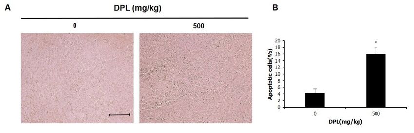
Yoo ES (2021) Extract of Dendropanax morbiferus H.Lév. Leaves Induces Apoptosis in Human Lung Carcinoma A549 Cells

fragmented, the cytoskeleton and nucleoproteins are decomposed, and          absorbance was measured at 595 nm with an ELISA reader (Bio-Rad
proteins are cross-linked to form an apoptotic body, which is finally        Laboratories, Hercules, CA, USA).
engulfed by a phagocyte [14]. Among apoptosis-inducing mechanisms,
the mitogen-activated protein kinase (MAPK) pathway plays an                 4′,6-diamidino-2-phenylindole (DAPI)
important role in tumor formation; activated MAPK regulates cell                 DAPI staining was conducted to demonstrate whether the decrease
growth, differentiation, apoptosis, and proliferation. Recently, small       in viability of A549 cells was caused by apoptosis. A549 cells were
molecule inhibitors targeting this pathway have been developed and           divided into 60 dishes with 2 × 10⁵ cells/mL per dish and cultured for
clinical studies are ongoing [15].                                           24 h. After 24 h, the culture medium was removed, and culture medium
   This study demonstrated induction of apoptosis by the extract of          containing DPL (0, 200, and 300 μg/mL) was added followed by culture
D. morbiferus leaves (DPL) through the MAPK pathway in A549 cells,           for another 24 h. Cells were washed twice with phosphate-buffered
a NSCLC cell line, and examined the effect of the extract on tumor           saline (PBS) and fixed in 4% paraformaldehyde solution for 15 min.
growth in vivo.                                                              Then, the paraformaldehyde was removed, cells were washed with
                                                                             PBS, and DAPI solution (2 mL) was added. DAPI-positive cells were
Material and methods                                                         observed at 200× magnification through an optical microscope (Zeiss
                                                                             fluorescence microscope, Carl Zeiss, Thornwood, NY, USA).
Chemicals, Drugs, and Antibodies
                                                                             Flow cytometry
    The A549 lung carcinoma cells (No. 10185) used in this study were
purchased from the Korean Cell Line Bank (Seoul, Korea). The WB-                  To analyze apoptosis, A549 cells were cultured in medium
F344 normal hepatic cells (No. JCRB0193) used in this study were             containing DPL (0, 200, and 300 μg/mL) in a 25 cm2 flask for 24 h.
purchased from the Japanese Collection of Research Bioresources              Following 24 h of incubation, trypsin-EDTA was used to suspend the
Cell Bank (Osaka, Japan). RPMI-1640 was purchased from Welgene               cells, which were then centrifuged at 1200 rpm for 5 min at 4°C. The
(Gyeongsan, Korea), and streptomycin/penicillin and fetal bovine             resulting pellet was resuspended at 1 × 10⁶ cells/mL with 1× annexin V
serum (FBS) were purchased from Gibco BRL (Grand Island, NY,                 binding buffer. Fluorescein isothiocyanate-conjugated annexin-V and
USA). Experimental reagents were purchased from Sigma-Aldrich                phycoerythrin-conjugated propidium iodide were added according
Co. (St. Louis, MO, USA). Anti-rabbit IgG, anti-Bcl-2, anti-Bax, anti-       to the manufacturer’s instructions of the annexin V staining kit (BD
poly (ADP-ribose) polymerase (PARP), anti-phospho (p)-extracellular          Pharmingen, San Diego, CA, USA). Cells were incubated for 15 min
signal-regulated kinase (ERK) 1/2, anti-p-c-Jun N-terminal kinase            and analyzed using flow cytometry.
(JNK), anti-p-p38, anti-ERK1/2, anti-JNK, anti-p38, and anti-β-actin
                                                                             Western blotting
were purchased from Cell Signaling Technology (Danvers, MA, USA).
                                                                                 Western blotting was conducted to assess the expression of
Cell culture                                                                 apoptosis-related proteins. A549 cells were cultured for 24 h in culture
    A549 lung carcinoma and normal WB hepatic cells were cultured            medium containing DPL (0, 200, and 300 μg/mL) in a 75 cm2 flask,
in RPMI-1640 medium containing 5% FBS and 1% penicillin/                     and the cells were suspended using trypsin-EDTA and centrifuged
streptomycin at 37°C in a humidified 5% CO2 atmosphere. When                 at 1200 rpm for 5 min at 4°C. The resulting pellets were lysed in lysis
the cells reached 80-90% confluence, they were sub-cultured, and the         buffer (Invitrogen, Carlsbad, CA, USA) and centrifuged at 13,000 rpm
culture medium was replaced every 2-3 days.                                  for 5 min at 4°C to obtain whole-cell lysates. The concentration of the
                                                                             extracted protein was determined using a Bradford protein assay kit
Plant materials and extraction                                               (Bio-Rad Laboratories), and proteins were separated by 5-12% sodium
     DPL were purchased from Sinbo Pharmacy (Seoul, Korea). The              dodecyl sulfate polyacrylamide gel electrophoresis and transferred to
origin of D. morbiferus was Jeju Island, Korea. DPL (100 g) were             a nitrocellulose membrane. The membrane was blocked for 1 h with
crushed, fermented with 70% ethanol and deionized water at a ratio of        Tris-buffered saline (TBS) containing 5% non-fat dry milk and 0.1%
4:6 for 10 days at room temperature, and then extracted with hot water.      Tween-20. After blocking, the membranes were incubated with anti-
The extracted solution was filtered using Whatman No. 1 disk paper.          Bax, anti-Bcl-2, anti-PARP, anti-ERK1/2, anti-JNK, anti-p38, anti-p-
The extracted liquid was vacuum-evaporated in a rotary evaporator (R-        ERK1/2, anti-p-JNK, anti-p-p38, and anti-β-actin antibodies overnight
220; BUCHI corporation, New Castle, DE, USA) to obtain the extract           at 4°C with gentle shaking. After washing with TBS containing 0.1%
through reheating [16]. The extract was then mixed with ethanol and          Tween-20, membranes were incubated with horseradish peroxidase-
refrigerated.                                                                conjugated secondary antibodies for 1 h at room temperature. After
                                                                             washing, bands were visualized using enhanced chemiluminescence
3-(4,5-dimethylthiazol-2-yl)-2,5-diphenyltetrazolium                         detection reagents (Pierce, Rockford, IL, USA) according to the
bromide (MTT) Assay                                                          manufacturer’s instructions. The density of each band was measured
                                                                             quantitatively using the ImageJ Launcher imaging program (provided
     The MTT assay was conducted to determine whether the viability
                                                                             by NCBI).
of A549 lung carcinoma and WB hepatic cells decreased following
exposure to DPL. A total of 2 × 10⁴ cells/mL was cultured on a 96-           Xenografts
well plate; after 24 h, the culture medium was discarded. WB cells were
added to culture medium containing 0-1000 μg/mL DPL, while A549                 Xenografts were performed to examine whether the in vitro anti-
cells were added to culture medium containing 0-500 μg/mL DPL and            tumor effects also occurred in vivo. BALB/c nude mice are commonly
cultured for 24 h. After 24 h, the culture medium containing DPL was         used in anticancer studies involving tumor models. Four-week-old
removed, MTT solution (1 mg/mL) was added, and cells were cultured           female BALB/c nude mice were purchased from Nara Biotec (Seoul,
in a CO2 incubator for 1 h 30 min. The MTT solution was removed after        Korea). Animal experiments were performed in accordance with the
1 h 30 min. To dissolve the formazan, 100 μL DMSO was added, and             Guidelines for the Care and Use of Animals of the Kongju National

Cancer Rep Rev, 2021     doi: 10.15761/CRR.1000227                                                                                     Volume 5: 2-8
Yoo ES (2021) Extract of Dendropanax morbiferus H.Lév. Leaves Induces Apoptosis in Human Lung Carcinoma A549 Cells

University Animals Care Committee (approval no. KNU_2019-02,                 the means of the DPL and control group were evaluated by one-way
Yesan, Korea). This animal experiment was conducted according to the         analysis of variance and Dunnett’s t-test. Differences with *P < 0.05 and
same protocol used in previously reported experiments [17,18]. The           **P
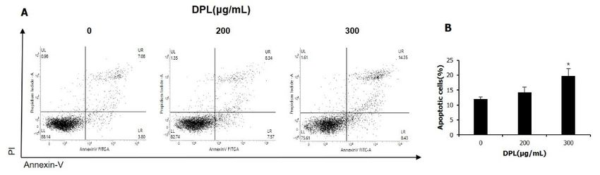
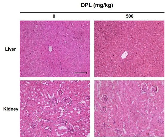
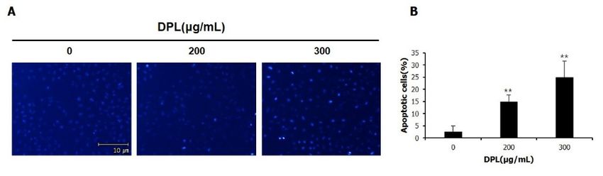
Yoo ES (2021) Extract of Dendropanax morbiferus H.Lév. Leaves Induces Apoptosis in Human Lung Carcinoma A549 Cells

Figure 1. Effects of Dendropanax morbifera leaves (DPL) on the viability of WB and A549 cells Cell viability was measured using the MTT assay. The results are presented as the mean ±
standard deviation (SD) from three independent experiments performed in triplicate. Significance was determined by Dunnett’s t-test. * P < 0.05, ** P < 0.01 versus control

Figure 2. Effects of DPL on apoptotic bodies of A549 cells. (A) Chromatin condensation was examined using a fluorescence microscope (200×). (B) Quantification of apoptotic cells. Scale
bar, 10 µm. Each bar represents the mean ± SD calculated from three independent experiments. * P < 0.05, ** P < 0.01 versus control

Figure 3. Effects of DPL on apoptosis of A549 cells. (A) Apoptotic cells were measured by flow cytometry. (B) Quantification of apoptotic cells. The results are presented as the mean ± SD
from three independent experiments performed in triplicate. * P < 0.05 versus control

tumor growth were examined: tumor size was measured twice a week                                DPL induces apoptosis in A549 tumor tissue
and tumor volume was compared to the control group, depending on
DPL administration. Tumor growth was inhibited beginning at day 15,                                  The anti-cancer effect of DPL on human lung carcinoma cells was
and significantly inhibited, down to 40.6%, by day 22 (Figure 5). Tumor                         examined using xenografts, and the induction of apoptosis in tumor
weight was reduced 22.2% in the DPL-treated group relative to the                               tissue was observed using a TUNEL assay. Apoptosis was significantly
control group (Figure 5). Body weight was similar between the DPL-                              increased in the group administered 500 mg/kg DPL compared with
treated and control groups (Figure 5). These results indicate that DPL                          the control group (Figure 6). Thus, administration of DPL induces
can inhibit tumor growth in vivo.                                                               apoptosis of tumor tissue.

Cancer Rep Rev, 2021           doi: 10.15761/CRR.1000227                                                                                                                Volume 5: 4-8
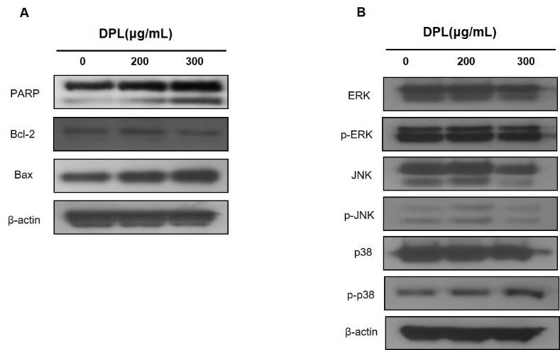
Yoo ES (2021) Extract of Dendropanax morbiferus H.Lév. Leaves Induces Apoptosis in Human Lung Carcinoma A549 Cells

Figure 4. Effects of DPL on apoptotic protein levels of A549 cells. The cells were harvested to measure the levels of apoptotic proteins (A) and MAPK protein (B) by western blotting.
β-actin was used as a loading control

Figure 5. Effects of DPL on lung cancer tumor growth in tumor tissues. Nude mice bearing A549 cells as xenograft models were treated with DPL for 22 days, and tumor volume (A),
weight (B) and body weight (C) were determined. Each value represents the mean ± standard error. Significance was determined by Dunnett’s t-test. * P < 0.05, ** P < 0.01 versus control

Cancer Rep Rev, 2021          doi: 10.15761/CRR.1000227                                                                                                               Volume 5: 5-8
Yoo ES (2021) Extract of Dendropanax morbiferus H.Lév. Leaves Induces Apoptosis in Human Lung Carcinoma A549 Cells

Figure 6. Effects of DPL on apoptosis in lung tumor tissues. Apoptosis was measured in tumor tissues using the TUNEL assay (A), and the number of apoptotic cells was quantified (B).
Scale bar, 10 µm. Significance was determined by the Dunnett’s t-test. * P < 0.05 versus control

Figure 7. Effects of DPL on phospho (p)-p38 expression in lung cancer tumor tissues. Excised tumors were analyzed by immunohistochemistry with antibodies specific for p-p38. Slides
were observed under a microscope (200×). Scale bar, 10 µm

DPL increases the expression of p-p38 in lung carcinoma                                      examining its effect on the viability of A549 cells, WB hepatic cells were
tissues                                                                                      treated with DPL to examine its effects in a normal cell line. Because
                                                                                             viability was shown to decrease when cells were treated with > 600 μg/
    Immunohistochemistry was conducted to determine the effect                               mL DPL for 24 h, cells were treated with 0-500 μg/mL in subsequent
of DPL on the expression of p-p38, an apoptosis-related protein, in
                                                                                             experiments. Further experiments revealed that the viability of A549
xenografts of human lung carcinoma tissues. The number of p-p38-
                                                                                             cells decreased following treatment with ≥ 300 μg/mL DPL in a
positive cells was increased compared to the control group (Figure
                                                                                             concentration-dependent manner (Figure 1). According to Aceituno
7). Thus, administration of DPL induced apoptosis by increasing
                                                                                             et al. [19], the viabilities of A549 lung carcinoma and HepG2 hepatic
expression of p-p38 in A549 carcinoma cells.
                                                                                             cells were reduced to 5 and 50 μg/mL, respectively, when silver-nano
DPL does not cause histopathological changes in hepatic and                                  particles of D. morbiferus were added at concentrations of 0, 1, 5, 10,
kidney tissues                                                                               and 50 μg/mL for 48 h. According to Hyun et al. [9], the viability
                                                                                             significantly decreased from 100 μg/mL when DPL was added to Huh-
    To examine organ toxicity induced by administration of DPL,                              7 liver carcinoma cells at concentrations of 0, 12.5, 25, 50, 100, and
hepatic and kidney tissues of xenografted mice were stained with
                                                                                             200 μg/well for 72 h. These findings are consistent with the results of
hematoxylin and eosin and observed using an optical microscope
                                                                                             this study, demonstrating the effect of DPL on the viability of various
(Figure 8). No histopathological changes were apparent, suggesting
                                                                                             carcinoma cells.
DPL did not cause detectable toxicity.
                                                                                                 As apoptosis occurs, DAPI and annexin V enter the nucleus
Discussion                                                                                   through membrane permeabilization and stain the DNA, allowing
    This study examined whether DPL induces apoptosis in A549                                visualization of apoptosis. Therefore, DAPI staining and flow
lung carcinoma cells as well as its anti-tumor effects in vivo. Prior to                     cytometry were conducted to demonstrate the induction of apoptosis

Cancer Rep Rev, 2021          doi: 10.15761/CRR.1000227                                                                                                            Volume 5: 6-8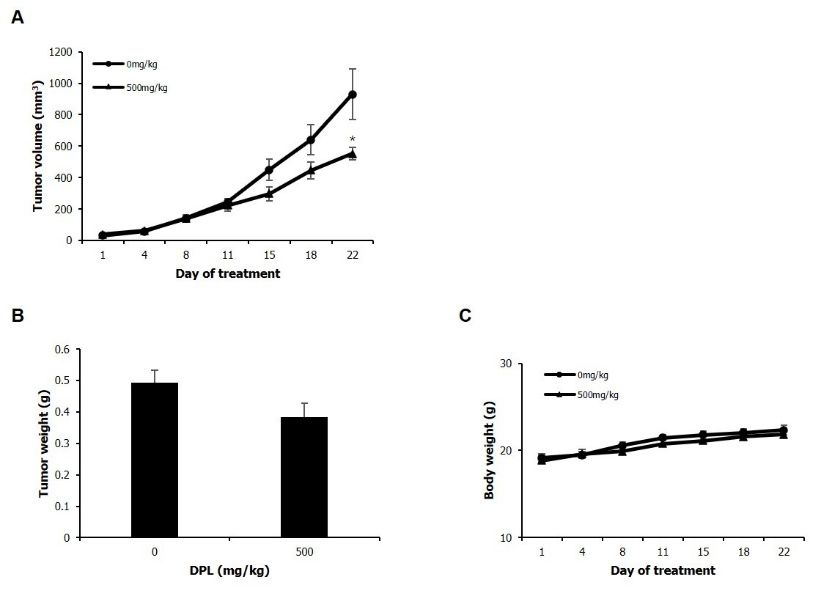
Yoo ES (2021) Extract of Dendropanax morbiferus H.Lév. Leaves Induces Apoptosis in Human Lung Carcinoma A549 Cells

Figure 8. Mice received an injection of A549 cells. Livers and kidneys were then excised and evaluated by hematoxylin and eosin staining (200×). Scale bar, 10 µm. DPL had no detectable
toxic effects on the nude mice

following treatment of A549 carcinoma cells with DPL for 24 h. DNA                            p-p38 increased in a concentration-dependent manner. Lee et al. [20]
fragmentation and apoptotic bodies were observed through DAPI                                 treated human myeloid leukemia U937 cells with D. morbiferus stem
staining. Flow cytometry showed a concentration-dependent increase                            extract for specific time periods (0, 0.5, 1, 2, 4, 6, 12, and 24 h) and
in apoptotic cells, consistent with the DAPI staining results. Lee et al.                     examined MAPK pathway-related proteins by western blotting. They
[20] observed concentration-dependent condensation of DAPI-stained                            observed that p-ERK, p-JNK, and p-p38 protein levels were increased
chromatin following treatment of human myeloid leukemia U937 cells                            at 0.5-6 h. The present study observed a concentration-dependent
with a D. morbiferus stem extract at various concentrations for 24 h.                         increase in p-p38 when A549 cells were treated with DPL for 24 h,
Hyun et al. [9] examined apoptosis using flow cytometry and reported                          whereas Lee et al. [20] observed an increase in p-p38 over 0.5-6 h
an increase in the number of apoptotic human hepatoma Huh-7 cells                             followed by a decrease at 24 h. These findings suggest that apoptosis
following treatment with 50 μg of yellow leaves and with 100 μg of                            was induced through the p38 MAPK pathway, because p-p38 increased
green leaves of D. morbiferus for 72 h. These results indicate that the                       in a concentration- and time-dependent manner in A549 cells when
leaves and stem of D. morbiferus induce apoptosis and cause anti-tumor                        DPL was administered.
effects in various carcinoma cells.
                                                                                                  DPL was orally administered in mice to examine whether the in
     In the present study, western blotting was performed to assess the                       vivo anti-tumor effects were as significant as the effects in vitro. Tumor
levels of apoptosis-related proteins in DPL-treated A549 cells. Levels of                     volume was inhibited beginning at day 15 after DPL administration and
the anti-apoptotic protein Bcl-2 were decreased compared to the control                       was significantly inhibited at day 22. Tumor weight decreased by 22.2%
group, while levels of the pro-apoptotic proteins Bax and cleaved PARP                        in the DPL-administered group relative to the control group. Apoptosis
were increased. Im et al. [16] treated breast carcinoma MCF-7 and                             was examined in A549 tumor tissue using the TUNEL assay; the
MDA-MB-231 cells with 0, 100, 150, and 300 μg/mL of D. morbiferus                             results showed that the number of TUNEL-positive cells was increased
stem extract for 24 h and examined apoptosis-related proteins. They                           compared with the control group. The number of p38-positive cells,
observed that the expression of Bcl-2 and Bcl-xL was significantly                            a protein involved in the MAPK signaling pathway, was increased in
decreased in MCF-7 cells following treatment with 300 μg/mL while                             tumor tissue compared with the control group.
the expression of Bad and Bax was significantly increased. In MDA-
                                                                                                  In summary, this study showed the induction of apoptosis in A549
MB-231 cells, the expression of Bcl-xL was significantly decreased
                                                                                              lung carcinoma cells by DPL both in vitro and in vivo through the
following treatment with 300 μg/mL, and the expression of Bax was
                                                                                              p-p38 MAPK pathway. Through these anti-cancer effects, DPL may be
significantly increased. Those authors observed that D. morbiferus leaf
                                                                                              a potential natural material for lung cancer treatment.
and stem extracts regulated Bax, Bcl-2, and PARP. This suggests that
DPL induces apoptosis at the protein level.                                                   Conclusion
   When A549 cells were treated with DPL, expression levels of                                    This study demonstrated the induction of apoptosis by the extract
p-ERK1/2 and p-JNK were similar to the level in the control group and                         of D. morbifera leaves (DPL) through the MAPK pathway in A549 cells,

Cancer Rep Rev, 2021          doi: 10.15761/CRR.1000227                                                                                                               Volume 5: 7-8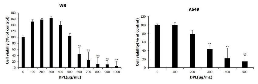
Yoo ES (2021) Extract of Dendropanax morbiferus H.Lév. Leaves Induces Apoptosis in Human Lung Carcinoma A549 Cells

a NSCLC cell line, and examined the effect of the extract on tumor                          7. Lee C, Yang M, Moon JO (2019) Antioxidant and hepatoprotective effects of the
                                                                                               ethanol extract of Dendropanax morbifera Leveille on the t-butyl hydroperoxide-
growth in vivo. In conclusion, DPL decreased the viability of A549 cells
                                                                                               induced HepG2 cell damages. Korean J Pharmacogn 50: 32-36.
and induced apoptosis by regulating p38 MAPK pathway in vitro. By
examining in vivo tumor growth following administration of DPL, we                          8. Lee SH, Lee HS, Park YS, Hwang B, Kim JH, et al. (2002) Screening of immune
                                                                                               activation activities in the leaves of Dendropanax morbifera Lev. Korean J Med Crop
showed that DPL significantly reduced tumor volume and increased                               Sci 10: 109-115.
apoptosis. DPL shows potential as a drug for lung cancer.
                                                                                            9. Hyun TK, Kim MO, Lee H, Kim YJ, Kim EK, et al. (2013) Evaluation of anti-oxidant
Author’ contributions                                                                          and anti-cancer properties of Dendropanax morbifera Léveille. Food Chem 141: 1947-
                                                                                               1955.
    Ji-Youn Jung and Eun-Seon Yoo conceptualized and designed the                           10. Park SA, Park J, Park CI, Jie YJ, Hwang YC, et al. (2013) Cellular antioxidant activity
study. Eun-Seon Yoo, Sung-Hyun Kim, Joong-Seok Woo, Jae-Han Lee,                                and whitening effects of Dendropanax morbifera leaf extracts. J Microbiol Biotechnol
So-Hee Han, and Soo-Hyun Jung acquired the data. Eun-Seon Yoo, Ji-                              41: 407-415.
Youn Jung, Sae Man Kim, Eun Gee Kim and Kwang Joong Kim analyzed                            11. Lee MK, Lee IS, Lee JS (2013) For the utilization of native plant resources as high-
and interpreted the data. Ji-Youn Jung and Eun-Seon Yoo drafted the                             value materials: evaluation on demelanizing activity of Dendropanax morbifera in
manuscript. All authors have read and approved the final manuscript.                            Bogildo. J Kor Island 25: 227-240.

                                                                                            12. Hyun TK, Ko YJ, Kim EH, Chung IM, Kim JS (2015) Anti-inflammatory activity and
Acknowledgment                                                                                  phenolic composition of Dendropanax morbifera leaf extracts. Ind Crops Prod 74: 263-
                                                                                                270.
    The present study was supported by Basic Science Research
Program through the National Research Foundation of Korea (NRF)                             13. An NY, Kim JE, Hwang D, Ryu HK (2014) Anti-diabetic effects of aqueous and ethanol
                                                                                                extract of Dendropanax morbifera Leveille in streptozotocin-induced diabetes model.
funded by the Ministry of Education, Science and Technology (grant                              J Nutr Health 47: 394-402.
no. NRF 2021R1A2C1010912).
                                                                                            14. Elmore S (2007) Apoptosis: a review of programmed cell death. Toxicol Pathol 35:
Conflict of interest                                                                            495-516.

                                                                                            15. Santarpia L, Lippman SM, El-Naggar AK (2012) Targeting the MAPK–RAS–RAF
    The authors declare that they have no competing interests.                                  signaling pathway in cancer therapy. Expert Opin Ther Targets 16: 103-119.

References                                                                                  16. Im KJ, Jang SB, Yoo DY (2015) Anti-cancer effects of Dendropanax Morbifera extract
                                                                                                in MCF-7 and MDA-MB-231 cells. J Korean Obstet Gynecol 28: 26-39.
1. Statistics Korea (2017) Cause of death statistics. Last accessed on 23 September 2019.
                                                                                            17. Kim SH, Choo GS, Yoo ES, Woo JS, Han SH, et al. (2019) Silymarin induces inhibition
2. National cancer information center. Last accessed on 23 September 2019.                      of growth and apoptosis through modulation of the MAPK signaling pathway in AGS
3. Rafei H, El-Bahesh E, Finianos A, Nassereddine S, Tabbara I (2017) Immune-based              human gastric cancer cells. Oncol Rep 42: 1904-1914.
   therapies for non-small cell lung cancer. Anticancer Res 37: 377-387. [Crossref]
                                                                                            18. Kim JM, Park JD, Park DC, Kim BO (2013) In vivo antitumor activity and acute,
4. Park SB, Kim HN, Kim JD, Park GH, Eo HJ, et al. (2019) Inhibitory effect of the              subacute toxicity of keumsa (Phellinus linteus) extracts. J Life Sci 23: 1388-1396.
   branch extracts from Taxillus yadoriki parasitic to Neolitsea sericea against the cell
                                                                                            19. Aceituno VC, Ahn S, Simu SY, Wang C, Mathiyalagan R, et al. (2016) Silver
   proliferation in human lung cancer cells, A549. Korean J Plant Res 32: 109-115.
                                                                                                nanoparticles from Dendropanax morbifera Léveille inhibit cell migration, induce
5. Lin L, Cheng K, Xie Z, Chen C, Chen L, et al. (2019) Purification and characterization       apoptosis, and increase generation of reactive oxygen species in A549 lung cancer
   a polysaccharide from Hedyotis diffusa and its apoptosis inducing activity toward            cells. In Vitro Cell Dev Biol Anim 52: 1012-1019.
   human lung cancer cell line A549. Int J Biol Macromol 122: 64-71. [Crossref]
                                                                                            20. Lee JW, Park C, Han MH, Hong SH, Lee TK, et al. (2013) Induction of human leukemia
6. Da Rocha AB, Lopes RM, Schwartsmann G (2001) Natural products in anticancer                  U937 cell apoptosis by an ethanol extract of Dendropanax morbifera Lev. through the
   therapy. Curr Opin Pharmacol 1: 364-369.                                                     caspase-dependent pathway. Oncol Rep 30: 1231-1238. [Crossref]

Copyright: ©2021 Yoo ES. This is an open-access article distributed under the terms of the Creative Commons Attribution License, which permits unrestricted use,
distribution, and reproduction in any medium, provided the original author and source are credited.

Cancer Rep Rev, 2021          doi: 10.15761/CRR.1000227                                                                                                             Volume 5: 8-8
You can also read