Paeonol alleviates lipopolysaccharide induced hepatocytes injury through alteration of mitochondrial
←
→
Page content transcription
If your browser does not render page correctly, please read the page content below
MOLECULAR MEDICINE REPORTS 24: 779, 2021

           Paeonol alleviates lipopolysaccharide‑induced hepatocytes
                  injury through alteration of mitochondrial
                       function and NF‑κB translocation
                          SHOUZHU XU1,2*, JIE XU2*, TING HAO2, YU YAN2, SHIHAO ZHANG2,
                            AIHONG LI1, CHUANDAO SHI2, QILING LIU2 and JING ZHAO3,4

              1
               Shaanxi Pharmaceutical Holding Grp Co. Ltd., Shaanxi Pharmaceutical Dev Ctr, Xian, Shaanxi 710075,
                           2
                             Department of Public Health; 3College of Acupuncture and Moxibustion and
                          4
                           Shaanxi Key Laboratory of Acupuncture and Medicine, Shaanxi University of
                                    Chinese Medicine, Xianyang, Shaanxi 712046, P.R. China

                                          Received April 1, 2021; Accepted July 26, 2021

                                                   DOI: 10.3892/mmr.2021.12419

Abstract. Sepsis is a severe disease, with high mortality.           NF‑κ B. In conclusion, PAE alleviated LPS‑induced liver
Permanent organ damage caused by sepsis reduces the quality          injury via alteration of mitochondrial function and NF‑κ B
of life of surviving patients. The liver is an easily damaged        translocation. Therefore, PAE has potential for the treatment
organ in sepsis and sepsis‑associated liver injury foretells a       of sepsis.
poor prognosis. Unfortunately, there are no effective treat‑
ments or drugs to solve this problem. Therefore, strategies          Introduction
or novel drugs are urgently required to protect against liver
dysfunction in sepsis. In the present study, lipopolysac‑            Sepsis is characterized by potentially fatal organ dysfunction,
charide (LPS) was used to establish a model of liver injury          resulting from the abnormal immune response to an infec‑
in vitro. The data demonstrated that pretreatment of L02             tion (1). In total, ~3 per 1,000 individuals are estimated to
human normal hepatocytes with paeonol (PAE) alleviated               be diagnosed with severe sepsis (2). In sepsis, the abnormal
LPS‑induced cell injury and decreased the levels of alanine          expression of proinflammatory cytokines and chemokines is
aminotransferase and aspartate transaminase, indicating a            the driving force behind the development of septic shock, which
protective effect of PAE. Further experiments demonstrated           results in an overt systemic inflammatory response and tissue
that PAE increased LPS‑decreased L02 cell viability,                 injury (3). In addition to causing high mortality, sepsis causes
the levels of superoxide dismutase and Bcl‑2 expression.             a reduction in the quality of life for surviving patients, due to
PAE decreased LPS‑increased cell apoptosis, intracel‑                the permanent organ damage (4). The liver serves a funda‑
lular reactive oxygen species and the expression levels of           mental role in hypermetabolism, generates acute phase proteins
Bax and cleaved‑caspase‑3. PAE decreased LPS‑promoted                during the inflammatory response and is of great importance
mitochondrial depolarization and nuclear translocation of            in bacteria clearance and host defense (5,6). Furthermore, the
                                                                     liver is an easily damaged organ in sepsis and sepsis‑associated
                                                                     liver injury forecasts a poor prognosis in patients with sepsis (7).
                                                                     Therefore, protection against liver injury during sepsis is of
                                                                     great importance.
Correspondence to: Mrs. Jing Zhao, College of Acupuncture and            Paeonol (PAE; 2'‑hydroxy‑4'‑methoxyacetophenone)
Moxibustion, Shaanxi University of Chinese Medicine, Shiji Ave,
                                                                     is one of the major components extracted from Dioscorea
Xi'an‑Xianyang New Economic Zone, Xianyang, Shaanxi 712046,
P.R. China
                                                                     japonica, Paeonia suffruticosa, Paeonia lactiflora and
E‑mail: zhaojing_1207@126.com                                        Arisaema erubescens (8‑10). PAE has numerous biological
                                                                     functions, such as antioxidant, anti‑inflammatory and cardio‑
Professor Aihong Li, Shaanxi Pharmaceutical Holding Grp Co.          vascular protection (11). In addition to the aforementioned
Ltd., Shaanxi Pharmaceutical Dev Ctr, 69 Keji 2nd Road, Xian,
                                                                     properties, PAE reduces the mortality by confining high
Shaanxi 710075, P.R. China
E‑mail: 408503572@qq.com
                                                                     mobility group box 1 to the nucleus in lipopolysaccharide
                                                                     (LPS)‑stimulated rats (12). However, the roles of PAE in liver
*
    Contributed equally                                              protection remain to be elucidated. It was hypothesized that
                                                                     PAE could exert an important protective role in LPS‑damaged
Key words: paeonol, sepsis, liver injury, reactive oxygen species,   hepatocytes. In the present study, LPS was used to induce injury
nuclear translocation of NF‑κB                                       in L02 human normal hepatocytes and the protective effect of
                                                                     PAE was observed using different methods as subsequently
                                                                     described.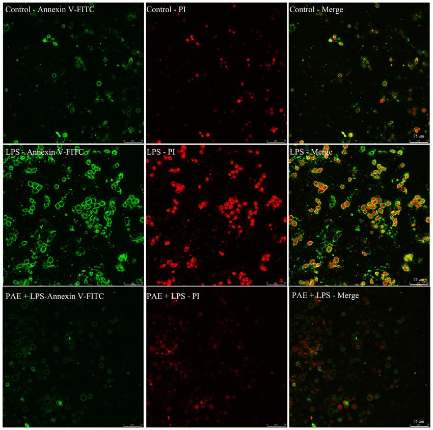
2                              XU et al: PAEONOL ALLEVIATES LPS-INDUCED HEPATOCYTES INJURY

Materials and methods                                              (10.0 µg/ml) or incubated with PAE (10.0 µg/ml) for 1 h and
                                                                   then co‑incubated with LPS (10 µg/ml) for 12 h at 37˚C in
Cell culture and drug treatment. L02 human normal hepato‑          a 5% CO2 atmosphere, separately. Following treatment, cells
cytes were purchased from BioVector NTCC Inc. and the cells        were washed with PBS three times and 5 µl FITC‑labeled
were cultured in a 5% CO2 atmosphere at 37˚C. The cells were       Annexin V and 195 µl binding buffer were added into each
maintained in DMEM (Gibco; Thermo Fisher Scientific, Inc.)         dish. The dishes were then incubated in the dark for 10 min at
supplemented with 10% (v/v) FBS (Cellmax; Lanzhou Minhai           25˚C. This was followed by incubation with 10 µl propidium
Bio‑engineering Co., Ltd.), 100 U/ml streptomycin and 100          iodide in the dark for another 20 min at 4˚C. Apoptotic cells
U/ml penicillin. The cells were seeded into 6‑, 24‑ or 96‑well     were observed using laser confocal microscopy (original
culture dishes and serum‑starved for 12 h in DMEM when the         magnification, x200). The cell population was separated into
cells had reached sub‑confluence. Cells treated with DMEM          three groups: Live cells with low fluorescence, early apoptotic
were used as a control group. Hepatocyte injury was induced        cells stained with green fluorescence and necrotic/advanced
by incubation with LPS (cat. no. ST1470‑10 mg; Beyotime            stage apoptotic cells stained with both red and green fluores‑
Institute of Biotechnology). PAE (cat. no. B20266; Shanghai        cence.
Yuanye Bio. Tech. Co., Ltd.) was added to the cells 1 h prior
to LPS stimulation. In the present study, a ScienCell HeLa         Western blotting. Cells (5‑7x10 6 /well) were seeded into
Cell Contamination Detection kit (cat. no. 8988; ScienCell         6‑well plates and serum‑starved for 12 h in DMEM when
Research Laboratories, Inc.) was used for the detection of         the cells had reached sub‑confluence. Subsequently, cells
HeLa cell contamination in cultured L02 cells by PCR. The          were incubated with LPS (10.0 µg/ml) or incubated with
data demonstrated that L02 cells had no detectable HeLa cell       PAE (10.0 µg/ml) for 1 h and then co‑incubated with LPS
contamination (Fig. 1).                                            (10 µg/ml) for 12 h, separately. Following treatment, cells
                                                                   were washed with ice‑cold PBS three times and solubilized
Cell viability assay. Cell viability was tested using a Cell       with 200 µl ice‑cold lyse buffer (cat. no. P0013C; Beyotime
Counting Kit‑8 (CCK‑8; cat. no. C0037; Beyotime Institute of       Institute of Biotechnology). Subsequently, supernatants were
Biotechnology) according to the manufacturer's instructions.       collected by centrifugation at 12,000 x g for 10 min at 4˚C,
A total of 1.5x10³ cells/well were seeded into a 96‑well plate     and the concentration of protein was identified by the BCA
and then serum‑starved for 12 h in DMEM when the cells             method (cat. no. AR1189; Boster Biological Technology).
had reached sub‑confluence. The cells were incubated with          Loading buffer was added into each supernatant followed
different concentrations of LPS (0.05, 0.5, 5.0 or 10.0 µg/ml),    by boiling for 8 min. Lysate (30 µg) was electrophoresed on
incubated with different concentrations of PAE (0.1, 1.0,          a 12% SDS‑PAGE gel and transferred onto a nitrocellulose
10.0 or 100.0 µg/ml) for 12 h or incubated with different          membrane. The membrane was blocked with 5% bovine
concentrations of PAE (0.1, 1.0 or 10.0 µg/ml) for 1 h and         serum albumin (Sigma‑Aldrich; Merck KGaA) overnight
then co‑cultured with LPS (10 µg/ml) for 12 h in a 5% CO2          at 4˚C. The blots were incubated with antibodies against
atmosphere. All incubations were at 37˚C. Following the            Bcl‑2 (dilution, 1:8,000; cat. no. AB112; Beyotime Institute
indicated treatment, 10 µl CCK‑8 solution was added to each        of Biotechnology), Bax (dilution, 1:5,000; cat. no. AF0054;
well. The cells were then incubated at 37˚C for 4 h and the        Beyotime Institute of Biotechnology), cleaved‑caspase‑3
absorbance value was determined at 450 nm using a micro‑           (dilution, 1:5,000; cat. no. 9661s; Cell Signaling Technology,
plate reader (Bio‑Rad Laboratories, Inc.). Each experiment         Inc.) or β‑actin (dilution, 1:10,000; cat. no. NC011; Zhuangzhi
was repeated six times.                                            Biotech.) overnight at 4˚C and then the membranes were
                                                                   incubated at 25˚C for 3 h with goat anti‑mouse IgG‑HRP
Measurements of alanine aminotransferase (ALT) and                 (1:5,000; cat. no. sc‑2005) or goat anti‑rabbit IgG‑HRP
aspartate transaminase (AST) activity. Hepatocyte damage           (1:5,000; cat. no. sc‑2004); these secondary antibodies
was examined by measuring the activities of ALT (U/g               were purchased from Santa Cruz Biotechnology, Inc. The
protein; cat. no. C010‑2‑1; Nanjing Jiancheng Bioengineering       immune complexes were enhanced by chemiluminescence
Institute) and AST (U/g protein; cat. no. C009; Nanjing            (cat. no. WBULS0500; Merck Life Science UK, Ltd.). The
Jiancheng Bioengineering Institute) using ALT and AST assay        results were analyzed using Quantity One software (Bio‑Rad
kits. Briefly, 1x10⁶ cells were seeded into 24‑well plates and     Laboratories, Inc.; version 6.0Inc.).
serum‑starved for 12 h in DMEM when the cells had reached
sub‑confluence. Subsequently, cells were incubated with LPS        Measurement of mitochondrial transmembrane potential.
(10.0 µg/ml) or incubated with PAE (10.0 µg/ml) for 1 h and        A JC‑1 probe (cat. no. C2006; Beyotime Institute of
then co‑incubated with LPS (10 µg/ml) for 12 h, separately.        Biotechnology) was used to measure mitochondrial depo‑
The activities of ALT and AST were assayed according to the        larization according to the manufacturer's protocol. Briefly,
manufacturer's instructions.                                       1x10 6 cells were seeded into Nunc glass bottom dishes
                                                                   and serum‑starved for 12 h in DMEM when the cells had
Cellular apoptosis assay. The apoptosis assay was carried          reached sub‑confluence. Subsequently, cells were incubated
out using an Annexin V‑FITC apoptosis detection kit                with LPS (10.0 µg/ml) or incubated with PAE (10.0 µg/ml)
(cat. no. C1067s; Beyotime Institute of Biotechnology). Briefly,   for 1 h and then co‑incubated with LPS (10 µg/ml) for 12 h
1x10⁶ cells were seeded in Nunc glass bottom dishes and then       at 37˚C in a 5% CO 2 atmosphere, separately. Following
serum‑starved for 12 h in DMEM when the cells had reached          treatment, cells were incubated at 37˚C for 20 min with
sub‑confluence. Subsequently, cells were incubated with LPS        25 µg/ml JC‑1, then washed twice with PBS and placed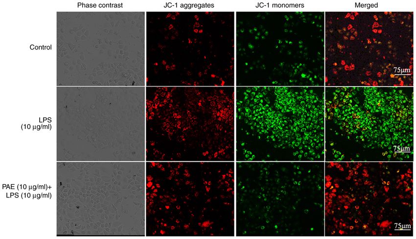
MOLECULAR MEDICINE REPORTS 24: 779, 2021                                                               3

in fresh DMEM without serum. Mitochondrial membrane
potentials were monitored by observing the relative amounts
of dual emissions from mitochondrial JC‑1 monomers or
aggregates using laser confocal microscopy. Mitochondrial
depolarization was indicated by an increase in the green
fluorescence intensity.

Detection of intracellular reactive oxygen species (ROS). ROS
were assayed with the probe 2',7'‑dichlorofluorescein diacetate
(DCFH‑DA). DCFH‑DA can easily diffuse into the cells and
is deacetylated to nonfluorescent 2',7'‑dichlorofluorescein
(DCFH) by esterases. DCFH reacts with intracellular ROS              Figure 1. Gel electrophoresis separation of PCR products. (A‑D) PCR prod‑
to form the fluorescent product DCF. Briefly, 1x10⁶ cells were       ucts of genomic DNA samples by using HeLa gDNA detection primer set.
                                                                     (E‑H) products of genomic DNA samples by using Human gDNA control
seeded into Nunc glass bottom dishes and serum‑starved for           primer set. (A and E) HeLa positive control PCR template, (B and F) HeLa cells,
12 h in DMEM when the cells had reached sub‑confluence.              (C and G) L02 cells mixed with 1% HeLa cells and (D and H) L02 cells. HLD,
Subsequently, cells were incubated with LPS (10.0 µg/ml) or          HeLa gDNA detection primer set; HGC, Human gDNA control primer set.
incubated with PAE (10.0 µg/ml) for 1 h and then co‑incubated
with LPS (10 µg/ml) for 4 h at 37˚C in a 5% CO2 atmosphere,
separately. Following treatment, the culture medium was              graph of known concentrations of MDA and presented as
removed and the cells were washed three times with PBS.              µmol/g protein.
DCFH‑DA (10 µM) was added into the dishes and the cells
were then incubated for an additional 20 min at 37˚C. The            Assay of the nuclear translocation of NF‑ κ B using
fluorescence was observed with a laser confocal microscope           immunofluorescence staining. The NF‑κ B activation nuclear
(original magnification, x200).                                      translocation assay kit (cat. no. SN368; Beyotime Institute of
                                                                     Biotechnology) was used to measure NF‑κ B activation. In
Determination of Cu/Zn superoxide dismutase (SOD) and Mn             brief, 1x10⁶ cells were seeded into Nunc glass bottom dishes
SOD activities. The enzyme activities of SOD were determined         and serum‑starved for 12 h in DMEM when the cells had
using a Cu/Zn SOD and Mn‑SOD assay kit (cat. no. S0103;              reached sub‑confluence. Then, cells were incubated with LPS
Beyotime Institute of Biotechnology) according to the manu‑          (10.0 µg/ml) or incubated with PAE (10.0 µg/ml) for 1 h and
facturer's instructions. In brief, 1x10⁶ cells/well were seeded in   then co‑incubated with LPS (10 µg/ml) for 12 h at 37˚C in
24‑well plates and serum‑starved for 12 h in DMEM when the           a 5% CO2 atmosphere, separately. Following treatment, cells
cells had reached sub‑confluence. Then, cells were incubated         on plates were washed with PBS once, fixed with 4% para‑
with LPS (10.0 µg/ml) or incubated with PAE (10.0 µg/ml)             formaldehyde for 20 min at 25˚C, washed three times with
for 1 h and then co‑incubated with LPS (10 µg/ml) for 12 h           PBS and blocked with 5% bovine serum albumin for 1.5 h
at 37˚C in a 5% CO2 atmosphere, separately. Following treat‑         at room temperature. Afterwards, they were incubated with
ment, Cu/Zn‑SOD inhibitors were added into cell lysates,             the primary rabbit anti‑NF‑κ B p65 antibody at 4˚C for 24 h,
followed by incubation for 30 min at 37˚C and mixing with            followed by incubation with Cy3‑labeled secondary antibody
WST‑8 enzyme for another 30 min at 37˚C. The absorbance              at 25˚C for 2 h. Subsequently, the cells were stained with DAPI
at 450 nm was assessed using a multimode microplate reader           solution for 8 min. Each aforementioned step was followed by
(Thermo Fisher Scientific, Inc.). The results were calculated        washing three times for 5 min. The activation or nuclear trans‑
and normalized to the protein concentration of each sample           location of NF‑κ B was observed by laser confocal microscopy
(U/g protein).                                                       (original magnification, x200).

Lipid peroxidation assay. Lipid peroxidation was measured by         Statistical analysis. The data are presented as the
detecting the content of thiobarbituric acid reactive substances     mean ± SEM. The normality and homogeneity of these data
using a lipid peroxidation malondialdehyde (MDA) assay               were tested by Shapiro‑Wilk tested, P>0.05 was considered
kit (cat. no. S0131S; Beyotime Institute of Biotechnology).          to normal distribution. The differences among groups were
In brief, 1x10 6 cells were seeded into 24‑well plates and           assessed by a one‑way ANOVA followed by a Dunnett's
serum‑starved for 12 h in DMEM when the cells had reached            post hoc test or non‑parametric Kruskal‑Wallis followed
sub‑confluence. The cells were then incubated with LPS               by Dunn's test analysis using GraphPad Prism 8.3 software
(10.0 µg/ml) or incubated with PAE (10.0 µg/ml) for 1 h and          (GraphPad Software, Inc.). P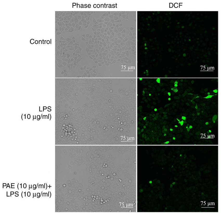
4                                    XU et al: PAEONOL ALLEVIATES LPS-INDUCED HEPATOCYTES INJURY

Figure 2. PAE increases LPS decreased cell viability of L02 cells. (A) The injury effect of LPS on cell viability of L02 cells. Cells were incu‑
bated with different concentration of LPS (0.05, 0.5, 5.0 or 10.0 µg/ml) for 12 h. (B) The effect of PAE alone on cell viability of L02 cells.
Cells were incubated with different concentration of PAE (0.1, 1.0, 10.0 or 100.0 µg/ml) for 12 h. (C) The protective effect of PAE on LPS damaged
L02 cells. Cells were incubated with different concentration of PAE (0.1, 1.0 or 10.0 µg/ml) prior 1 h and then co‑incubated with LPS (10 µg/ml) for 12 h. Cell
viability was assessed by CCK‑8. Data represent mean ± standard error of the mean of six independent experiments. *P
MOLECULAR MEDICINE REPORTS 24: 779, 2021 5
Figure 4. PAE inhibits LPS induced the apoptosis of L02 cells. Cells were incubated with LPS at 10.0 µg/ml for 12 h or cells were incubated with PAE at
10.0 µg/ml prior 1 h and then co‑incubated with LPS (10 µg/ml) for 12 h. Cell apoptosis was observed with laser confocal microscopy (scale bar=75 µm). The
population was separated into three groups: Live cells with a low level of fluorescence, apoptotic cells in the earlier period with green fluorescence and necrotic
and advanced stage apoptotic cells with both red and green fluorescence. PAE, paeonol; LPS, lipopolysaccharide.
intensity of fluorescence. However, pretreatment with PAE mitochondrial depolarization. The control cells stained with
(10 µg/ml) markedly decreased the intensity of fluorescence JC‑1 emitted mitochondrial red fluorescence with weak green
(Fig. 6), which demonstrated the anti‑oxidative effects of fluorescence (Fig. 8), indicating hyperpolarized mitochondria.
PAE. However, cells treated with LPS exhibited increased green
fluorescence and weakened red fluorescence, indicating the
PAE decreases LPS‑increased MDA levels and increases depolarization of mitochondria. Compared with that in the
LPS‑decreased SOD levels. Given the important role of LPS‑damaged group, PAE relieved the LPS‑induced mito‑
oxygen free radicals in the pathogenesis of liver injury, MDA chondrial depolarization, as indicated by the fluorescent color
and SOD levels were examined. As shown in Fig. 7, LPS changes from green to red.
markedly increased MDA levels and decreased SOD levels.
Furthermore, pretreatment of the cells with PAE could mark‑ PAE inhibits LPS‑induced nuclear translocation of NF‑ κ B.
edly decrease the levels of MDA and tended to increase the To further investigate the protective effect of PAE on
SOD levels (P=0.182). LPS‑induced cell injury, the NF‑κ B levels in the nuclei of L02
cells were detected using immunofluorescence staining. The
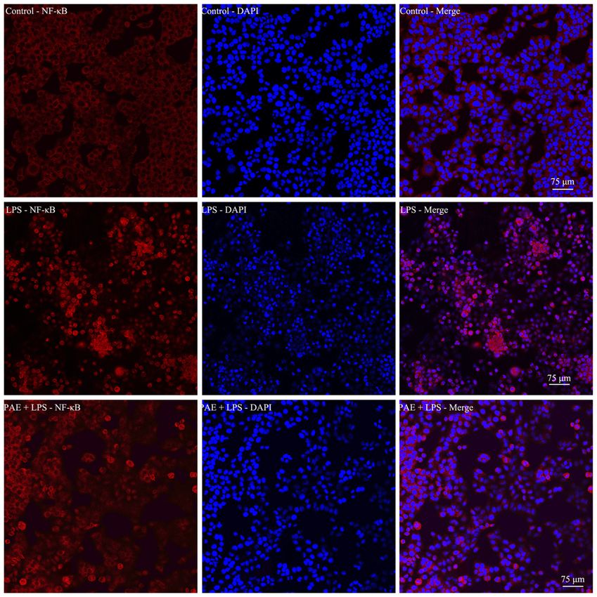
PA E increases the L PS ‑ decreased mitochon drial immunofluorescence staining revealed that LPS promoted
transmembrane potential. The protective effect of PAE on NF‑κ B translocation into the nuclei of L02 cells. As shown
LPS‑induced L02 cell injury was further assayed by detecting in Fig. 9, the p65 subunit was translocated to the nucleus (the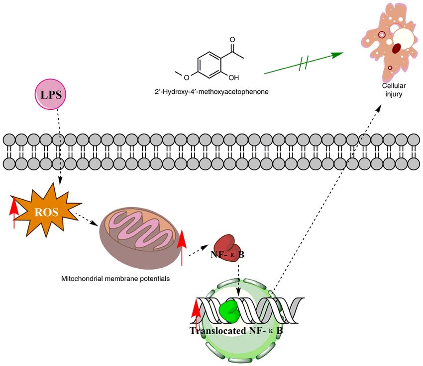
6 XU et al: PAEONOL ALLEVIATES LPS-INDUCED HEPATOCYTES INJURY
Figure 6. PAE decreases LPS induced ROS generation of L02 cells
(bar=75µm). Cells were incubated with LPS at 10.0 µg/ml for 12 h or cells
were incubated with PAE at 10.0 µg/ml prior 1 h and then co‑incubated with
LPS (10 mg/ml) for 4 h. Intracellular ROS was observed with laser confocal
microscopy. The population was separated into two groups: Left panels were
cells observed by phase contrast and right panels were cells observed by fluo‑
rescence microscopy. PAE, paeonol; LPS, lipopolysaccharide; ROS, reactive
oxygen species; DCF, dichlorofluorescein.
of which suggested an established in vitro liver injury model
of sepsis.
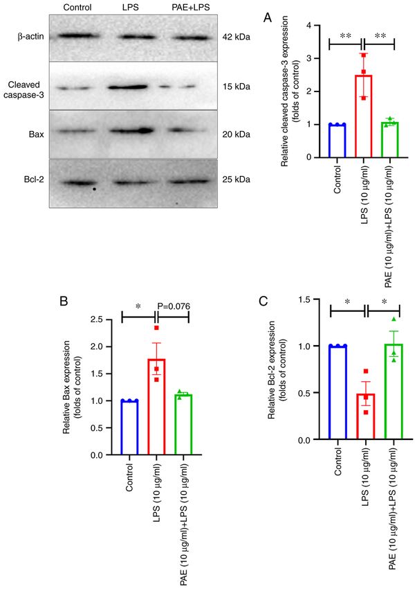
Figure 5. PAE decreases LPS‑increased the expression of Bax and caspase‑3,
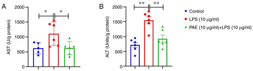
ALT and AST levels tend to increase when liver damage
increases the level of Bcl‑2. Cells were incubated with LPS at 10.0 µg/ml
for 12 h or cells were incubated with PAE at 10.0 µg/ml prior 1 h and then occurs (17). The present results indicated that PAE pretreat‑
co‑incubated with LPS (10 µg/ml) for 12 h. (A) Cleaved‑casapse‑3, (B) Bax ment considerably reduced the levels of liver enzymes (ALT
and (C) Bcl‑2 were assayed by western blotting. Data represent mean ± stan‑ and AST). MDA is the final product of lipid peroxidation
dard error of the mean of six independent experiments. *P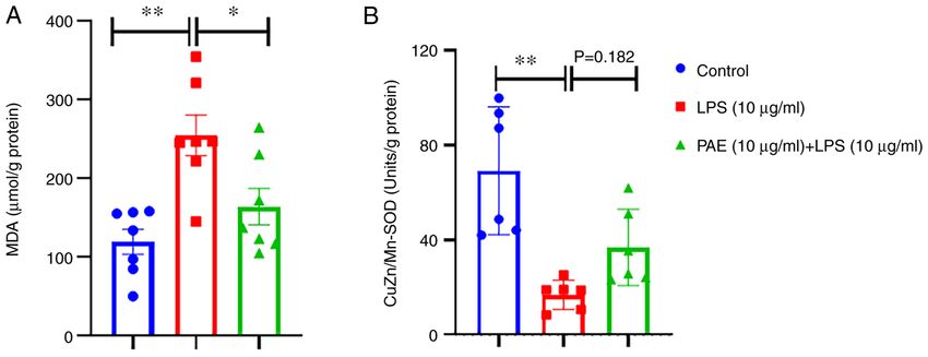
MOLECULAR MEDICINE REPORTS 24: 779, 2021                                                                7

Figure 7. PAE inhibits LPS increased MDA and increases LPS decreased SOD level of L02 cells. Cells were incubated with LPS at 10.0 µg/ml for 12 h or cells
were incubated with PAE at 10.0 µg/ml prior 1 h and then co‑incubated with LPS (10 µg/ml) for 12 h. (A) MDA and (B) SOD were assayed by respective kits.
Data represent mean ± standard error of the mean of six independent experiments. *P
8                                     XU et al: PAEONOL ALLEVIATES LPS-INDUCED HEPATOCYTES INJURY

Figure 9. PAE inhibits LPS‑promoted nuclear translocation of NF‑κB (scale bar=75 µm). Cells were incubated with LPS at 10.0 µg/ml for 12 h or cells were incubated
with PAE at 10.0 µg/ml prior 1 h and then co‑incubated with LPS (10 µg/ml) for 12 h. The NF‑κB level in the nuclei of L02 cells was detected by immunofluorescence
staining. The right panels were the merged field of red (for NF‑κB subunit p 65) and blue color (for cell nucleus). PAE, paeonol; LPS, lipopolysaccharide.

Figure 10. PAE alleviates LPS‑induced hepatocytes injury through alteration of mitochondrial function and NF‑κB translocation. PAE, paeonol; LPS, lipo‑
polysaccharide; ROS, reactive oxygen species.
MOLECULAR MEDICINE REPORTS 24: 779, 2021                                                            9

Acknowledgements                                                           6. Hensley MK and Deng JC: Acute on chronic liver failure and
                                                                              immune dysfunction: A mimic of sepsis. Semin Respir Crit Care
                                                                              Med 39: 588‑597, 2018.
Not applicable.                                                            7. Woznica EA, Inglot M, Woznica RK and Lysenko L: Liver
                                                                              dysfunction in sepsis. Adv Clin Exp Med 27: 547‑551, 2018.
                                                                           8. Wu CF: A review on the pharmacology of Paeonia lactiflora and
Funding                                                                       its chemical components. Zhong Yao Tong Bao 10: 43‑45, 1985
                                                                              (Ιn Chinese).
The present study was sponsored by Key Research and                        9. Ducki S, Hadfield JA, Lawrence NJ, Zhang X and McGown AT:
                                                                              Isolation of paeonol from Arisaema erubescens. Planta Med 61:
Development Program of Shaanxi (program no. 2021SF‑071). The                  586‑587, 1995.
National Training Program of Innovation and Entrepreneurship for          10. Adki KM and Kulkarni YA: Chemistry, pharmacokinetics, phar‑
Students of China (grant nos. 201910716019 and 201910716020),                 macology and recent novel drug delivery systems of paeonol.
                                                                              Life Sci 250: 117544, 2020.
the Research Project of the Shaanxi University of Chinese                 11. Zhang L, Li DC and Liu LF: Paeonol: Pharmacological effects and
Medicine (grant no. 2020GP19), the Scientific Research Fund                   mechanisms of action. Int Immunopharmacol 72: 413‑421, 2019.
Project of Shaanxi Province Department of Education (grant                12. Liu X, Xu Q, Mei L, Lei H, Wen Q, Miao J, Huang H, Chen D,
                                                                              Du S, Zhang S, et al: Paeonol attenuates acute lung injury by
no. 19JK0228), Subject Innovation Team of Shaanxi University                  inhibiting HMGB1 in lipopolysaccharide‑induced shock rats. Int
of Chinese Medicine (grant no. 2019‑QN07/132041933) and                       Immunopharmacol 61: 169‑177, 2018.
Project of the Extension of Science and Technology of Xianyang            13. Opal SM: Severe sepsis and septic shock: Defining the clinical
                                                                              problem. Scand J Infect Dis 35: 529‑534, 2003.
City (grant no. 2019KT‑11).                                               14. Oswari H, Widjaja RK, Rohsiswatmo R and Cleghorn G:
                                                                              Prognostic value of biochemical liver parameters in neonatal
Availability of data and materials                                            sepsis‑associated cholestasis. J Paediatr Child Health 49: E6‑E11,
                                                                              2013.
                                                                          15. Kim TS and Choi DH: Liver dysfunction in sepsis. Korean
The datasets used and/or analyzed during the current study are                J Gastroenterol 75: 182‑187, 2020.
available from the corresponding author on reasonable request.            16. Iketani M, Ohshiro J, Urushibara T, Takahashi M, Arai T,
                                                                              Kawaguchi H and Ohsawa I: Preadministration of hydrogen‑rich
                                                                              water protects against Lipopolysaccharide‑induced sepsis and
Authors' contributions                                                        attenuates liver injury. Shock 48: 85‑93, 2017.
                                                                          17. Webster GF, Webster TG and Grimes LR: Laboratory tests in
SX and JZ made substantial contributions to the conception                    patients treated with isotretinoin: Occurrence of liver and muscle
                                                                              abnormalities and failure of AST and ALT to predict liver
and design of the study. YY, JX, TH and SZ acquired,                          abnormality. Dermatol Online J 23: 2017.
analyzed and interpreted the data. JZ and AL confirmed the                18. Long J, Wang X, Gao H, Liu Z, Liu C, Miao M and Liu J:
authenticity of all the raw data, and with CS and QL, further                 Malonaldehyde acts as a mitochondrial toxin: Inhibitory effects
                                                                              on respiratory function and enzyme activities in isolated rat liver
interpreted the data, drafted the article and revised it criti‑               mitochondria. Life Sci 79: 1466‑1472, 2006.
cally for important intellectual content. All authors read and            19. El‑Shabrawi MH, Kamal NM, Halawa FA, El‑Guindi MA and
approved the final manuscript.                                                Sobhy GA: Serum superoxide dismutase activity in acute and
                                                                              chronic paediatric liver diseases. Arab J Gastroenterol 15: 72‑75,
                                                                              2014.
Ethics approval and consent to participate                                20. Silwal P, Kim JK, Kim YJ and Jo EK: Mitochondrial reactive
                                                                              oxygen species: Double‑edged weapon in host defense and
                                                                              pathological inflammation during infection. Front Immunol 11:
Not applicable.                                                               1649, 2020.
                                                                          21. Forrester SJ, Kikuchi DS, Hernandes MS, Xu Q and
Patient consent for publication                                               Griendling KK: Reactive oxygen species in metabolic and
                                                                              inflammatory signaling. Circ Res 122: 877‑902, 2018.
                                                                          22. Sanderson TH, Reynolds CA, Kumar R, Przyklenk K and
Not applicable.                                                               Huttemann M: Molecular mechanisms of ischemia‑reperfusion
                                                                              injury in brain: Pivotal role of the mitochondrial membrane
                                                                              potential in reactive oxygen species generation. Mol Neurobiol 47:
Competing interests                                                           9‑23, 2013.
                                                                          23. Abu‑Qare AW and Abou‑Donia MB: Biomarkers of apoptosis:
The authors declare that they have no competing interests. In                 Release of cytochrome c, activation of caspase‑3, induction of
                                                                              8‑hydroxy‑2'‑deoxyguanosine, increased 3‑nitrotyrosine and
the present study, the cell culture of LO2 was performed in                   alteration of p53 gene. J Toxicol Environ Health B Crit Rev 4:
Shaanxi Pharmaceutical Holding Grp Co Ltd.                                    313‑332, 2001.
                                                                          24. D'Orsi B, Mateyka J and Prehn JH: Control of mitochondrial
                                                                              physiology and cell death by the Bcl‑2 family proteins Bax and
References                                                                    Bok. Neurochem Int 109: 162‑170, 2017.
                                                                          25. Baker RG, Hayden MS and Ghosh S: NF‑κ B, inflammation and
 1. Theertha M, Sanju S, Priya VV, Jain P, Varma PK and Mony U:               metabolic disease. Cell Metab 13: 11‑22, 2011.
    Innate lymphoid cells: Potent early mediators of the host immune      26. Zhang G and Ghosh S: Molecular mechanisms of NF‑kappaB
    response during sepsis. Cell Mol Immunol 17: 1114‑1116, 2020.             activation induced by bacterial lipopolysaccharide through
 2. Vakkalanka JP, Harland KK, Swanson MB and Mohr NM: Clinical               Toll‑like receptors. J Endotoxin Res 6: 453‑457, 2000.
    and epidemiological variability in severe sepsis: An ecological       27. Hwang CJ, Park MH, Hwang JY, Kim JH, Yun NY, Oh SY,
    study. J Epidemiol Community Health 72: 741‑745, 2018.                    Song JK, Seo HO, Kim YB, Hwang DY, et al: CCR5
 3. Cohen J: Mechanisms of tissue injury in sepsis: Contrasts between         deficiency accelerates lipopolysaccharide‑induced astro‑
    gram positive and gram negative infection. J Chemother 13 Spec            gliosis, amyloid‑beta deposit and impaired memory function.
    No 1: 153‑158, 2001.                                                      Oncotarget 7: 11984‑11999, 2016.
 4. Sygitowicz G and Sitkiewicz D: Molecular mechanisms of organ
    damage in sepsis: An overview. Braz J Infect Dis 24: 552‑560, 2020.                    This work is licensed under a Creative Commons
 5. Yan J, Li S and Li S: The role of the liver in sepsis. Int Rev                         Attribution 4.0 International (CC BY-NC 4.0) License
    Immunol 33: 498‑510, 2014.
You can also read