Differences in Callosal and Forniceal Diffusion between Patients with and without Postconcussive Migraine
←
→
Page content transcription
If your browser does not render page correctly, please read the page content below
Published January 26, 2017 as 10.3174/ajnr.A5073

                                                                                                                                ORIGINAL RESEARCH
                                                                                                                                        ADULT BRAIN

                      Differences in Callosal and Forniceal Diffusion between
                          Patients with and without Postconcussive Migraine
                                                                                                             X L.M. Alhilali, X J. Delic, and X S. Fakhran

      ABSTRACT
      BACKGROUND AND PURPOSE: Posttraumatic migraines are common after mild traumatic brain injury. The purpose of this study was to
      determine if a specific axonal injury pattern underlies posttraumatic migraines after mild traumatic brain injury utilizing Tract-Based Spatial
      Statistics analysis of diffusion tensor imaging.

      MATERIALS AND METHODS: DTI was performed in 58 patients with mild traumatic brain injury with posttraumatic migraines. Controls
      consisted of 17 patients with mild traumatic brain injury without posttraumatic migraines. Fractional anisotropy and diffusivity maps were
      generated to measure white matter integrity and were evaluated by using Tract-Based Spatial Statistics regression analysis with a general
      linear model. DTI findings were correlated with symptom severity, neurocognitive test scores, and time to recovery with the Pearson
      correlation coefficient.

      RESULTS: Patients with mild traumatic brain injury with posttraumatic migraines were not significantly different from controls in terms of
      age, sex, type of injury, or neurocognitive test performance. Patients with posttraumatic migraines had higher initial symptom severity (P ⫽
      .01) than controls. Compared with controls, patients with mild traumatic brain injury with posttraumatic migraines had decreased fractional
      anisotropy in the corpus callosum (P ⫽ .03) and fornix/septohippocampal circuit (P ⫽ .045). Injury to the fornix/septohippocampal circuit
      correlated with decreased visual memory (r ⫽ 0.325, P ⫽ .01). Injury to corpus callosum trended toward inverse correlation with recovery
      (r ⫽ ⫺0.260, P ⫽ .05).

      CONCLUSIONS: Injuries to the corpus callosum and fornix/septohippocampal circuit were seen in patients with mild traumatic brain injury with
      posttraumatic migraines, with injuries in the fornix/septohippocampal circuit correlating with decreased performance on neurocognitive testing.

      ABBREVIATIONS: CC ⫽ corpus callosum; FA ⫽ fractional anisotropy; FSHC ⫽ fornix/septohippocampal circuit; 5-HT ⫽ serotonin; mTBI ⫽ mild traumatic brain
      injury; PTH ⫽ posttraumatic headache; PTM ⫽ posttraumatic migraine

M      ild traumatic brain injury (mTBI), often referred to as
       “concussion,” affects nearly 42 million individuals world-
wide, annually.1 Headaches are the most common, persistent, and
                                                                                      tomatology, with patients presenting with tension-type, migraine
                                                                                      and clusterlike, cervicogenic, and mixed headaches.5 Despite the
                                                                                      different symptoms associated with the various types of PTH,
debilitating sequelae of mTBI, with estimates of up to 90% prev-                      most studies evaluating PTH did not distinguish among the dif-
alence following mTBI.2 The costs associated with posttraumatic                       ferent PTH groups, focusing merely on the presence or absence of
headache (PTH) are high, with poorer performance on neurocog-                         PTH.5-7 However, there is mounting evidence that important dif-
nitive testing, increased risk for compounded disability, and a                       ferences exist in the pathophysiology underlying posttraumatic
protracted recovery.2-4                                                               migraines (PTMs) and the remaining types of PTH.2,8 Disrupted
    PTH can be quite variable in clinical characteristics and symp-                   ionic homeostasis and altered electrophysiologic patterns are
                                                                                      unique to PTM.8 Furthermore, PTM alone demonstrates a strong
                                                                                      correlation with both decreased performance on neurocognitive
Received August 31, 2016; accepted after revision November 16.                        testing and a delayed recovery.2,3
From the Department of Neuroradiology (L.M.A.), Barrow Neurological Institute,            The mechanism underlying PTM following mTBI is poorly
Phoenix, Arizona; Department of Radiology (J.D.), Division of Neuroradiology, Uni-
versity of Pittsburgh Medical Center, Pittsburgh, Pennsylvania; and East Valley Di-   understood. At a molecular level, there are similarities between
agnostic Imaging (S.F.), Banner Health and Hospital System, Mesa, Arizona.            the pathophysiologic changes after mTBI and those seen with
Please address correspondence to Lea Alhilali, MD, Barrow Neurological Institute,
Department of Neuroradiology, 350 W Thomas Rd, Phoenix, AZ 85013; e-mail:
                                                                                      atraumatic migraines, including abnormalities of cellular sodium/
lalhilali@sniweb.net                                                                  potassium homeostasis and increases in cellular markers of hyp-
http://dx.doi.org/10.3174/ajnr.A5073                                                  oxia.9 At a macroscopic level, abnormalities in cerebral blood flow
                                                                                                   AJNR Am J Neuroradiol ●:●   ● 2017   www.ajnr.org       1

                                       Copyright 2017 by American Society of Neuroradiology.
and subcortical connectivity2,10 after mTBI mirror changes seen Table 1: International Headache Society guidelines for
classification of migraines and definition of migraine aura18
in patients with nontraumatic migraine. Structurally, similar re-
Definition of migraine
gions of gray matter volume loss have been found in the frontal
At least 5 attacks fulfilling the following criteria:
regions in patients with both nontraumatic migraines and 1) Headache lasting 4–72 hr
chronic PTM.7,11 However, no studies have yet evaluated the role 2) Headache with at least 2 of the following characteristics:
of white matter injury in the development of PTM, despite white Unilateral location
matter injuries being seen with nontraumatic migraine, especially Pulsating quality
Moderate or severe pain intensity
in the corpus callosum (CC).12,13 Aggravation by or causing avoidance of routine physical
Given increasing awareness of white matter injuries underly- activity
ing nontraumatic migraine, we sought to determine whether 3) During the headache, at least 1 of the following:
there was a relationship between white matter injury and PTM in Nausea and/or vomiting
Photophobia or phonophobia
patients with mTBI. Previous studies have demonstrated that dis-
4) Not attributed to another disorder
tinct white matter injuries after mTBI result in different and Definition of aura
unique postconcussion symptoms, including sleep-wake distur- At least 1 of the following, but no motor weakness:
bances, vestibulopathy, and ocular convergence insufficiency.14,15 1) Fully reversible visual symptoms, including positive
features (eg, flickering lights) and/or negative features
We therefore hypothesized that a unique axonal injury pattern also
(ie, loss of vision)
underlies PTM, with a distinct injury pattern in patients with mTBI 2) Fully reversible sensory symptoms, including positive
with posttraumatic migraine symptoms. Thus, the purpose of this features (ie, pins and needles) and/or negative features
study was to determine whether a central axonal injury underlies (ie, numbness)
PTM after mTBI by using a Tract-Based Spatial Statistics (TBSS; 3) Fully reversible dysphasic speech disturbance
http://fsl.fmrib.ox.ac.uk/fsl/fslwiki/TBSS) analysis of diffusion ten-
sor imaging. Added Qualification– certified neuroradiologists [L.M.A., S.F.]
with 5 years of experience).
MATERIALS AND METHODS
Subjects DTI and Conventional MR Imaging Assessments
Our institutional review board approved this study, with a waiver Conventional MR imaging and DTI were performed with a 1.5T
of informed consent. All studies included were performed as the unit (Signa; GE Healthcare, Milwaukee, Wisconsin) and a stan-
standard of care, and retrospectively reviewed. dard head coil. Despite the relatively long span of this study, all
We searched our electronic medical records to retrospectively patients and controls included in this study underwent the same
identify MR imaging studies performed with DTI for mTBI. Ra- imaging sequences on the same system as follows: sagittal and
diology reports from March 1, 2006, to January 1, 2014, were axial T1-weighted (TR, 600 ms; TE, minimum; section thickness,
searched with the keywords “concussion” and “diffusion-tensor 5 mm; NEX, 1), axial proton-density–weighted (TR, 2000 –2500
imaging.” Inclusion criteria for patients and controls were the ms; TE, minimum; section thickness, 5 mm; NEX, 1), T2-
weighted (TR/TE, 2000 –2500/84 –102 ms; section thickness, 5
following: 10 –50 years of age, witnessed closed head trauma, no
mm; NEX, 1), fluid-attenuated inversion recovery (TR/TE, 9000 –
focal neurologic deficit, no loss of consciousness of ⬎1 minute,
10,000/149 ms; TI, 2200 ms), and diffusion-weighted (single-shot
posttraumatic amnesia of ⬍30 minutes, and English language
echo-planar sequence; TR, 10,000 ms; TE, minimum; section
proficiency. Exclusion criteria for patients and controls were the
thickness, 5 mm; matrix, 128 ⫻ 128). T2*-weighted gradient re-
following: a history of a neuropsychiatric illness (2 patients) or
called-echo (TR/TE, 4400/21 ms; NEX, 1; flip angle, 90°; section
substance abuse (3 patients), abnormal CT or conventional MR
thickness, 3 mm) or susceptibility-weighted (TR/TE, 37/23 ms;
imaging findings (3 patients), lack of DTI (4 patients) or neuro-
NEX, 1; flip angle, 15°; section thickness, 2.4 mm) sequences were
cognitive assessment (6 patients), or the initial Total Symptom
performed. FOV ranged from 200 to 240 mm.
Score being zero (3 patients).
DTI was performed with a single-shot echo-planar sequence
Patients underwent neurocognitive testing with the Immedi-
(TR/TE, 4000/80 ms; NEX, 2; section thickness, 5 mm; matrix,
ate Post-Concussion Assessment Cognitive Test, which also pro-
128 ⫻ 128; FOV, 260 mm). Diffusion gradients were set in 25
duces a Total Symptom Score by using a 7-point Likert survey noncollinear directions by using 2 b-values (b⫽0 and 1000
over 22 categories. Individuals were classified as patients if they s/mm2).
had migraine headaches based on the International Headache So-
ciety guidelines16 following a postconcussion clinical examina- TBSS Analysis
tion (Table 1). The remaining patients with mTBI were selected as TBSS from FSL (http://www.fmrib.ox.ac.uk/fsl) was used to ana-
controls. Postconcussive headache was not an exclusion criterion lyze DTI by using a skeleton-based approach to resolve alignment
for controls as long as it did not meet the International Headache inaccuracies.17 TBSS works well for studies that include both
Society guidelines for migraines. “Time to recovery” was defined adults and children, in which variability in head size or FOV may
as the patient stating that he or she was asymptomatic or the Total hinder other voxelwise techniques. Accordingly, TBSS has been used
Symptom Score being zero (clinical examination performed by a in studies of white matter development spanning pediatric and adult
clinical neuropsychologist with 14 years of experience in treating populations18 and to evaluate pathologies in both children and
patients with concussion and chart review by 2 Certificate of adults, such as mTBI.14,15 Analysis was performed as follows: frac-
2 Alhilali ● 2017 www.ajnr.orgTable 2: Comparison of demographic and clinical characteristics of patients with mTBI Additional Statistical Analyses
with and without PTMs Statistical analysis of proportions and
+ PTM − PTM P Valuea means in the demographic data was per-
Mean age (range) (yr) 17.6 (10–38) 19.7 (12–47) .26 formed with the Fisher exact test and the
No. of males 39 (67%) 12 (71%) 1.00
unpaired 2-tailed t test, respectively. P ⬍
Median time to presentation (range) (day) 22 (2–506) 24 (1–261) .79
Prior concussion (No.) 24 (41%) 8 (47%) .56 .05 was considered a statistically signifi-
Sports injury (No.) 34 (59%) 8 (47%) .42 cant difference. Analysis was performed
Mean ImPACTb Total Symptom Score 36.1 (1–97) 20.8 (1–74) .01 by a physician with postgraduate statistics
(percentile) (range) training (L.M.A.).
Mean Verbal Memory Score (percentile) (range) 30.2 (1–99) 37.5 (7–92) .20
Mean Visual Memory Score (percentile) (range) 28.1 (1–97) 36.5 (1–88) .13 RESULTS
Mean reaction time (percentile) (range) 34.6 (1–95) 43.1 (1–94) .18 Subjects
Mean processing speed (percentile) (range) 33.5 (1–98) 47.7 (1–94) .05
Seventy-four subjects with mTBI were in-
Median time to recovery (range) (week) 51.9 (1–252) 39.4 (3–194) .50
a
cluded (51 males, 23 females; mean age,18
P values were 2-tailed and calculated with the use of an unpaired t test for continuous variables and a Fisher exact
test for categoric variables. years; range, 10 – 47 years). The median
b
ImPACT indicates Immediate Post-Concussion Assessment and Cognitive Test. Scores are percentiles determined time from injury to presentation was 20
by normative data from baseline testing of ⬎17,000 athletes as part of their presport participation with percentile days (range, 0 –506 days). Sports injury
information accounting for both sex and age.
was the most common mechanism of
trauma (43 patients, 57%), followed by
tional anisotropy (FA) or diffusivity images, including mean diffu- motor vehicle collision (9 patients, 12%). Of the 74 patients with
sivity, axial diffusivity, and radial diffusivity, were aligned into a com- mTBI included, 58 had PTMs. Controls consisted of 17 patients
mon space by using the FMRIB Nonlinear Registration Tool with mTBI without PTMs.
(FNIRT; http://fsl.fmrib.ox.ac.uk/fsl/fslwiki/FNIRT), which uses a Patients with mTBI with migraines had a significantly higher
B-spline representation of the registration warp field. A mean FA Total Symptom Score than those without migraines (P ⫽ .01).
image was then created and thinned to create a mean FA skeleton. Patients with PTMs trended toward poorer performance on tests
The FA skeleton was thresholded for FA ⱖ 0.2 to suppress areas of of processing speed (P ⫽ .05). Comparison of the demographic
extremely low FA or regions with considerable variability. Each and clinical characteristics between subjects with mTBI with and
subject’s aligned FA data were then projected onto this skeleton, without PTMs is summarized in Table 2.
and the resulting data were fed into voxelwise cross-subject
statistics. DTI Assessment with TBSS
A Monte Carlo permutation test (5000 permutations) was used Voxelwise analysis in patients with mTBI with and without PTM
with a general linear model and threshold-free cluster enhancement demonstrated that those with PTM had significantly lower FA in
(significance at P ⬍ .05, family-wise error– corrected for multiple the CC and fornix/septohippocampal circuit (FSHC) than those
comparisons). We used a general linear model to determine areas of without PTM (Fig 1). There was a trend toward increased mean
significantly different DTI metrics in patients with mTBI compared diffusivity (P ⫽ .10) in the CC of those with compared with those
with controls, adjusting for covariates of age, sex, and prior concus- without PTM. There were no regions where patients with mTBI
sion. Processing was performed by 2 Certificate of Added Qualifica- without PTM had lower FA or higher mean diffusivity than those
tion– certified neuroradiologists with 5 years of neuroradiology ex- with mTBI with PTM. No significant differences were seen be-
perience (L.M.A., S.F.) and a physician with 3 years of image analysis tween the 2 groups in axial or radial diffusivity.
experience (J.D.).
DTI Assessment with ROI Analysis
ROI Data Analysis FA in regions of abnormality in the FSHC identified by TBSS
ROI analysis was performed to quantify DTI values in regions of correlated with performance on neurocognitive tests of visual
significant difference localized with TBSS. ROI analysis was based on memory (r ⫽ 0.325, P ⫽ .01). Comparison of FA in ROIs for
the original TBSS mean skeleton overlaid with regions of significant patients with mTBI with and without PTM are summarized in
difference in DTI metrics between patients and control subjects (cor- Tables 3 and 4.
rected, voxelwise). The clusters with significant differences between
patients and controls were used as ROIs for further analyses. FA or DISCUSSION
diffusivity values of patients and control subjects were then extracted Using voxel-based analysis of DTI, we found injury to the CC and
in an automated fashion from each of these ROIs. Data extraction FSHC in patients with mTBI with PTM compared with mTBI
was limited to the voxels contributing to the TBSS skeleton to mini- without PTM. Injuries to the FSHC correlated with decreased
mize partial volume effects. Values between patients and controls performance on neurocognitive testing in the realm of visual
were compared with a 2-sample t test. Correlation of FA or diffusivity memory.
values extracted from the ROI with continuous variables was per- Previous studies have used spectroscopy or volumetric analy-
formed with the Pearson correlation coefficient. The Cohen d was sis of gray matter to evaluate changes associated with PTH. These
used to assess effect size. Analysis was performed by 2 Certificate of studies have found evidence of multifocal neuronal injury in the
Added Qualification– certified neuroradiologists with 4 years of ex- frontal and parietal regions, with spectroscopy and gray matter
perience in image analysis (L.M.A., S.F.). volume loss in the dorsolateral prefrontal cortex.7,19 However,
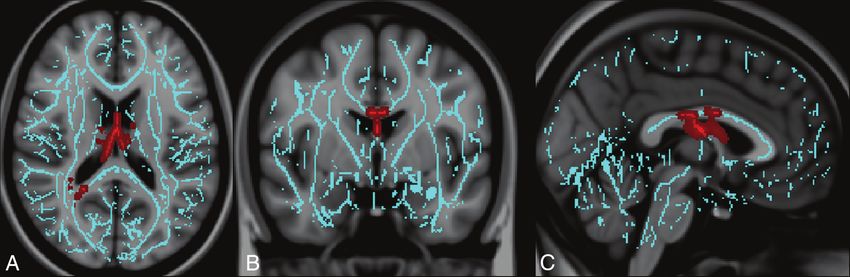
AJNR Am J Neuroradiol ●:● ● 2017 www.ajnr.org 3FIG 1. Regions of injury involving the corpus callosum and fornix/septohippocampal circuit among patients with posttraumatic migraine
headaches. Images derived from Tract-Based Spatial Statistics results and rendered on T1-weighted images from the Montreal Neurological
Institute atlas show common regions of injury involving the corpus callosum and fornix/septohippocampal circuit in patients with posttrau-
matic migraine headaches. ROIs involving the FSHC and CC corresponding to voxels of significant difference in FA (P ⬍ .05 corrected for multiple
comparisons) between controls (subjects with mTBI without PTM) and patients with mTBI with PTM in are shown overlaid on the white matter
skeleton (blue). ROIs corresponding to voxels of significantly decreased FA in patients with mTBI with PTM are shown in red in the axial (A),
coronal (B), and sagittal (C) planes.
Table 3: Comparison of FA in ROIs for patients with mTBI with implicated in cortical spreading depression, a propagating, tem-
and without PTMs porary loss of membrane potential in neurons thought to play a
Patients with Patients with P Valuea
part in the pathophysiology of migraines.20,22
mTBI + PTMs mTBI − PTMs (db)
The FSHC contains serotonergic pathways. Multiple studies
Mean FA 0.688 (0.581–0.793) 0.721 (0.615–0.823) .028 (.680)
value, CC have documented the important role of central serotonergic sys-
(95% CI) tems in the neurobiology of migraine, with abnormalities seen in
Mean FA 0.315 (0.284–0.344) 0.327 (0.298–0.356) .045 (.827) serotonin (5-HT) reuptake/metabolism and 5-HT-related signal-
value, FSHC ing both during and between migraine episodes. In fact, inade-
(95% CI)
quate signaling along important pain and limbic 5-HT pathways
a
P values were 2-tailed and calculated with an unpaired t test.
b
The value of the Cohen d.
is thought to be a primary component of the pathobiology of
migraine headache.23 The FSHC is part of the serotonin projec-
Table 4: Correlation of FA with clinical findings for ROIs in tions along the median raphé nucleus– dorsal hippocampal path-
patients with mTBI and PTMs
way. Fibers of this tract that run through the FSHC are very thin,
Correlation Correlation
with Mean with Mean with small varicosities, and are the most susceptible to injury.24
FA Value FA Value Treatment of migraine headaches in the nontraumatic setting
Variable CC ROI FSHC ROI is based mainly on the assumption that abnormalities of serotonin
Agea (P value)b 0.092 (.49) ⫺0.141 (.29) signaling result in an inflammatory process that can be inhibited
Verbal Memory Score (P value) ⫺0.120 (.37) ⫺0.167 (021) with serotonin receptor agonists. The discovery of sumatriptan, a
Visual Memory Score (P value) ⫺0.122 (.36) 0.325 (.01)
full agonist of the serotonin receptors (5-HT1b,1d,1f) revolution-
Processing-speed score (P value) ⫺0.133 (.32) ⫺0.143 (.28)
Reaction time score (P value) ⫺0.072 (.59) ⫺0.113 (.40) ized the treatment of migraines in the general population.23 Pilot
Symptom Severity Score (P value) 0.156 (.24) ⫺0.015 (.91) studies of sumatriptan use in PTM have demonstrated efficacy in
Time to recovery (P value) ⫺0.260 (.05) 0.099 (.46) the mTBI population in the acute setting.25,26 The FSHC and CC
a
Correlation was performed with the Pearson correlation coefficient. both express the 5-HT1a receptor,23,27-29 for which serotonin is a
b
Two-tailed P value for the Pearson correlation coefficient.
partial agonist.
these studies did not directly interrogate the microstructural in- Although quantitative use of DTI metrics in the clinical setting
tegrity of the white matter, despite evidence that white matter is currently limited by the lack of universally accepted normative
injury is a common feature of both mTBI and migraines.11,14,15 data, demonstrating injured regions associated with PTM is the
Our DTI-based evaluation of white matter microstructure in first step toward an individual diagnostic tool. Furthermore,
PTM demonstrated common abnormalities in 2 major regions: knowledge that injuries to these pathways result in migraines may
the CC and FSHC. Both of these regions have been implicated in help radiologists recognize the relationship between macroscopic
the pathogenesis of nontraumatic migraines. DTI abnormalities lesions in these areas (eg, macroscopic trauma, multiple sclerosis)
in the CC are commonly seen in nontraumatic migraine, in which and patient headache symptomatology.
they are associated with a more chronic disease course, greater Our study has limitations. Our evaluation was a retrospective,
headache frequency, and comorbid neuropsychiatric condi- single-institution study with a moderate sample size. Addition-
tions.21 It is therefore not surprising that FA in the CC in our ally, the number of controls in our population was small, largely
study trended toward an inverse correlation with time to recov- because our controls were not healthy controls, but rather pa-
ery. The role of the FSHC in migraines is less clear, but it has been tients with mTBI without migraine symptoms. As a result of the
4 Alhilali ● 2017 www.ajnr.orgvery high prevalence of posttraumatic migraines among patients the visual pathway in patients with migraine: a 3-T tractography
with mTBI,30 it is very difficult to obtain a large cohort of patients study. Cephalgia 2008;28:1061– 68 CrossRef Medline
with mTBI without posttraumatic migraines. Accordingly, the 14. Fakhran S, Yaeger K, Alhilali L. Symptomatic white matter changes
in mild traumatic brain injury resemble pathologic features of early
findings should be corroborated with a multicenter, prospective
Alzheimer dementia. Radiology 2013;269:249 –57 CrossRef Medline
study. Furthermore, because most patients with mTBI do not 15. Alhilali LM, Yaeger K, Collins M, et al. Detection of central white
undergo imaging, there is a possible selection bias toward patients matter injury underlying vestibulopathy after mild traumatic brain
with more severe injuries. However, these are also the more symp- injury. Radiology 2014;272:224 –32 CrossRef Medline
tomatic patients, in whom imaging biomarkers are most needed. 16. Headache Classification Subcommittee of the International Headache
Society. The International Classification of Headache Disorders: 2nd
edition. Cephalalgia 2004;24(suppl 1):9 –160 CrossRef Medline
CONCLUSIONS 17. Smith SM, Jenkinson M, Johansen-Berg H, et al. Tract-based spatial
White matter injuries in patients with mTBI with PTM may indi- statistics: voxelwise analysis of multi-subject diffusion data. Neuro-
cate that axonal injury can result in migraine symptomatology. image 2006;31:1487–505 CrossRef Medline
White matter injuries in patients with PTM were in regions im- 18. Giorgio A, Watkins KE, Chadwick M, et al. Longitudinal changes in
plicated in the pathophysiology of nontraumatic migraines, grey and white matter during adolescence. Neuroimage 2010;49:94 –
which may help elucidate the biologic underpinning of disease 103 CrossRef Medline
19. Sarmento E, Moreira P, Brito C, et al. Proton spectroscopy in pa-
processes, direct treatment, and guide future research.
tients with post-traumatic headache attributed to mild head injury.
Headache 2009;49:1345–52 CrossRef Medline
REFERENCES 20. Bartsch T, Goadsby PJ. The trigeminocervical complex and
1. Gardner RC, Yaffe K. Epidemiology of mild traumatic brain injury migraine: current concepts and synthesis. Curr Pain Headache Rep
and neurodegenerative disease. Mol Cell Neurosci 2015;66(pt B): 2003;7:371–76 CrossRef Medline
75– 80 CrossRef Medline 21. Li XL, Fang YN, Gao QC, et al. A diffusion tensor magnetic reso-
2. Kontos A, Elbin R, Lau B, et al. Posttraumatic migraine as a predic- nance imaging study of corpus callosum from adult patients with
tor of recovery and cognitive impairment after sports-related con- migraine complicated with depressive/anxious disorder. Headache
cussion. Am J Sports Med 2013;41:1497–504 CrossRef Medline 2011;51:237– 45 CrossRef Medline
3. Mihalik JP, Register-Mihalik J, Kerr ZY, et al. Recovery of posttrau- 22. Moskowitz MA, Nozaki K, Kraig RP. Neocortical spreading depres-
matic migraine characteristics in patients after mild traumatic sion provokes the expression of c-fos protein-like immunoreactiv-
brain injury. Am J Sports Med 2013;41:1490 –96 CrossRef Medline ity within trigeminal nucleus caudalis via trigeminovascular mech-
4. Carlson K, Taylor B, Hagel E, et al. Headache diagnoses among Iraq anisms. J Neurosci 1993;13:1167–77 Medline
and Afghanistan war veterans enrolled in VA: a gender comparison. 23. Hamel E. Serotonin and migraine: biology and clinical implica-
Headache 2013;53:1573– 82 CrossRef Medline tions. Cephalalgia 2007;27:1293–300 CrossRef Medline
5. D’Onofrio F, Russo A, Conte F, et al. Post-traumatic headaches: an 24. Murphy DL, Andrews AM, Wichems CH, et al. Brain serotonin
epidemiological overview. Neurol Sci 2014;35(suppl 1):203– 06 neurotransmission: an overview and update with an emphasis on
CrossRef Medline serotonin subsystem heterogeneity, multiple receptors, interac-
6. Packard RC. Chronic post-traumatic headache: associations with tions with other neurotransmitter systems, and consequent impli-
mild traumatic brain injury, concussion, and post-concussive dis- cations for understanding the actions of serotonergic drugs. J Clin
order. Curr Pain Headache Rep 2008;12:67–73 CrossRef Medline Psychiatry 1998;59(suppl 15):4 –12 Medline
7. Obermann M, Nebel K, Schumann C, et al. Gray matter changes
25. McCrory P, Heywood J, Ugoni A. Open label study of intranasal
related to chronic posttraumatic headache. Neurology 2009;73:
sumatriptan (Imigran) for footballer’s headache. Br J Sports Med
978 – 83 CrossRef Medline
2005;39:552–54 CrossRef Medline
8. Kontos AP, Reches A, Elbin RJ, et al. Preliminary evidence of re-
26. Abend NS, Nance ML, Bonnemann C. Subcutaneous sumatriptan in
duced brain network activation in patients with post-traumatic mi-
an adolescent with acute posttraumatic headache. J Child Neurol
graine following concussion. Brain Imaging Behav 2016;10:594 – 603
2008;23:438 – 40 CrossRef Medline
CrossRef Medline
9. Harris JL, Yeh HW, Choi IY, et al. Altered neurochemical profile 27. Hawthorne AL, Hu H, Kundu B, et al. The unusual response of se-
after traumatic brain injury: (1)H-MRS biomarkers of pathological rotonergic neurons after CNS injury: lack of axonal dieback and
mechanisms. J Cereb Blood Flow Metab 2012;32:2122–34 CrossRef enhanced sprouting within the inhibitory environment of the glial
Medline scar. J Neurosci 2011;31:5605–16 CrossRef Medline
10. Tang L, Ge Y, Sodickson DK, et al. Thalamic resting-state functional 28. Pacheco J, Beevers CG, Benavides C, et al. Frontal-limbic white mat-
networks: disruption in patients with mild traumatic brain injury. ter pathway associations with the serotonin transporter gene pro-
Radiology 2011;260:831– 40 CrossRef Medline moter region (5-HTTLPR) polymorphism. J Neurosci 2009;29:
11. Bashir A, Lipton RB, Ashina S, et al. Migraine and structural changes 6229 –33 CrossRef Medline
in the brain: a systemic review and meta-analysis. Neurology 2013; 29. Caspers S, Schleicher A, Bacha-Trams M, et al. Organization of the
81:1260 – 68 CrossRef Medline human inferior parietal lobule based on receptor architectonics.
12. Yu D, Yuan K, Qin W, et al. Axonal loss of white matter in migraine Cereb Cortex 2013;23:615–28 CrossRef Medline
without aura: a tract-based spatial statistics study. Cephalgia 2013; 30. Lucas S, Hoffman JM, Bell KR, et al. A prospective study of preva-
33:34 – 42 CrossRef Medline lence and characterization of headache following mild traumatic
13. Rocca MA, Pagani E, Colombo B, et al. Selective diffusion changes of brain injury. Cephalalgia 2014;34:93–102 CrossRef Medline
AJNR Am J Neuroradiol ●:● ● 2017 www.ajnr.org 5You can also read