Agreement of novel hemodynamic imaging parameters for the acute and chronic stages of ischemic stroke: a matched-pair cohort study
←
→
Page content transcription
If your browser does not render page correctly, please read the page content below
NEUROSURGICAL
            FOCUS                                                                                                              Neurosurg Focus 51 (1):E12, 2021

                          Agreement of novel hemodynamic imaging parameters
                          for the acute and chronic stages of ischemic stroke: a
                          matched-pair cohort study
                          *Martina Sebök, MD,1,2 Christiaan Hendrik Bas van Niftrik, MD, PhD,1,2 Susanne Wegener, MD,2,3
                          Andreas Luft, MD,2,3 Luca Regli, MD,1,2 and Jorn Fierstra, MD, PhD1,2
                          Departments of 1Neurosurgery and 3Neurology, and 2Clinical Neuroscience Center, University Hospital Zurich, University of
                          Zurich, Switzerland

                          OBJECTIVE In symptomatic patients with cerebrovascular steno-occlusive disease, impaired blood oxygenation level–
                          dependent cerebrovascular reactivity (BOLD-CVR) and increased flow velocity of the P2 segment of the posterior cere-
                          bral artery (PCA-P2) on transcranial Doppler (TCD) ultrasonography have been introduced as emerging clinical imaging
                          parameters to identify patients at high risk for recurrent ischemic events. Since hemodynamic physiology differs between
                          the acute and chronic stages of ischemic stroke, the authors sought to investigate whether those parameters have merit
                          for both the acute and chronic stages of ischemic stroke.
                          METHODS From a prospective database, patients who underwent BOLD-CVR and TCD examinations in the acute
                          stroke stage (< 10 days) were matched to patients in the chronic stroke stage (> 3 months). A linear regression analysis
                          for both groups was performed between ipsilateral PCA-P2 systolic flow velocity and BOLD-CVR of the ipsilateral (af-
                          fected) hemisphere, the ipsilateral middle cerebral artery (MCA) territory, and the ipsilateral steal volume (i.e., para-
                          doxical BOLD-CVR response). The resulting slopes and intercepts were statistically compared to evaluate differences
                          between groups.
                          RESULTS Forty matched patient pairs were included. Regression analysis showed no significant difference for either
                          the intercept (p = 0.84) or the slope (p = 0.85) between PCA-P2 flow velocity and BOLD-CVR as measured for the ipsilat-
                          eral (affected) hemisphere. Similarly, no significant difference was seen between PCA-P2 flow velocity and BOLD-CVR
                          of the ipsilateral MCA territory (intercept, p = 0.72; slope, p = 0.36) or between PCA-P2 flow velocity and steal volume
                          (intercept, p = 0.59; slope, p = 0.34).
                          CONCLUSIONS The study results indicated that the relationship between ipsilateral PCA-P2 systolic flow velocity and
                          BOLD-CVR remains the same during the acute and chronic stages of ischemic stroke. This provides further support
                          that these novel hemodynamic imaging parameters may have merit to assess the risk for recurrent ischemic events for
                          a wide ischemic stroke population. PCA-P2 systolic flow velocity, in particular, may be a highly practical screening tool,
                          independent of ischemic stroke stage.
                          https://thejns.org/doi/abs/10.3171/2021.4.FOCUS21125
                          KEYWORDS BOLD-CVR; PCA-P2 flow; hemodynamic; steno-occlusive disease; stroke

T
       o advance ischemic stroke care, clinically versatile                            To address this well-known challenge, we have recently
       imaging parameters to identify patients with symp-                           validated two emerging imaging parameters derived from
       tomatic cerebrovascular steno-occlusive disease                              existing clinical imaging techniques that have ample avail-
at highest risk for recurrent stroke are strongly desired.1                         ability in routine ischemic stroke management in patients
Although increased oxygen extraction fraction from PET                              with symptomatic cerebrovascular steno-occlusive disease
and impaired cerebrovascular reactivity (CVR) from xe-                              as follows:
non-CT and SPECT have been strongly associated with                                 1. Increased ipsilateral systolic flow of the P2 segment
recurrent stroke risk,2–5 these imaging modalities are not                             of the posterior cerebral artery (PCA-P2) from trans-
feasible for routine clinical stroke imaging.                                          cranial Doppler (TCD) ultrasonography.6,7 TCD ul-

ABBREVIATIONS BOLD = blood oxygenation level–dependent; CVR = cerebrovascular reactivity; ICA = internal carotid artery; MCA = middle cerebral artery; MP-RAGE =
magnetization-prepared rapid acquisition gradient echo; PCA-P2 = P2 segment of the posterior cerebral artery; TCD = transcranial Doppler.
SUBMITTED February 28, 2021. ACCEPTED April 7, 2021.
INCLUDE WHEN CITING DOI: 10.3171/2021.4.FOCUS21125.
* M.S. and C.H.B.v.N. contributed equally to this work.

©AANS 2021, except where prohibited by US copyright law                                                        Neurosurg Focus Volume 51 • July 2021           1

                                                                                                               Unauthenticated | Downloaded 10/10/21 07:56 PM UTC
Sebök et al.
trasonography is routinely used in the diagnosis and older with symptomatic unilateral steno-occlusive disease
management of symptomatic steno-occlusive disease.8 and 2) who exhibited focal neurological symptoms that
Increased PCA-P2 systolic flow velocity is a surrogate were sudden in onset and referable to the appropriate ante-
marker for the necessity of leptomeningeal collateral rior circulation large-artery distribution (ipsilateral to the
activation to compensate a state of hypoperfusion6,9 and significant large-vessel atherosclerotic pathology), includ-
has been linked to increased risk for recurrent ischemia ing one or more transient ischemic attacks, characterized
and increased steal volume (i.e., paradoxical CVR).6,10 by focal neurological dysfunction or transient monocular
2. CVR measured quantitatively with blood oxygenation blindness, or one or more minor (nondisabling) ischemic
level–dependent (BOLD) functional MRI during a stan- strokes.21 Unilateral disease was considered as a maximal
dardized hypercapnic—CO2—stimulus (BOLD-CVR) stenosis of 50% on the contralateral side graded by duplex
has demonstrated a good agreement with hemodynam- sonography according to the NASCET (North American
ic failure derived from acetazolamide-challenged (15O-) Symptomatic Carotid Endarterectomy Trial) criteria.22
H2O-PET.7,11,12 CVR describes the remaining vasodila- First, all symptomatic patients with unilateral steno-
tory capacity at the brain tissue level, whereas impaired occlusive disease who underwent a separate clinical TCD
CVR,12–14 especially paradoxical BOLD-CVR (i.e., ultrasonography investigation were selected from the
steal volume), has been associated with an increased database. For the acute group (scanning performed < 10
risk for recurrent ischemic events.1,15,16 days from the stroke event), only patients who underwent
Recently, we have found a strong agreement between both investigations within 1 week were included in this
PCA-P2 systolic flow velocity and BOLD-CVR, with study. For the chronic group (scanning performed > 90
higher PCA-P2 systolic flow velocity values correlating to days from the stroke event), we allowed for an interval of
impaired BOLD-CVR and steal volume.7,17 This makes 8 weeks, and TCD ultrasonography could not have been
PCA-P2 flow velocity, which can even be performed at the performed in the acute stroke phase. Patients with an in-
bedside,8 eligible as a clinical screening tool for symptom- sufficient insonation bone window by TCD examination, a
atic steno-occlusive patients prone to ischemic stroke re- vascular pathology of the posterior circulation, or bilateral
currence who are in need of a further hemodynamic work- anterior circulation steno-occlusive disease (inclusive of
up to assess brain tissue perfusion, typically obtained with moyamoya disease) were excluded from further analysis.
advanced neuroimaging techniques such as PET, SPECT, After evaluation, 165 patients (96 with acute stroke and
or BOLD-CVR. 69 with chronic stroke) qualified for inclusion. Based on
Hemodynamic adaptations, such as collateral flow ac- the average BOLD-CVR values of the ipsilateral hemi-
tivation and compensatory autoregulation of the cerebro- sphere, we were able to match 80 patients (40 per group)
vascular reserve capacity, however, may follow a different and include them for further analysis. Patients who under-
pattern for various stages of ischemic stroke.14,18–20 Most went both BOLD-CVR and TCD ultrasonography studies
studies investigating ischemic stroke hemodynamics in- in the acute stage of ischemic stroke (< 10 days) composed
terchangeably merge patient data from acute stages with the acute hemodynamic cohort, whereas patients who un-
chronic stages after ischemic stroke. Therefore, it is un- derwent both examinations in the chronic stroke stage (> 3
known whether increased ipsilateral PCA-P2 flow activa- months) made up the chronic hemodynamic cohort. Some
tion and ipsilateral BOLD-CVR impairment have clinical patient data from this cohort have been reported previ-
merit for both the acute and chronic stages of ischemic ously.7,17
stroke.7,12,17
Hence, our aim was to compare symptomatic patients Image Acquisition and Analysis
with steno-occlusive disease who underwent BOLD-CVR BOLD functional MRI images were obtained using a
and TCD ultrasonography in the acute stage of ischemic 3-T Skyra VD13 (Siemens Healthcare) scanner with CO2
stroke (< 10 days) with those who underwent imaging in as the vasoactive stimulus modulated by a computer-
the chronic stage (> 3 months) using a matched-pair co- controlled gas blender with prospective gas-targeting al-
hort study design. gorithms (RespirAct, Thornhill Research Institute). This
allowed for a short 80-second duration of iso-oxic hyper-
capnia to induce a vascular response. A high-resolution
Methods T1-weighted magnetization-prepared rapid acquisition
Patient Selection gradient echo (MP-RAGE) image was obtained for ana-
The data that support the findings of this study are tomical overlay of the BOLD-CVR images. All BOLD-
available on reasonable request from the corresponding CVR images were obtained using a previously published
author (M.S.). This project is part of an ongoing BOLD- method,23 which has been used in multiple studies.14,17,20
CVR study in patients with symptomatic cerebrovascular BOLD-CVR, defined as the percentage of the BOLD sig-
steno-occlusive disease that has been approved by the lo- nal change/mm Hg CO2, was calculated from the slope of
cal research ethics board (Kantonale Ethikkommission a linear least-square fit of the BOLD signal time course to
Zurich). Written informed consent was obtained from the CO2 time course during the BOLD scan.23
each participant before inclusion in the database. The From the T1-weighted MP-RAGE scan, a probabil-
study was conducted in accordance with the ethical stan- ity map for the gray matter, white matter, and CSF was
dards as defined in the 1964 Declaration of Helsinki and obtained. Each T1-weighted MP-RAGE image was then
its later amendments. manually masked for the affected hemisphere, and in com-
The inclusion criteria were 1) patient age 18 years or bination with a gray-white matter probability map (> 80%
2 Neurosurg Focus Volume 51 • July 2021
Unauthenticated | Downloaded 10/10/21 07:56 PM UTCSebök et al.
FIG. 1. Study flowchart. From 217 patients with symptomatic steno-occlusive disease included in our prospective BOLD-CVR
database, 165 patients were diagnosed with unilateral steno-occlusive disease and had corroborating BOLD-CVR and TCD
ultrasonography examination findings. Ninety-six of 165 patients underwent both examinations in the acute ischemic stroke stage,
whereas 69 patients underwent both examinations in the chronic stroke stage. After a matching process, based on the ipsilateral
hemisphere BOLD-CVR values, we were able to include 80 patients (40 per group). *Unilateral disease was considered as maxi-
mal stenosis of 50% of the contralateral side.
probability), CVR of the affected hemisphere was calcu- egorical ordinal variables are presented as median (IQR),
lated. whereas dichotomous variables are shown as frequency
(%). To evaluate the comparability of both groups, the chi-
Steal Volume Analysis square test and the Student t-test were used. A 2-sided p
Steal volume, describing the number of voxels with value < 0.05 was considered significant. A linear regres-
paradoxical (i.e., negative) BOLD-CVR and measured sion analysis for both groups was performed between the
in milliliters, was determined by measuring the number ipsilateral PCA-P2 systolic flow velocity and BOLD-CVR
of voxels in the whole brain as well as in the ipsilateral of the ipsilateral hemisphere, and BOLD-CVR of the ipsi-
and contralateral hemispheres, with < 0% BOLD signal lateral MCA territory and the ipsilateral steal volume. The
change/mm Hg CO2 after exclusion of voxels around the resulting slopes and intercepts were statistically compared
frontal sinus in order to exclude artifacts. to evaluate differences between groups.
Analysis of the MCA Territory Results
Quantitative BOLD-CVR values of the middle cerebral Study Population Characteristics
artery (MCA) territory of the ipsilateral and contralateral A flowchart illustrating patient screening and inclu-
hemispheres were determined by applying a vascular atlas sion is shown in Fig. 1. In the acute hemodynamic cohort,
to the normalized CVR maps. This vascular atlas was de- which included patients in an acute ischemic stroke stage,
rived from the predefined brain regions listed in the stand- 20 patients with internal carotid artery (ICA) occlusion, 1
ard N30R83 atlas by Hammers et al.24 and Kuhn et al.25 patient with MCA occlusion, 4 patients with tandem (ICA
and MCA) occlusion, 13 patients with ICA stenosis, and
Statistical Analysis 2 patients with MCA stenosis were included. The chronic
Statistical analysis was performed using IBM SPSS hemodynamic cohort consisted of 23 patients with ICA
Statistics version 26 (IBM Corp.). All normally distrib- occlusion, 1 patient with MCA occlusion, 15 patients with
uted continuous variables are reported as mean ± SD. Cat- ICA stenosis, and 1 patient with MCA stenosis.
Neurosurg Focus Volume 51 • July 2021 3
Unauthenticated | Downloaded 10/10/21 07:56 PM UTCSebök et al.
TABLE 1. Baseline characteristics of included symptomatic steno-occlusive patients
All Patients (n = 80) Acute Cohort (n = 40) Chronic Cohort (n = 40) p Value
Mean age, yrs 67.89 ± 10.41 69.51 ± 10.90 66.26 ± 9.77 0.16
Male sex 66 (82.5) 31 (77.5) 35 (87.5) 0.25
Mean vol of ischemic lesions, ml 10.81 ± 23.37 12.00 ± 17.70 9.61 ± 28.11 0.65
Smoking 43 (53.8) 16 (40) 27 (67.5) 0.01
Hypertension 51 (63.7) 26 (65.0) 25 (62.5) 0.82
Hypercholesterolemia 36 (45) 20 (50) 16 (40) 0.38
Obesity 14 (17.5) 10 (25) 4 (10) 0.08
Diabetes 12 (15) 5 (12.5) 7 (17.5) 0.54
Positive family history for cerebral ischemic events 8 (10) 2 (5) 6 (15) 0.14
Values represent the number of patients (%) unless stated otherwise. Mean values are presented as the mean ± SD. Boldface type indicates statistical significance.
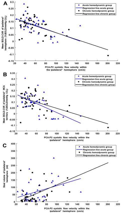
Table 1 shows the relevant clinical and baseline char- a moderate association between PCA-P2 flow velocity and
acteristics of the enrolled patients. As shown in Table 2, BOLD-CVR for the acute phase (R = −0.53, R2 = 0.28, p
no significant differences were found in relevant BOLD- < 0.001) and a good correlation for the chronic phase (R =
CVR values of the whole brain, hemispheres, MCA ter- −0.60, R2 = 0.36, p < 0.001). Regression analysis (Fig. 2A)
ritories, steal volumes, or PCA-P2 systolic flow velocities. showed no significant difference for either the intercept (p
= 0.84) or the slope (p = 0.85), indicating that a difference
Linear Regression Analysis for the Acute and Chronic in the poststroke phase does not change the relationship
Hemodynamic Cohorts between PCA-P2 flow velocity and BOLD-CVR as mea-
PCA-P2 Systolic Flow Velocity and Ipsilateral Hemisphere sured for the ipsilateral (affected) hemisphere.
BOLD-CVR
PCA-P2 Systolic Flow Velocity and Ipsilateral MCA Territory
The linear regression curve between the quantitative BOLD-CVR
ipsilateral PCA-P2 systolic flow velocity values and the
mean ipsilateral hemisphere BOLD-CVR values showed Similarly, the linear regression curve between the quan-
titative ipsilateral PCA-P2 systolic flow velocity values
and the mean ipsilateral (affected) MCA territory BOLD-
CVR values showed a strong association in the acute (R =
−0.61, R2 = 0.36, p < 0.001) and chronic (R = −0.54, R2 =
TABLE 2. BOLD-CVR and TCD values of acute and chronic
0.29, p < 0.001) stroke phases. Regression analysis showed
symptomatic steno-occlusive patient cohorts
that the stroke phase had no influence on this relationship
Acute Cohort Chronic Cohort p (intercept, p = 0.72; slope, p = 0.36) (Fig. 2B).
(n = 40) (n = 40) Value
PCA-P2 Systolic Flow Velocity and Affected Hemisphere
Mean CVR, % BOLD/mm Steal Volume
Hg CO2
Lastly, we tested the relationship between the quantita-
Whole brain 0.13 ± 0.06 0.14 ± 0.06 0.74 tive ipsilateral PCA-P2 systolic flow velocity values and
Gray matter 0.17 ± 0.11 0.16 ± 0.06 0.54 the steal volume of the ipsilateral (affected) hemisphere.
White matter 0.10 ± 0.05 0.10 ± 0.05 0.89 Both the acute and chronic stages, separately, showed a
Contralat hemisphere 0.15 ± 0.06 0.16 ± 0.05 0.72 significant correlation (acute cohort: R = −0.39, R2 = 0.15,
Ipsilat MCA territory 0.08 ± 0.08 0.10 ± 0.08 0.39 p = 0.013; and chronic cohort: R = −0.64, R2 = 0.40, p <
Contralat MCA territory 0.15 ± 0.06 0.16 ± 0.05 0.31
0.001). The correlation for the chronic stroke phase was
not significantly different than the acute stroke phase (in-
Mean steal vol, ml tercept, p = 0.59; slope, p = 0.34) (Fig. 2C).
Whole brain 109.00 ± 113.32 86.42 ± 115.49 0.38
Ipsilat hemisphere 70.40 ± 69.28 54.73 ± 76.36 0.34
Discussion
Contralat hemisphere 38.61 ± 58.75 31.69 ± 45.21 0.56
The current matched-pair cohort study shows that the
Median SFV of PCA-P2, relationship between ipsilateral PCA-P2 systolic flow ve-
cm/sec locity and BOLD-CVR does not change for the acute and
Ipsilat 57 (34) 52 (33) 0.70 chronic stages of ischemic stroke. This may have clini-
Contralat 55 (28) 51 (30) 0.85 cal merit for a wide stroke population in order to identify
SFV = systolic flow velocity.
symptomatic steno-occlusive patients at risk for recurrent
The ipsilateral hemisphere is considered the hemisphere on the side of the ischemic events. Increased ipsilateral systolic PCA-P2 flow
symptomatic steno-occlusive disease. Values are presented as the mean ± SD velocity, in particular, has been shown to independently
unless otherwise stated. Median values are presented as the median (IQR). correlate with impaired BOLD-CVR7 and steal volume17
4 Neurosurg Focus Volume 51 • July 2021
Unauthenticated | Downloaded 10/10/21 07:56 PM UTCSebök et al.
FIG. 2. Regression analysis of PCA-P2 systolic flow velocity and different BOLD-CVR parameters. A: Regression analysis showing no significant
difference for both the intercept (p = 0.84) and slope (p = 0.85), indicating that the difference in the poststroke phase does not change the relationship
between ipsilateral PCA-P2 systolic flow velocity and BOLD-CVR of the ipsilateral hemisphere. FIG. 2. (continued)→
Neurosurg Focus Volume 51 • July 2021 5
Unauthenticated | Downloaded 10/10/21 07:56 PM UTCSebök et al.
FIG. 2. B: Regression analysis showing no influence (intercept, p = 0.72; slope, p = 0.36) of the stroke phase on the relationship between ipsilateral
PCA-P2 systolic flow velocity and BOLD-CVR of the ipsilateral MCA territory. C: Regression analysis showing no significant difference (intercept, p =
0.59; slope, p = 0.34) between the acute and chronic hemodynamic groups for the relationship between ipsilateral PCA-P2 systolic flow velocity and steal
volume of the ipsilateral hemisphere. *The ipsilateral hemisphere is considered the hemisphere on the side of the symptomatic steno-occlusive disease.
and is increased in patients with recurrent stroke.6 This acute and chronic stages of stroke. Such studies are lack-
association describes the necessity of leptomeningeal ac- ing, as advanced hemodynamic imaging studies with PET
tivation to compensate for a state of hypoperfusion,6,9 and or SPECT are usually difficult and expensive to obtain in a
makes PCA-P2 systolic flow velocity an ideal screening large cohort of stroke patients with varying stroke stages.
method for stroke patients requiring further hemodynamic
workup. BOLD-CVR, the measurement of intracranial he- Hemodynamic Impairment and Its Correlation With PCA-P2
modynamic status at the brain tissue level, is a quantitative Flow Velocity
and highly reproducible imaging method and has shown a Previous studies highlighted the importance of sys-
good agreement with CVR as measured using the clinical tolic PCA-P2 flow velocity, as measured by TCD, as an
gold-standard (15O-)H2O-PET examination.12 To allow for independent marker of intracranial hemodynamic status
an optimal investigation of the relationship between PCA- in patients with steno-occlusive disease.6,17 Since TCD is a
P2 flow velocities and BOLD-CVR during different stroke noninvasive bedside method, it can be repeatedly used to
stages, patients were matched based on the mean BOLD- evaluate the flow velocity of intra- and extracranial vessels,
CVR of the ipsilateral hemisphere. We found that the sys- as well as the presence of primary and secondary collater-
tolic PCA-P2 flow velocity of the ipsilateral hemisphere als, without the need for an exogenous contrast agent.8,33
did correlate during both the acute and chronic stroke
A recent study showed that in patients with symptomatic
stages with different BOLD-CVR measurements (ipsilat-
unilateral steno-occlusive disease, increased ipsilateral
eral hemisphere BOLD-CVR, BOLD-CVR of the ipsilat-
eral MCA territory, and volume of the region with the steal TCD PCA-P2 systolic flow velocity is a strong independent
phenomenon [i.e., paradoxical BOLD-CVR]). Moreover, predictor of hemodynamic impairment in the ipsilateral
no differences in the intercept and slope between each of hemisphere and MCA territory.7 A flow increase > 30%
the correlations were found, meaning that the influence in the ipsilateral PCA-P2 segment compared with the con-
of the stroke phase (acute vs chronic) on this relationship tralateral PCA-P2 segment indicates leptomeningeal col-
has been indiscernible. This indicates that systolic PCA-P2 lateral flow, known as a secondary collateral pathway.6,9,34
flow velocity can be used as an independent hemodynamic Such pathways are recruited once primary collaterals19,35
parameter in both acute and chronic hemodynamic stages have failed to sufficiently compensate, indicating a more
in patients with symptomatic unilateral anterior circula- severe impairment.10,36 Therefore, a necessary activation of
tion steno-occlusive disease. secondary collaterals (e.g., leptomeningeal pial branches
over the posterior circulation) has been associated with an
Hemodynamic Features of Anterior Circulation increased risk of recurrent stroke.6,10,18
Steno-Occlusive Disease Using the systolic PCA-P2 flow velocity of the ipsilat-
eral hemisphere, we have found a significant correlation
Atherosclerotic steno-occlusive disease describes a vas- between different quantitative BOLD-CVR measurements
cular state in which the extracranial or intracranial arteries on the brain tissue level without an influence of the hemo-
supplying the brain show either a stenosis (abnormal nar- dynamic stage (acute vs chronic) on this correlation. The
rowing of a blood vessel) or an occlusion (complete block- positive correlation between PCA-P2 flow velocity and
age of a blood vessel). A stenosis or occlusion of the ap- steal volume is especially interesting. Steal volume (i.e., a
propriate anterior circulation large artery is an important paradoxical, negative BOLD-CVR response to hypercap-
cause of anterior circulation ischemic stroke and stroke nia) is a prime hemodynamic parameter or hemodynamic
recurrence and has been linked to chronic deficiencies in
failure type 212 and describes the classic state of hypoper-
regional cerebral blood flow.26,27
fusion (i.e., tissue at risk for recurrent ischemic stroke).37
Acute ischemia results in irreversible loss of brain
tissue and function,28 whereas the presence of a chronic This indicates that despite hemodynamic changes over
cerebrovascular steno-occlusive disease can alter cerebral time, PCA-P2 flow velocity remains linked to the present
hemodynamics up to a point where brain tissue perfu- hemodynamic state of patients with anterior circulation
sion becomes insufficient (chronic hypoperfusion).11,29–31 steno-occlusive disease.
The consequences of chronic intermittent hypoperfusion
are still not completely understood. It is supposed that the Clinical Implications and Future Direction
perfusion of brain tissue may be just sufficient enough to The good agreement found between increased ipsilat-
prevent gross ischemia but may fail to respond adequately eral P2 TCD flow velocity and impaired BOLD-CVR for
to increases in demand such as those normally seen dur- the acute and chronic stages of ischemic stroke has a po-
ing neuronal activation.1,27 If hemodynamic impairment tentially important clinical application for a wide stroke
is present, it is hypothesized to alter brain structure and population, since both novel parameters have individu-
function.27,32 Between acute and chronic stages of stroke, ally been associated with hemodynamic failure6,7,12 (i.e.,
an evolution in collateral flow occurs either parallel to or the imaging parameter correlated with recurrent ischemic
as a response to these changes;18,33 therefore, the efficacy stroke1,15,16).
of hemodynamic parameters should be tested for both the Furthermore, this study has underlined the value of
6 Neurosurg Focus Volume 51 • July 2021
Unauthenticated | Downloaded 10/10/21 07:56 PM UTCSebök et al.
TCD ultrasonography in combination with a quantifiable cerebral blood flow reactivity is a predictor of stroke in
hemodynamic parameter (i.e., increased ipsilateral P2 flow patients with symptomatic carotid artery occlusive disease. J
velocity) as a screening tool for a wide stroke population Vasc Surg. 1995;21(2):338–345.
4. Powers WJ, Zazulia AR. PET in cerebrovascular disease.
in order to identify patients with potential hemodynamic PET Clin. 2010;5(1):83–106.
failure6,7 that will need further imaging workup with ei- 5. Grubb RL Jr, Powers WJ, Clarke WR, et al. Surgical results
ther BOLD-CVR, acetazolamide-challenged H2O-PET, of the Carotid Occlusion Surgery Study. J Neurosurg. 2013;
SPECT, MR perfusion, or whichever modality is consid- 118(1):25–33.
ered the institutional clinical standard to assess brain tis- 6. Schneider J, Sick B, Luft AR, Wegener S. Ultrasound and
sue perfusion. clinical predictors of recurrent ischemia in symptomatic
Finally, although randomized controlled clinical tri- internal carotid artery occlusion. Stroke. 2015;46(11):
als38,39 have failed to show a benefit of cerebral bypass 3274–3276.
7. van Niftrik CHB, Sebök M, Wegener S, et al. Increased
revascularization for stroke prevention, routine clinical ex- ipsilateral posterior cerebral artery P2-segment flow veloc-
perience has indicated that a subset of patients with both ity predicts hemodynamic impairment. Stroke. 2021;52:
acute and chronic ischemic stroke exhibiting impaired or 1469–1472.
even exhausted cerebral hemodynamic status could ben- 8. Bonow RH, Young CC, Bass DI, et al. Transcranial Doppler
efit from surgical or endovascular recanalization proce- ultrasonography in neurological surgery and neurocritical
dures.40–42 To detect at-risk patients requiring further and care. Neurosurg Focus. 2019;47(6):E2.
more complex hemodynamic investigations, a noninvasive 9. Reinhard M, Müller T, Guschlbauer B, et al. Dynamic cere-
bral autoregulation and collateral flow patterns in patients
and bedside-available TCD ultrasonography–derived sys- with severe carotid stenosis or occlusion. Ultrasound Med
tolic PCA-P2 flow velocity can be widely implemented as Biol. 2003;29(8):1105–1113.
an additional triage investigation.8 10. Klijn CJ, Kappelle LJ, van Huffelen AC, et al. Recurrent is-
chemia in symptomatic carotid occlusion:prognostic value of
Limitations hemodynamic factors. Neurology. 2000;55(12):1806–1812.
11. Fierstra J, Poublanc J, Han JS, et al. Steal physiology is spa-
The inclusion of patients with different unilateral symp- tially associated with cortical thinning. J Neurol Neurosurg
tomatic anterior circulation ischemic stroke pathology Psychiatry. 2010;81(3):290–293.
may have influenced our findings. Specifically, our study 12. Fierstra J, van Niftrik C, Warnock G, et al. Staging hemody-
enrolled a mixed cohort of symptomatic patients with namic failure with blood oxygen-level-dependent functional
steno-occlusive disease; that is, patients with occlusion as magnetic resonance imaging cerebrovascular reactivity:a
well as stenosis were included. Cerebral autoregulation comparison versus gold standard (15O-)H2O-positron emis-
was reported to worsen between the acute and subacute sion tomography. Stroke. 2018;49(3):621–629.
stroke phase; therefore, this may have influenced our re- 13. Mandell DM, Han JS, Poublanc J, et al. Mapping cerebro-
vascular reactivity using blood oxygen level-dependent MRI
sults, as BOLD-CVR and TCD ultrasonography investiga- in Patients with arterial steno-occlusive disease:comparison
tions were not always done on the same day.43,44 with arterial spin labeling MRI. Stroke. 2008;39(7):2021–
2028.
Conclusions 14. Sebök M, van Niftrik CHB, Piccirelli M, et al. BOLD cere-
brovascular reactivity as a novel marker for crossed cerebel-
Our study indicates that the relationship between ip- lar diaschisis. Neurology. 2018;91(14):e1328–e1337.
silateral PCA-P2 systolic flow velocity and BOLD-CVR 15. Silvestrini M, Vernieri F, Pasqualetti P, et al. Impaired
does not change for the acute and chronic stages of is- cerebral vasoreactivity and risk of stroke in patients with
chemic stroke. This provides further support that these asymptomatic carotid artery stenosis. JAMA. 2000;283(16):
novel hemodynamic imaging parameters may have merit 2122–2127.
16. Schoof J, Lubahn W, Baeumer M, et al. Impaired cerebral
to assess the risk for recurrent ischemic events for a wide autoregulation distal to carotid stenosis/occlusion is associ-
ischemic stroke population. PCA-P2 systolic flow velocity, ated with increased risk of stroke at cardiac surgery with
in particular, may be a highly practical screening tool, in- cardiopulmonary bypass. J Thorac Cardiovasc Surg. 2007;
dependent of ischemic stroke stage. 134(3):690–696.
17. von Bieberstein L, van Niftrik CHB, Sebök M, et al. Crossed
cerebellar diaschisis indicates hemodynamic compromise
Acknowledgments in ischemic stroke patients. Transl Stroke Res. 2021;12(1):
This project was funded by the Clinical Research Priority 39–48.
Program of the University of Zurich (UZH CRPP Stroke). Dr. 18. Klijn CJ, Kappelle LJ. Haemodynamic stroke:clinical
Susanne Wegener received funding from the Swiss National features, prognosis, and management. Lancet Neurol. 2010;
Science Foundation (SNSF PP00P3_170683). 9(10):1008–1017.
19. van Everdingen KJ, Visser GH, Klijn CJ, et al. Role of col-
lateral flow on cerebral hemodynamics in patients with uni-
References lateral internal carotid artery occlusion. Ann Neurol. 1998;
1. Yonas H, Smith HA, Durham SR, et al. Increased stroke risk 44(2):167–176.
predicted by compromised cerebral blood flow reactivity. J 20. Hendrik Bas van Niftrik C, Sebok M, Muscas G, et al.
Neurosurg. 1993;79(4):483–489. Characterizing ipsilateral thalamic diaschisis in symptomatic
2. Markus H, Cullinane M. Severely impaired cerebrovascu- cerebrovascular steno-occlusive patients. J Cereb Blood Flow
lar reactivity predicts stroke and TIA risk in patients with Metab. 2020;40(3):563–573.
carotid artery stenosis and occlusion. Brain. 2001;124(pt 3): 21. Barnett HJM, Taylor DW, Haynes RB, et al. Beneficial effect
457–467. of carotid endarterectomy in symptomatic patients with high-
3. Webster MW, Makaroun MS, Steed DL, et al. Compromised grade carotid stenosis. N Engl J Med. 1991;325(7):445–453.
Neurosurg Focus Volume 51 • July 2021 7
Unauthenticated | Downloaded 10/10/21 07:56 PM UTCSebök et al.
22. North American Symptomatic Carotid Endarterectomy Trial. 38. Powers WJ, Clarke WR, Grubb RL Jr, et al. Extracranial-
Methods, patient characteristics, and progress. Stroke. 1991; intracranial bypass surgery for stroke prevention in hemo-
22(6):711–720. dynamic cerebral ischemia:the Carotid Occlusion Surgery
23. van Niftrik CHB, Piccirelli M, Bozinov O, et al. Iterative Study randomized trial. JAMA. 2011;306(18):1983–1992.
analysis of cerebrovascular reactivity dynamic response by 39. EC/IC Bypass Study Group. Failure of extracranial-intra-
temporal decomposition. Brain Behav. 2017;7(9):e00705. cranial arterial bypass to reduce the risk of ischemic stroke.
24. Hammers A, Allom R, Koepp MJ, et al. Three-dimensional Results of an international randomized trial. N Engl J Med.
maximum probability atlas of the human brain, with particu- 1985;313(19):1191–1200.
lar reference to the temporal lobe. Hum Brain Mapp. 2003; 40. Burkhardt JK, Winklhofer S, Fierstra J, et al. Emergency
19(4):224–247. extracranial-intracranial bypass to revascularize salvageable
25. Kuhn FP, Warnock G, Schweingruber T, et al. Quantitative brain tissue in acute ischemic stroke patients. World Neuro-
H2[(15)O]-PET in pediatric moyamoya disease:evaluat- surg. 2018;109:e476–e485.
ing perfusion before and after cerebral revascularization. J 41. Nussbaum ES, Erickson DL. Extracranial-intracranial bypass
Stroke Cerebrovasc Dis. 2015;24(5):965–971. for ischemic cerebrovascular disease refractory to maximal
26. Arenillas JF. Intracranial atherosclerosis:current concepts. medical therapy. Neurosurgery. 2000;46(1):37–43.
Stroke. 2011;42(1)(suppl):S20–S23. 42. Nussbaum ES, Janjua TM, Defillo A, et al. Emergency
27. Fierstra J, Maclean DB, Fisher JA, et al. Surgical revascular- extracranial-intracranial bypass surgery for acute ischemic
ization reverses cerebral cortical thinning in patients with stroke. J Neurosurg. 2010;112(3):666–673.
severe cerebrovascular steno-occlusive disease. Stroke. 2011; 43. Reinhard M, Roth M, Guschlbauer B, et al. Dynamic cere-
42(6):1631–1637. bral autoregulation in acute ischemic stroke assessed from
28. Desai SM, Rocha M, Jovin TG, Jadhav AP. High variability spontaneous blood pressure fluctuations. Stroke. 2005;36(8):
in neuronal loss. Stroke. 2019;50(1):34–37. 1684–1689.
29. Sekhon LH, Morgan MK, Spence I, Weber NC. Chronic ce- 44. Reinhard M, Wihler C, Roth M, et al. Cerebral autoregulation
rebral hypoperfusion and impaired neuronal function in rats. dynamics in acute ischemic stroke after rtPA thrombolysis.
Stroke. 1994;25(5):1022–1027. Cerebrovasc Dis. 2008;26(2):147–155.
30. Muller M, van der Graaf Y, Algra A, et al. Carotid athero-
sclerosis and progression of brain atrophy:the SMART-MR
study. Ann Neurol. 2011;70(2):237–244. Disclosures
31. Bakker FC, Klijn CJ, Jennekens-Schinkel A, et al. Cogni- Dr. Luft: honoraria from Moleac, and speakers bureau for Bayer
tive impairment in patients with carotid artery occlusion and and Amgen.
ipsilateral transient ischemic attacks. J Neurol. 2003;250(11):
1340–1347. Author Contributions
32. Jussen D, Zdunczyk A, Schmidt S, et al. Motor plasticity
after extra-intracranial bypass surgery in occlusive cerebro- Conception and design: all authors. Acquisition of data: Sebök,
vascular disease. Neurology. 2016;87(1):27–35. van Niftrik. Analysis and interpretation of data: Sebök, van
33. Liebeskind DS. Collaterals in acute stroke:beyond the clot. Niftrik. Drafting the article: Sebök, van Niftrik, Regli, Fierstra.
Neuroimaging Clin N Am. 2005;15(3):553–573, x. Critically revising the article: all authors. Reviewed submitted
34. Baumgartner RW, Baumgartner I, Mattle HP, Schroth G. version of manuscript: all authors. Approved the final version of
Transcranial color-coded duplex sonography in unilateral the manuscript on behalf of all authors: Sebök. Statistical analy-
flow-restrictive extracranial carotid artery disease. AJNR Am sis: Sebök, van Niftrik. Administrative/technical/material support:
J Neuroradiol. 1996;17(4):777–783. Sebök, van Niftrik. Study supervision: Regli, Fierstra.
35. Badacz R, Przewlocki T, Karch I, et al. Low prevalence of
collateral cerebral circulation in the circle of Willis in pa- Correspondence
tients with severe carotid artery stenosis and recent ischemic Martina Sebök: University Hospital Zurich, Switzerland. martina.
stroke. Postep Kardiol Inter. 2015;11(4):312–317. seboek@usz.ch.
36. Liebeskind DS. Collateral circulation. Stroke. 2003;34(9):
2279–2284.
37. Kataoka H, Miyamoto S, Ogasawara K, et al. Results of
prospective cohort study on symptomatic cerebrovascular
occlusive disease showing mild hemodynamic compromise
[Japanese Extracranial-Intracranial Bypass Trial (JET)-2
Study]. Neurol Med Chir (Tokyo). 2015;55(6):460–468.
8 Neurosurg Focus Volume 51 • July 2021
Unauthenticated | Downloaded 10/10/21 07:56 PM UTCYou can also read