Tattoo-Associated Uveitis

Page created by Jacqueline Parsons
 
CONTINUE READING
Tattoo-Associated Uveitis

         TRUCIAN A. OSTHEIMER, BRYN M. BURKHOLDER, THERESA G. LEUNG, NICHOLAS J. BUTLER,
                             JAMES P. DUNN, AND JENNIFER E. THORNE

 PURPOSE:    To describe the clinical presentation of uve-                 of systemic sarcoidosis.1 This was followed, in 1969, by the
itis with coincident onset of raised and indurated tattooed                 first case series to describe bilateral intraocular inflamma-
skin.                                                                       tion with the simultaneous development of tattoo granu-
 DESIGN: Case series.                                                      lomas in 3 patients felt to have no evidence of systemic
 METHODS: Seven consecutive patients were evaluated                        sarcoidosis at the time of presentation.2 The pathologic
at a tertiary ophthalmologic facility with coincident uve-                  hallmark of sarcoidosis is the noncaseating granuloma;
itis and cutaneous tattoo induration over an 18-month                       however, it remains a diagnosis of exclusion because of its
period. All subjects underwent complete ophthalmic                          lack of pathognomonic histopathology, imaging, or sero-
examination and a focused systemic medical evaluation                       logic studies.3 Among patients with sarcoidosis, anywhere
including serologic testing and imaging studies. Two par-                   from 25% to 80% may suffer from ocular or adnexal
ticipants underwent biopsy of their tattoos. The patients’                  involvement,4 and approximately 25%–35% of patients
clinical courses and responses to treatment over a follow-                  develop cutaneous findings.3 Anterior uveitis is the most
up period of 1–20 months are reported (mean follow-                         common ocular manifestation of sarcoidosis, occurring in
up [ 9 months). Main outcome measures included degree                       65% of patients with ophthalmic involvement.3
of intraocular inflammation, ocular complications, visual                      We present 7 patients with no prior diagnosis of sarcoid-
acuity, clinically observable tattooed skin changes, and                    osis who developed bilateral uveitis in temporal association
biopsy results.                                                             with inflammation of tattooed skin.
 RESULTS: Five of 7 patients had bilateral nongranulom-
atous anterior uveitis: 4 with chronic and 1 with recurrent
disease. The remaining 2 patients had bilateral chronic
granulomatous panuveitis. Biopsies of raised and indurated                                           METHODS
tattoos were performed in 2 patients and demonstrated
                                                                            A RETROSPECTIVE REVIEW OF 7 CONSECUTIVE PATIENTS
noncaseating granulomatous inflammation surrounding
                                                                            with bilateral uveitis and associated cutaneous changes sug-
tattoo ink in the dermis. The skin changes resolved in all
                                                                            gestive of tattoo inflammation evaluated over a 20-month
patients, with a faster response noted in those treated
                                                                            period was conducted at the Division of Ocular Immu-
with high-dose oral prednisone for intraocular inflamma-
                                                                            nology, Wilmer Eye Institute. The study was approved by
tion. Five patients subsequently experienced recurrent
                                                                            the Johns Hopkins School of Medicine Institutional
flares of intraocular inflammation in conjunction with
                                                                            Review Board and adhered to all tenets of the Declaration
the recurrence of raised and indurated tattoos.
 CONCLUSIONS: These cases represent a subset of
                                                                            of Helsinki. All patient data were handled in accordance
                                                                            with the Health Information Portability and Account-
patients in whom skin tattooing may have incited an
                                                                            ability Act.
immune response leading to simultaneous inflammation
                                                                               All patients underwent a complete ophthalmologic
of the eyes and tattooed skin. (Am J Ophthalmol
                                                                            examination and received a medical evaluation (Table)
2014;158:637–643. Ó 2014 by Elsevier Inc. All rights
                                                                            in an attempt to rule out syphilis (fluorescent treponemal
reserved.)
                                                                            antibody-absorption [FTA-ABS] and rapid plasma reagin
                                                                            testing [RPR]) and sarcoidosis (chest x-ray and/or
                                                                            computed tomography [CT] chest, serum angiotensin-

I
    N 1952, LUBECK AND EPSTEIN PUBLISHED THE FIRST
    report of a patient with bilateral intraocular inflamma-                converting enzyme [ACE] and/or serum lysozyme). Testing
    tion and simultaneous tattoo granulomas in the setting                  for HLA-B27 positivity and infectious etiologies such as
                                                                            Mycobacterium tuberculosis, Toxoplasma gondii, Bartonella
                                                                            henselae, and Borrelia burdorferi was performed in selected
Accepted for publication May 16, 2014.                                      patients. Two of the 7 patients underwent biopsy of their
  From Wilmer Eye Institute, Johns Hopkins University School of             inflamed tattoos. The patients’ clinical courses and
Medicine (T.A.O., B.M.B., T.G.L., N.J.B., J.P.D., J.E.T.); and
Department of Epidemiology, Bloomberg School of Public Health, Johns        responses to treatment were reviewed over a follow-up
Hopkins University (J.E.T.), Baltimore, Maryland.                           period of 1–20 months.
   James P. Dunn is currently employed at the Wills Eye Institute, Phila-
delphia, Pennsylvania.
  Inquiries to Trucian A. Ostheimer, Wilmer Eye Institute, 600 N Wolfe       SELECTED CASE REPORT: PATIENT 1:   Patient 1 was a 20-
St, Woods 476, Baltimore, MD 21287; e-mail: tosthei1@jhmi.edu               year-old African-American man who initially presented for

0002-9394/$36.00                                Ó   2014 BY   ELSEVIER INC. ALL   RIGHTS RESERVED.                                  637
http://dx.doi.org/10.1016/j.ajo.2014.05.019
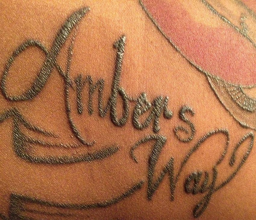
Tattoo-Associated Uveitis
638

                                                                                                             TABLE. Patient Demographics and Findings in Tattoo-Associated Uveitis

                                                                                           Intraocular                                       Sarcoidosis                                                                                                           Follow-up
                                    Patients Demographics          Initial VA             Inflammation           Uveitic Evaluationa         Evaluationa        Tattoo Findings           Tattoo Biopsy       Ocular Complications            Treatment             Duration

                                    1       20yo AAM        OD(sc): 20/100, OS(sc): Bilateral                  RPR, FTA-ABS, HLA-       ACE, chest          Elevation/induration of   Noncaseating            OU: posterior             Mycophenolate mofetil, 13 months
                                                              20/400; PHNI OU           granulomatous            B27, IGRA, Lyme          x-ray (32)          skin tattooed with        granulomatous           synechiae,                oral prednisone,
                                                                                        chronic panuveitis       serology,                                    black pigment             reaction associated     glaucoma s/p              topical
                                                                                                                 Toxoplasma IgG,                              (predominantly            with tattoo ink         Baerveldt glaucoma        corticosteroids, and
                                                                                                                 CMP, CBC                                     black tattoos on                                  implants                  IOP-lowering drops
                                                                                                                                                              arms, chest, and
                                                                                                                                                              abdomen)
                                    2       31yo WM         OU(sc): 20/25; PHNI      Bilateral                 RPR, FTA-ABS, HLA-       ACE, lysozyme,      Elevation/induration of   Noncaseating            OU: steroid-associated Topical corticosteroids, 17 months
                                                              OU                        nongranulomatous         B27, PPD, CMP,           chest x-ray, CT     skin tattooed with        granulomatous           ocular hypertension       IOP-lowering drop
                                                                                        recurrent anterior       CBC                      chest, lymph        black pigment             reaction associated
AMERICAN JOURNAL OF OPHTHALMOLOGY

                                                                                        uveitis                                           node biopsyb        (multicolored,            with tattoo ink
                                                                                                                                                              extensive tattoos on
                                                                                                                                                              both arms)
                                    3       32yo AAM        OU(sc): 20/25-2; PHNI    Bilateral                 RPR, FTA-ABS, HLA-       ACE, chest x-ray    Elevation/induration of   Not performed           OU: pupillary             Methotrexate, oral       20 months
                                                              OU                        nongranulomatous         B27, Lyme serology,                          skin tattooed with                                mebranes,                 prednisone, topical
                                                                                        chronic anterior         CMP, CBC                                     black pigment on                                  cataracts, CME            corticosteroids,
                                                                                        uveitis                                                               both arms (black                                OD: posterior               IOP-lowering drops
                                                                                                                                                              tattoos on chest                                  synechiae
                                                                                                                                                              never involved)                                 OS: iris bombe, uveitic
                                                                                                                                                                                                                glaucoma
                                    4       23yo AAF        OD(sc): 20/40, OS(sc):   Bilateral                 RPR, FTA-ABS, HLA-       Lysozyme,           Elevation, induration,    Not performed           OU: posterior             Mycophenolate mofetil, 7 months
                                                              20/200-2; PHNI OU         nongranulomatous         B27, Lyme serology,      chest x-ray         and scaling of                                    synechiae, pupillary      oral prednisone,
                                                                                        chronic anterior         CMP, CBC                                     tattoos with black                                membranes, iris           topical
                                                                                        uveitis                                                               ink (multicolored                                 bombe, severe CME         corticosteroids,
                                                                                                                                                              tattoos on face,                                OS: uveitic glaucoma        IOP-lowering drops
                                                                                                                                                              neck, torso, back,
                                                                                                                                                              and all limbs)
                                    5       23yo AAM        OD(sc): 20/25, OS(sc):   Bilateral                 RPR, FTA-ABS, HLA-       ACE, chest x-ray    Elevation/induration of   Not performed           OS: posterior             Topical corticosteroids 4 months
                                                              20/20þ1; PHNI OU          nongranulomatous         B27, Lyme serology,                          skin tattooed with                                synechiae
                                                                                        chronic anterior                                                      black pigment (black
                                                                                        uveitis                                                               tattoos on arms/
                                                                                                                                                              chest)
                                    6       21yo AAF        OD(sc): 20/100, OS(sc): Bilateral                  RPR, FTA-ABS, HLA-       ACE,b chest x-ray   Elevation/induration of   Not performed           OU: severe optic nerve Oral prednisone,            2 months
                                                              20/40; PH 20/80 OD,       nongranulomatous         B27, PPD, Lyme                               skin tattooed with                                elevation and             topical
                                                              PHNI OS                   chronic panuveitis       serology, Bartonella                         black pigment                                     hyperemia with            corticosteroids
                                                                                        with hypopyon            antibody panel,                              (multicolored tattoos                             papillomacular
                                                                                                                 CMP, CBC                                     on back)                                          exudates, ERM
                                                                                                                                                                                                              OD: subfoveal RPE
SEPTEMBER 2014

                                                                                                                                                                                                                detachment

                                                                                                                                                                                                                                                    Continued on next page
Tattoo-Associated Uveitis
evaluation of a 1-week history of blurred vision, photo-
                                                                                                                                                                                                                                                                                                                                                                                                                                              phobia, and pain in both eyes. He had experienced similar

                                                                                                                          1 month (lost to

                                                                                                                                                                                                                                         Patient 2 underwent biopsy of an enlarged axillary lymph node, which displayed a noncaseating granulomatous reaction. Patient 6 had an elevated ACE value of 85 (reference range: 9–67 U/L).
                                                                                                                                                                                                                                       AAF ¼ African-American female; AAM ¼ African-American male; ACE ¼ serum angiotensin-converting enzyme; CBC ¼ complete blood count; CME ¼ cystoid macular edema; CMP ¼ complete

                                                                                                                                                                                                                                     G; IGRA ¼ interferon gamma release assay (QuantiFERON–TB Gold); IOP ¼ intraocular pressure; PH ¼ visual acuity measured with pinhole occluder; PHNI ¼ visual acuity measured with pinhole
                                                                                                                                                                                                                                     occluder offered no improvement in vision; PPD ¼ tuberculosis purified protein derivative skin testing; RPE ¼ retinal pigment epithelium; RPR ¼ syphilis rapid plasma regain; sc ¼ without correction;
                                                                                                                                                                                                                                     metabolic panel including liver function testing; CT ¼ computed tomography; ERM ¼ epiretinal membrane; FTA-ABS ¼ syphilis fluorescent treponemal antibody-absorption; IgG ¼ immunoglobulin
                                                                                       Follow-up

                                                                                                                                                    follow-up)
                                                                                        Duration                                                                                                                                                                                                                                                                                                                                              symptoms 6 months earlier, which lasted approximately
                                                                                                                                                                                                                                                                                                                                                                                                                                              1 month before spontaneously resolving. Review of systems
                                                                                                                                                                                                                                                                                                                                                                                                                                              was notable for elevation and swelling of 8 tattoos on his
                                                                                                                                                                         immunosuppression                                                                                                                                                                                                                                                    arms and chest that occurred in conjunction with his ocular
                                                                                                                                                                                               recommended                                                                                                                                                                                                                                    symptoms on both occasions (Figure 1). All of his tattoos
                                                                                                Treatment

                                                                                                                          Oral prednisone;

                                                                                                                                                                                                                                                                                                                                                                                                                                              were performed during a 1-year period, approximately
                                                                                                                                                    systemic

                                                                                                                                                                                                                                                                                                                                                                                                                                              6 months prior to his initial ocular complaints.
                                                                                                                                                                                                                                                                                                                                                                                                                                                 Upon presentation, best-corrected visual acuity
                                                                                                                                                                                                                                                                                                                                                                                                                                              (BCVA) was 20/100 in the right eye and 20/400 in the
                                                                                                Ocular Complications

                                                                                                                                                                                               retinal detachment

                                                                                                                                                                                                                                                                                                                                                                                                                                              left eye, with an intraocular pressure (IOP) of 11 mm Hg
                                                                                                                                                                         OD: neurosensory

                                                                                                                                                                                                                    OS: severe CME

                                                                                                                                                                                                                                                                                                                                                                                                                                              in the right eye and 10 mm Hg in the left eye. Slit-lamp
                                                                                                                          OU: posterior
                                                                                                                                                    synechiae

                                                                                                                                                                                                                                                                                                                                                                                                                                              examination revealed diffuse conjunctival injection, dense
                                                                                                                                                                                                                                                                                                                                                                                                                                              mutton-fat keratic precipitates with overlying corneal
                                                                                                                                                                                                                                                                                                                                                                                                                                              microcystic edema, and posterior synechiae bilaterally.
  TABLE. Patient Demographics and Findings in Tattoo-Associated Uveitis (Continued )

                                                                                                                                                                                                                                                                                                                                                                                                                                              Both eyes displayed anterior chamber inflammation of 3þ
                                                                                                Tattoo Biopsy

                                                                                                                                                                                                                                                                                                                                                                                                                                              cells and 1þ flare, with 2þ anterior vitreous cells. Both
                                                                                                                          Not performed

                                                                                                                                                                                                                                                                                                                                                                                                                                              optic nerves appeared hyperemic and edematous, but a
                                                                                                                                                                                                                                                                                                                                                                                                                                              more detailed posterior segment examination, including
                                                                                                                                                                                                                                                                                                                                                                                                                                              an assessment of vitreous haze, was limited by bilateral
                                                                                                                                                                                                                                                                                                                                                                                                                                              corneal edema and posterior synechiae.
                                                                                                                                                                         black pigment (arm)
                                                                                                                          Elevation/induration of

                                                                                                                                                                                                                                                                                                                                                                                                                                                 An initial evaluation, consisting of FTA-ABS, RPR,
                                                                                                                                                    skin tattooed with

                                                                                                                                                                                                                                     Patient 7 had an elevated lysozyme value of 32 (reference range: 9–17 mg/mL) and a normal serum ACE value of 47.
                                                                                                Tattoo Findings

                                                                                                                                                                                                                                                                                                                                                                                                                                              QuantiFERON-TB Gold, chest x-ray, and Lyme antibody,
                                                                                                                                                                                                                                                                                                                                                                                                                                              was unrevealing. He was initially treated with intensive
                                                                                                                                                                                                                                                                                                                                                                                                                                              topical corticosteroids and cycloplegic drops. Three days
                                                                                                                                                                                                                                                                                                                                                                                                                                              after presentation, BCVA improved to 20/50 in the right
                                                                                                                                                                                                                                                                                                                                                                                                                                              eye and 20/60 in the left eye, with an IOP of 20 mm Hg
                                                                                                                                                                                                                                                                                                                                                                                                                                              in the right eye and 22 mm Hg in the left eye. However,
                                                                                                                                                    chest x-ray, CT
                                                                                       Sarcoidosis
                                                                                       Evaluationa

                                                                                                                        ACE, lysozyme,b

                                                                                                                                                                                                                                                                                                                                                                                                                                              his intraocular inflammation persisted and high-dose
                                                                                                                                                                                                                                                                                                                                                                                                                                              (1 mg/kg/day) oral prednisone therapy was initiated. The
                                                                                                                                                                         chest

                                                                                                                                                                                                                                                                                                                                                                                                                                              bilateral anterior chamber inflammation and elevated tat-
                                                                                                                                                                                                                                                                                                                                                                                                                                              toos resolved over the course of 3 weeks; however, his
                                                                                                                                                                                                                                                                                                                                                                                                                                              IOP became elevated at this time (33 mm Hg in the right
                                                                                             Uveitic Evaluationa

                                                                                                                          FTA-ABS, HLA-B27,

                                                                                                                                                                                                                                                                                                                                                                                                                                              eye and 27 mm Hg in the left eye). Topical IOP-lowering
                                                                                                                                                    CMP, CBC

                                                                                                                                                                                                                                                                                                                                                                                                                                              therapy was initiated in a stepwise manner, which escalated
                                                                                                                                                                                                                                                                                                                                                                                                                                              to maximal topical therapy. Topical prednisolone acetate
                                                                                                                                                                                                                                         All results unremarkable/negative unless otherwise indicated.

                                                                                                                                                                                                                                                                                                                                                                                                                                              1% was changed to loteprednol etabonate 0.5% approxi-
                                                                                                                                                                                                                                                                                                                                                                                                                                              mately 8 weeks after presentation in an attempt to mini-
                                                                                                                                                    nongranulomatous

                                                                                                                                                                                                                                                                                                                                                                                                                                              mize any steroid-response component; however, oral
                                                                                                                                                                         chronic anterior
                                                                                       Inflammation
                                                                                        Intraocular

                                                                                                                                                                                                                                     VA ¼ visual acuity; WM ¼ white male; yo ¼ year old.

                                                                                                                                                                                                                                                                                                                                                                                                                                              acetazolamide was necessary. A prolonged attempt to taper
                                                                                                                                                                                                                                                                                                                                                                                                                                              his oral prednisone led to a recurrence of intraocular
                                                                                                                                                                                               uveitis
                                                                                                                          Bilateral

                                                                                                                                                                                                                                                                                                                                                                                                                                              inflammation and tattoo elevation, and 3 months after
                                                                                                                                                                                                                                                                                                                                                                                                                                              his initial presentation he was referred for dermatologic
                                                                                                                                                    20/40; PHNI OD, PH
                                                                                                                          OD(sc): 20/20, OS(sc):

                                                                                                                                                                                                                                                                                                                                                                                                                                              evaluation. His oral prednisone dose was tapered from
                                                                                                                                                                                                                                                                                                                                                                                                                                              40 mg/day to 30 mg/day 6 days prior to this dermatology
                                                                                                Initial VA

                                                                                                                                                                                                                                                                                                                                                                                                                                              appointment, and a biopsy was taken from an area of previ-
                                                                                                                                                                         OS 20/32

                                                                                                                                                                                                                                                                                                                                                                                                                                              ously affected skin on his chest. Histologic sections of this
                                                                                                                                                                                                                                                                                                                                                                                                                                              biopsy displayed macrophages in clusters around the dermal
                                                                                                                                                                                                                                                                                                                                                                                                                                              superficial vascular plexus that contained tattoo pigment
                                                                                                Patients Demographics

                                                                                                                                                                                                                                                                                                                                                                                                                                              with surrounding granulomatous inflammation. A repeat
                                                                                                                          42yo AAM

                                                                                                                                                                                                                                                                                                                                                                                                                                              chest x-ray, comprehensive chemistry panel, and serum
                                                                                                                                                                                                                                                                                                                                                                                                                                              ACE were ordered by dermatology, with all results inter-
                                                                                                                                                                                                                                                                                                                                                                                                                                              preted as normal. He then returned for routine follow-up
                                                                                                                                                                                                                                       b
                                                                                                                                                                                                                                       a

                                                                                                                                                                                                                                                                                                                                                                                                                                              nearly 3 weeks later on 20 mg/day of prednisone, at which
                                                                                                                          7

                                                                                                                                                                                                                                                                                                                                                                                                                                              time he was noted to have diffuse elevation of all tattoos

VOL. 158, NO. 3                                                                                                                                                                                                                                                                                                                                                                                                          TATTOO-ASSOCIATED UVEITIS                                                    639
FIGURE 1. Tattoo inflammation in a 20-year-old African-American male subject with simultaneous bilateral, chronic, granulomatous
panuveitis (Patient 1). (Upper left) Nodular elevation of focal areas of tattooed skin on the patient’s left shoulder. (Upper right) Diffuse
elevation of black tattoos on the chest, with involvement of adjacent nontattooed skin. Hematoxylin-eosin stain (original magnifica-
tion 3100) of biopsies from the chest and shoulder display a granulomatous reaction in the dermis with aggregates of epithelioid his-
tiocytes, giant cells, and a sparse mononuclear infiltrate (Bottom left); all granulomas contained tattoo pigment (inset, Bottom right).

containing black ink. Punch biopsies of the affected skin               panuveitis. One patient with anterior uveitis had recurrent
were taken from his left shoulder and chest. Histology slides           disease, while all other patients had chronic uveitis.
of these biopsy specimens revealed aggregates of epithelioid               A medical examination was performed on all patients
histiocytes and giant cells containing pigment particles                and interpreted as unremarkable, with the exception of a
with sparse mononuclear cell infiltrate (Figure 1).                     single elevated serum ACE (Patient 6) and lysozyme value
   Ultimately, his refractory ocular hypertension resulting             (Patient 7). Patient 4 presented with submandibular and
from a combination of uveitic and steroid response mecha-               occipital lymphadenopathy not associated with overlying
nisms required placement of glaucoma drainage devices                   tattooing, though she did have extensive head and neck
4 months and 6 months after presentation in the right                   tattoos. Patient 2 developed an enlarged left axillary lymph
and left eye, respectively. The patient agreed to initiation            node near the area of heavy tattooing on the same arm.
of immunosuppressive therapy with mycophenolate mofe-                      All patients denied abnormal cutaneous reactions imme-
til, which enabled successful tapering of oral prednisone.              diately after tattooing, and all had their most recent tattoo
At the most recent examination, 13 months after presenta-               placed at least 6 months prior to the onset of cutaneous and
tion, BCVA was 20/25 bilaterally with controlled IOP and                intraocular symptoms. Five of the patients had extensive
intraocular inflammation on mycophenolate mofetil 1.5 g                 tattoos, all of which consisted entirely of only black
twice daily and prednisone 5 mg once daily.                             pigment or multicolored tattoos containing black ink.
                                                                        The remaining 2 patients had more limited tattooing
                                                                        (Patients 6 and 7). Only tattoos or portions of tattoos
                                                                        containing black pigment were affected in all 7 patients.
                          RESULTS                                       Involvement of the affected tattoos varied from focal
SELECTED CLINICAL CHARACTERISTICS OF ALL 7 PATIENTS                     nodular elevation in regions of black pigment to uniform
are summarized in the Table. Six patients were African                  elevation and induration of all portions of the tattoo
Americans, 4 of whom were male. The remaining patient                   containing black ink (Figure 2). In Patient 1, striking
was a white man. The age range of all patients was 20–42                elevation and induration of the surrounding skin was also
years at the time of presentation. Five patients presented              observed. Five patients (Patient 1 and Patients 3–6) expe-
with bilateral nongranulomatous anterior uveitis, while                 rienced at least 1 recurrence of intraocular inflammation
the remaining 2 patients presented with granulomatous                   with simultaneous tattoo involvement.

640                                             AMERICAN JOURNAL OF OPHTHALMOLOGY                                          SEPTEMBER 2014
DISCUSSION
                                                                WE DESCRIBE A SERIES OF 7 CONSECUTIVE PATIENTS WHO
                                                                demonstrated simultaneous onset of bilateral uveitis and
                                                                tattoo elevation with induration. Five other cases
                                                                published in 3 articles reported similar findings and no
                                                                additional evidence of sarcoidosis on examination or chest
                                                                imaging (chest x-ray and/or CT studies) at the time of pre-
                                                                sentation.2,5,6 Similar to our series, these 5 patients
                                                                exhibited bilateral uveitis, ranging from iridocyclitis to
                                                                panuveitis. During observed follow-up, 1 patient developed
                                                                ‘‘red infiltrates resembling erythema nodosum’’ on both
                                                                shins that were not associated with tattooed skin.2 In our
                                                                series, Patient 2 developed noncaseating granulomatous
                                                                inflammation of an axillary lymph node adjacent to an
                                                                area of dense tattooing, and Patient 3 developed subman-
                                                                dibular and occipital lymphadenopathy, which was suspi-
FIGURE 2. Tattoo elevation in a 23-year-old African-            cious for sarcoidosis. Furthermore, there have been cases
American female subject with simultaneous bilateral, chronic,   reported in the literature of patients with a diagnosis of sys-
nongranulomatous anterior uveitis (Patient 4). All areas of     temic sarcoidosis at the time of presentation with bilateral
her abdominal tattoo containing black pigment were raised and   uveitis and tattoo induration.1,7,8
indurated, and similar tattoo involvement was noted on her          Each of the patients in our series had extensive areas of
head, neck, and back.                                           tattooing that contained or consisted entirely of black
                                                                ink. Other published reports describe skin inflammation
                                                                associated with blue,2 red,5 and black ink.6–8 Tattoo size
   Tattoo biopsies were performed on Patients 1 and 2. A        and color varied, although 1 case report described a
tattoo biopsy on Patient 2 was performed 2 weeks prior          patient with extensive black tattooing,6 which was similar
to presentation and displayed noncaseating granulomas           to the patients in our series. All of our patients received the
composed of histiocytes surrounding black tattoo pigment        majority of their tattoos over a relatively short period of
in the dermis. His enlarged axillary lymph node also was        time—approximately 1 year or less—but none experienced
biopsied and displayed noncaseating granulomatous               induration or inflammation of the tattoos at the time of
inflammation. Similar findings were demonstrated in the         tattoo placement. In the majority of reports, the inflamma-
tattoo biopsies of Patient 1. Patients 4 and 5 were referred    tion of tattooed skin coincided with the onset of uveitis
for biopsy of their tattoos and enlarged lymph nodes (Pa-       except for 2 of 3 patients reported by Rorsman and associ-
tient 4) immediately after their initial evaluation, but        ates, in whom tattoo inflammation occurred immediately
both failed to follow up with dermatology and had rapid         after placement and did not correlate temporally with their
resolution of these findings following initiation of high-      delayed ocular involvement.2
dose oral prednisone. The remaining patients (Patients 3,           Of the 8 previously reported patients with tattoo-
6, and 7) were not sent for biopsy of their involved tattoos    associated uveitis, 4 underwent complete excision of the
because they were immediately treated with high-dose oral       affected tattoo(s).2,5 Interestingly, 3 of these 4 patients
prednisone, which coincided with rapid resolution of their      were reported to have improvement in their intraocular
tattoo findings over the course of approximately 1 week.        inflammation following tattoo excision, with 2 achieving
Owing to military service obligations requiring him to relo-    complete resolution off of all medications. As with our
cate, Patient 7 was lost to follow-up after 2 visits.           series, the follow-up periods in these cases were limited,
   Five of 7 patients suffered potentially vision-threatening   and it remains unclear whether tattoo excision had a role
ocular complications at some point during observation. Pa-      in disease resolution or simply coincided with spontaneous
tients 3 and 4 presented with iris bombe, pupillary mem-        disease remission. Unlike these previously reported cases,
branes, and cystoid macular edema, which subsequently           tattoo excision was not offered to the patients we continue
required treatment with immunosuppressive therapy.              to follow at our facility, as the percentage of body surface
Patient 7 presented to our institution with a neurosensory      area encompassed by their involved tattoos was deemed
retinal detachment in the right eye and severe cystoid mac-     too extensive for subsequent skin grafting.
ular edema in the left eye. All patients treated with oral          Various patterns of histologic reactions have been reported
prednisone (Patients 1, 3, 4, 6, and 7) displayed simulta-      to occur in tattooed skin, and one of the more common find-
neous improvement in their ocular inflammation and reso-        ings is granulomatous inflammation.9 Histologically, this can
lution of their tattoo findings.                                be classified as a foreign body or sarcoid-type reaction, and the

VOL. 158, NO. 3                                  TATTOO-ASSOCIATED UVEITIS                                                  641
differentiation of these 2 types of granulomas may be both                         exposure in lung cell lines13 and mice14 have shown
challenging and open to controversy.9,10 Allergic reactions                        that these particles ‘‘induce inflammation, oxidize
to tattoo pigment can also occur, which may exhibit a                              DNA, cause DNA strand breaks and increase the mutant
variety of histologic forms, some of which are also                                frequency following long-term exposure at a subcytotoxic
consistent with sarcoidosis.9 Granulomatous reactions                              concentration.’’15 All of our patients received the major-
confined to single tattoo colors typically represent a local                       ity of their tattoos over a relatively short period of
hypersensitivity reaction to specific components of tattoo                         time—approximately 1 year or less—which may have
pigment, but they may also represent a manifestation of                            conferred an increased risk of disease development as a
systemic sarcoidosis.9 Interestingly, reports of allergic reac-                    result of the relatively large antigenic and/or toxic
tions to black tattoo pigment are very rare.11 The histologic                      load. Interestingly, the US Food and Drug Administra-
appearance of tattoo biopsy specimens obtained from 2 of                           tion has not approved any tattoo pigments for injection
the 7 patients in our series were interpreted as noncaseating                      into the skin, and many pigments used in tattoo inks are
granulomatous inflammation in association with dermal                              industrial-grade colors suitable for printers’ ink or auto-
tattoo pigment, which is consistent with but not specific for                      mobile paint.16 Among adults 18–50 years of age in
sarcoidosis.                                                                       this country, the prevalence of tattoos may be as high
   Although the etiology of sarcoidosis remains unclear, it is                     as 24%.17
hypothesized that the disease process is initiated when a genet-                      Ultimately, the patients in our series seem to represent a
ically susceptible host is exposed to an inciting environmental                    subset of patients in whom some component of tattoo
antigen(s).3,10 In such an event, an exaggerated immune                            pigment initiated a localized cutaneous response that by
response characterized by the activation of macrophages                            some means also played a role in the simultaneous develop-
and CD4þ T lymphocytes occurs, resulting in cytokine                               ment of ocular inflammation. Whether the pathophysi-
production consistent with a TH1-type immune response,                             ology of this process is similar to systemic sarcoidosis, is
ultimately leading to granuloma formation.3,10 Considering                         the result of a hypersensitivity response, or is attributble
the multiple environmental risk factors reported to date, it                       to some other mechanism is not yet known. Continued
seems reasonable to conclude that the development of                               observation for the development of additional organ system
sarcoidosis is likely the end result of immune responses to a                      involvement consistent with sarcoidosis, and the potential
potentially large variety of environmental triggers.3,4                            benefit of tattoo removal, if performed, may be useful
   The production of black tattoo ink is based on soot,                            knowledge. Altogether, the clinical presentation of the
which may ‘‘contain toxic, mutagenic or carcinogenic                               patients collected for this series nearly equals the cumula-
compounds such as carbon black and polycyclic aromatic                             tive number of previously reported cases, suggesting that
hydrocarbons or phenol.’’12 Carbon black nanoparticle                              this association is likely underappreciated.

ALL AUTHORS HAVE COMPLETED AND SUBMITTED THE ICMJE FORM FOR DISCLOSURE OF POTENTIAL CONFLICTS OF INTEREST.
Jennifer E. Thorne discloses the following: grant funding from the National Institutes of Health (Bethesda, Maryland, USA), Research to Prevent Blindness
Sybil B. Harrington Special Scholars Award (New York, New York, USA), Allergan (Irvine, California, USA); consultant for AbbVie (North Chicago,
Illinois, USA), Gilead (Foster City, California, USA), Navigant (Chicago, Illinois, USA), Xoma (Berkeley, California, USA). The authors indicate no
funding support. Contributions of authors: all listed authors made substantial contributions regarding (1) data acquisition (T.O., B.B., T.L., N.B., J.D., J.T.);
(2) drafting (T.O.) or revising the article (T.O., B.B., T.L., N.B., J.D., J.T.); and (3) approval of submitted version (T.O., B.B., T.L., N.B., J.D., J.T.).
    Pathology consultation was provided by Gulsun Erdag, MD, Johns Hopkins School of Medicine, Baltimore, Maryland, USA.

                          REFERENCES                                                7. Anolik R, Mandal R, Franks A. Sarcoidal tattoo granu-
                                                                                       loma. Dermatol Online J 2010;16(11):19. Available at,
 1. Lubeck G, Epstein E. Complications of tattooing. Calif Med                         http://escholarship.org/uc/item/1fm1d840. Accessed May
    1952;76(2):83–85.                                                                  6, 2014.
 2. Rorsman H, Brehmer-Andersson E, Dahlquist I, et al. Tattoo                      8. Post J, Hull P. Tattoo reactions as a sign of sarcoidosis. CMAJ
    granuloma and uveitis. Lancet 1969;294(7610):27–28.                                2012;184(4):432.
 3. Iannuzzi MC, Rybicki BA, Teirstein AS. Sarcoidosis. N Engl J                    9. Morales-Callaghan AM, Aquilar-Bernier M, Martinez-
    Med 2007;357(21):2153–2165.                                                        Garcia G, Miranda-Romero A. Sarcoid granuloma on black
 4. Bonfioli AA, Orefice F. Sarcoidosis. Semin Ophthalmol 2005;                        tattoo. J Am Acad Dermatol 2006;55(5 Suppl):S71–S73.
    20(3):177–182.                                                                 10. Marcoval J, Mana J, Moreno A, Gallego I, Fortuno Y, Peyri J.
 5. Barbarasi Z, Kiss E, Balaton G, Vajo Z. Cutaneous granuloma                        Foreign bodies in granulomatous cutaneous lesions of patients
    and uveitis caused by a tattoo. Wien Klin Wochenschr 2008;                         with systemic sarcoidosis. Arch Dermatol 2001;137(4):
    120(1-2):18.                                                                       427–430.
 6. Saliba N, Owen ME, Beare N. Tattoo-associated uveitis. Eye                     11. Kaur RR, Kirby W, Maibach H. Cutaneous allergic reactions
    2010;24(8):1406.                                                                   to tattoo ink. J Cosmet Dermatol 2009;8(4):295–300.

642                                                    AMERICAN JOURNAL OF OPHTHALMOLOGY                                                      SEPTEMBER 2014
12. Wenzel SM, Rittmann I, Landthaler M, Baumler W. Adverse        15. Hogsberg T, Jacobsen NR, Clausen PA, Serup J. Black tattoo
    reactions after tattooing: review of the literature and com-       inks induce reactive oxygen species production correlating
    parison to results of a survey. Dermatology 2013;226(2):           with aggregation of pigment nanoparticles and product brand
    138–147.                                                           but not with the polycyclic aromatic hydrocarbon content.
13. Jacobsen NR, Saber AT, White P, et al. Increased mutant fre-       Exp Dermatol 2013;22(7):464–469.
    quency by carbon black, but not quartz, in the lacZ and cll    16. U.S. Food and Drug Administration. Consumer Health Informa-
    transgenes of muta mouse lung epithelial cells. Environ Mol        tion. Think before you ink: are tattoos safe? Available at, http://
    Mutagen 2007;48(6):451–461.                                        www.fda.gov/forconsumers/consumerupdates/ucm048919.htm.
14. Jacobsen NR, Moller P, Jensen KA, et al. Lung inflamma-            Accessed May 6, 2014.
    tion and genotoxicity following pulmonary exposure             17. Laumann AE, Derick AJ. Tattoos and body piercings in the
    to nanoparticles in ApoE-/- mice. Part Fibre Toxicol               United States: a national data set. J Am Acad Dermatol
    2009;6:2.                                                          2006;55(3):413–421.

VOL. 158, NO. 3                                     TATTOO-ASSOCIATED UVEITIS                                                        643
Biosketch
Trucian Ostheimer, MD, graduated from the Ohio State University College of Medicine in 2008, and completed his
ophthalmology residency at the Illinois Eye & Ear Infirmary in 2012. He is currently completing a two-year ocular
immunology fellowship at the Wilmer Eye Institute, and has a special interest in birdshot chorioretinopathy. Dr
Ostheimer will begin a fellowship in vitreoretinal surgery at the University of Washington in July, 2014.

643.e1                                AMERICAN JOURNAL OF OPHTHALMOLOGY                             SEPTEMBER 2014
You can also read