Short Running title: Diagnostic Approach for Allergic Reactions to Tattoos Servicio de Dermatología, Hospital General Universitario de Alicante ...
←
→
Page content transcription
If your browser does not render page correctly, please read the page content below
1

Diagnostic Approach for Suspected Allergic Cutaneous Reaction to a Permanent
Tattoo

Short Running title: Diagnostic Approach for Allergic Reactions to Tattoos

Silvestre JF, González-Villanueva I

Servicio de Dermatología, Hospital General Universitario de Alicante, Alicante,
Spain

This article has been accepted for publication and undergone full peer review but has
not been through the copyediting, typesetting, pagination and proofreading process,
which may lead to differences between this version and the Version of Record. Please
cite this article as doi: 10.18176/jiaci.0383

J Investig Allergol Clin Immunol 2019; Vol. 29(6)           © 2019 Esmon Publicidad
doi: 10.18176/jiaci.0383
2

Abstract
       The last few decades have seen a notable increase in the number of people who
have a tattoo. This practice is not free from complications. Most adverse effects appear
early and are temporary, although they can occasionally develop later and be permanent
and serious. Recent research has generated new knowledge on the composition of inks,
their degradation over time, the immune activity that is stimulated, and the various
clinical disorders that can arise. This information enables better approaches to diagnosis
and management when complications arise.
       Diagnosing allergic reactions to permanent tattoo ink can be very challenging.
This review aims to identify clinical and histological clues to help practitioners
differentiate allergic reactions from other complications. We discuss the yield and
appropriateness of skin tests and biopsies and propose an algorithm to guide the
diagnostic process.

Key words: Tattoo. Adverse effect. Skin. Allergy. Patch testing. Biopsy. Diagnosis.

Resumen

       En las últimas décadas ha aumentado ostensiblemente el número de personas
que se realiza un tatuaje en la piel. Esta técnica no está exenta de complicaciones. la
mayoría son precoces y transitorias, pero, en ocasiones, son tardías, permanentes y
graves. En los últimos años, los avances en el conocimiento de la composición de tintas,
de los procesos de degradación de las mismas tras el tatuado, de la actividad inmunitaria
que se desencadena y la descripción de una clasificación en patrones clínicos, nos han
permitido realizar un mejor enfoque diagnóstico y manejo terapéutico de estas
complicaciones.
       El diagnóstico de las reacciones alérgicas a las tintas de los tatuajes permanentes
supone un verdadero reto. En esta revisión intentaremos dar claves clínicas e
histológicas que nos ayudan a diferenciar las reacciones alérgicas del resto de
complicaciones. Discutiremos la rentabilidad e idoneidad de las pruebas epicutáneas y
de la biopsia cutánea, y propondremos un algoritmo diagnóstico.

Palabras clave: Tatuaje. Complicación. Piel. Alergia. Pruebas epicutáneas. Biopsia.
Diagnóstico.

J Investig Allergol Clin Immunol 2019; Vol. 29(6)            © 2019 Esmon Publicidad
doi: 10.18176/jiaci.0383
3

Introduction
      Tattooing is a decorative skin technique that has been used with various
objectives for millennia. Tattoos are common in a number of cultures, and the practice
has risen in popularity since the 1970s, especially in young people [1]. By some
estimates, 10% to 20% of people in Western countries now have at least 1 tattoo [2].
The deposition of pigment particles and other components of ink in the dermis induces a
response. Most reactions are due either to an inflammatory defense response to foreign
substances or local toxicity following exposure to irritants. In clinical practice, this
translates into the appearance of mild symptoms within a month of receiving the tattoo
(pruritus, swelling, or hair loss) in about 1 in every 5 individuals [3]. Most of these
complaints do not require a visit to a doctor, as tattooists are familiar with and know
how to manage them. Other reactions are serious, persistent, and chronic and tend to
appear later and more rarely. Some studies have estimated that 6% to 8% of people who
have a tattoo are affected by this type of complication [4,5] and seek specialist care.
However, the right diagnostic approach is difficult, as it must take into account multiple
clinical entities ranging from infections, tumors, allergic processes, and foreign body
reactions to manifestations of systemic diseases or isomorphic phenomena arising from
other skin conditions.
       This review aims to provide clinical and histological clues that can help
clinicians to differentiate between allergic reactions to tattoos and other complications.
We also discuss the yield and appropriateness of skin tests and biopsies and propose an
algorithm to guide the diagnostic process based on our clinical experience [6].

Clinical Manifestations
       A detailed history is essential for reaching a diagnosis and should include the
following: an investigation of signs and symptoms related to lesions in the tattooed
area; questions on the patient’s personal medical history, with an emphasis on previous
dermatoses; and a comprehensive account of the conditions in which the patient
received the tattoo (Table 1) [7].
       An important detail that can orient the diagnosis is the time between getting the
tattoo and onset of symptoms [6]. This can help clinicians to classify the reaction as
early (within a month of being tattooed) and late (appearing months or even years

J Investig Allergol Clin Immunol 2019; Vol. 29(6)            © 2019 Esmon Publicidad
doi: 10.18176/jiaci.0383
4
afterward) [8]. The most common early complications are inflammatory reactions,
which occur secondary to cutaneous exposure to a foreign substance (ink). These
usually occur within the space of a few weeks and rarely require medical care, as the
tattooists themselves can manage them. However, early reactions to tattoos can involve
an infectious etiology [9]. Clinical data that are suggestive of infection include pain, a
rise in local temperature, oozing, and ulceration (Figure 1). If these signs are present,
the purulent discharge should be cultured, and patients should initiate empirical
antibiotic treatment.
Allergic reactions to tattoo ink are late reactions. These are permanent lesions
that do not respond to treatment with topical corticosteroids. The differential diagnosis
should always consider other late reactions such as granulomatous foreign body
reactions, other dermatoses, systemic disease (sarcoidosis) (Figure 2), microbial
infections, pseudolymphomas, and neuropathic reactions. Establishing a diagnosis for
an allergic reaction is not simple, as the skin lesions are nonspecific and the histological
findings are not characteristic.
Serup et al [9,10] developed a classification system for the clinical presentations
associated with permanent tattoos to facilitate the practitioner’s approach to these
patients. Rather than specific patterns, they are clinical forms that can orient clinicians
toward various complications and include papulonodular, plaque-like, hyperkeratotic,
ulceronecrotic, lymphopathic, neurosensory, and scar patterns. Advanced stages of any
of these clinical patterns produce patches of scarring, with the disappearance of pigment
or changes to its color.
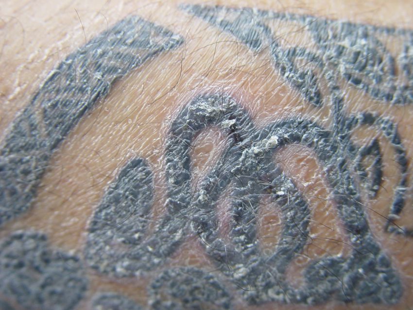
The clinical patterns that are most frequently associated with allergic reactions
are plaque-like, hyperkeratotic, and ulceronecrotic patterns. The plaque-like
presentation is characterized by raised tissue in the areas tattooed with the problem
color; the surface may be scaly (eczematous) (Figure 3) or smooth and shiny (lichenoid)
(Figure 4) [11]. This pattern is uniform and homogeneous and is often seen in tattoos
with red ink, which is the most frequent cause of allergic reactions. Green and blue ink
also tend to produce this pattern and may give way to color changes, ie, from blue to
green or vice versa. The hyperkeratotic pattern entails thickening of the skin until it
resembles sandpaper, with a flat surface that may ulcerate or necrotize. This pattern
could be considered a variant of the plaque-like pattern, albeit with an excessive
J Investig Allergol Clin Immunol 2019; Vol. 29(6) © 2019 Esmon Publicidad
doi: 10.18176/jiaci.0383
5
epidermal reaction caused by an inflammatory dermal cascade in response to the ink.
This induces leakage of the pigment into the epidermis, thus activating the process of
epidermal proliferation that gives rise to hyperkeratosis. The histological study reveals
pseudoepitheliomatous hyperplasia. Finally, the last pattern that Serup et al [9,10] link
to an allergic reaction is the ulceronecrotic pattern. This manifestation arises from an
aggressive inflammatory process resulting in ulceration of the areas tattooed with the
culprit ink. This ulceration can affect the entire dermal layer and even the hypodermis.
If the pigment spreads deep enough to reach the fascia of the muscle underneath or the
regional lymphatic nodes, the inflammatory cascade can eventually provoke severe deep
tissue necrosis or systemic autoimmune processes such as vasculitis or blistering
diseases. All these patterns can evolve to a final stage in which there is loss of pigment,
changes in color, and scar areas (Figure 5).
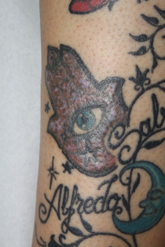
Regardless of the clinical pattern, an essential finding to confirm a suspected
allergic reaction is a uniform, monomorphic appearance throughout the area tattooed
with the problem ink (Figure 6). Clinicians need not suspect allergic reactions when
there are localized spots of thickening (papulonodular pattern) in some areas but not
others tattooed with a specific color of ink. Likewise, complaints of intense pruritus,
pain, or dysesthesia on the tattoo—in the absence of skin lesions—do not indicate an
allergic reaction. These manifestations correspond to the neuropathic pattern, which is
thought to be secondary to the stimulation of the C-fiber sensory nerves by degraded ink
components (Figure 7) [12].
Another very useful clinical sign is the simultaneous appearance of similar
reactions in both the problem tattoo and a previous tattoo of the same color. This
finding is very suggestive of an allergic reaction (Figure 8). Table 2 shows all of the
clinical signs suggesting an allergic reaction to tattoo inks [11].
Allergic reactions tend to present locally, but some authors have described cases
of generalized rashes or eczemas, especially in previously sensitized patients. These
reactions appear early (within 1-2 days of being tattooed) and tend to resolve without
treatment after a few weeks or months [8]. Other authors have described cases of
generalized reactions upon attempting to eliminate the pigment with laser treatment
[13], and cases of photoallergic reactions in tattoos with yellow ink containing cadmium
[1].
J Investig Allergol Clin Immunol 2019; Vol. 29(6) © 2019 Esmon Publicidad
doi: 10.18176/jiaci.0383
6

       A final consideration is the fact that tattooing is an inherently aggressive
process, and this can trigger the appearance of certain dermatoses on the tattooed skin
(isomorphic phenomenon of Koebner), including psoriasis, lichen planus, cutaneous
lupus erythematous, and pyoderma gangrenosum (Figure 9). Recognizing these
situations is straightforward when a patient has a prior diagnosis, although they may
also present as new-onset lesions.

Histology Findings
       Skin biopsy is a crucial part of the study of any late or persistent reaction in the
tattooed area. Histopathology of the samples can rule out serious entities such as
cutaneous infections by atypical microorganisms, systemic diseases, or lymphomatous
tumor infiltrations [14]. It is important to remember that since allergic reactions have no
pathognomonic histological findings, clinicians should not expect to see a histological
pattern of spongiotic dermatitis, as with allergic contact eczema, because the allergen is
directly inoculated into the dermis. The indication that is most suggestive of allergy is
interface dermatitis, with predominantly lymphocytic inflammatory infiltrate, with or
without eosinophils. This band-like inflammatory infiltrate occupies the basal layer of
the epidermis and the papillary dermis (Figure 10). It is made up of mononuclear cells
and macrophages loaded with intracellular pigment. Dermal fibrosis is apparent to some
extent, especially in advanced stages. The immunohistochemical study shows a
predominance of T lymphocytes that are CD3+ and also CD4+ or CD8+; both
populations are frequent in allergic inflammatory infiltrates [15,16].
       In many cases, attempted transepidermal elimination of the pigment and even
pseudoepitheliomatous hyperplasia can be observed (Figure 11). Given that this has
been described as a pattern of response to various infectious processes, stains are
recommended as a means of ruling out the presence of fungi, bacteria, and microbes [1].
       More rarely, allergic reactions to tattoos can present with histological patterns
that are granulomatous or pseudolymphomatous (both B- and T-cell types). Thus,
biopsies showing 1 of these patterns are insufficient to exclude the diagnosis of an
allergic reaction. In the presence of a granulomatous pattern, clinicians should rule out
systemic granulomatous diseases and microbial or fungal infections. In the case of

J Investig Allergol Clin Immunol 2019; Vol. 29(6)             © 2019 Esmon Publicidad
doi: 10.18176/jiaci.0383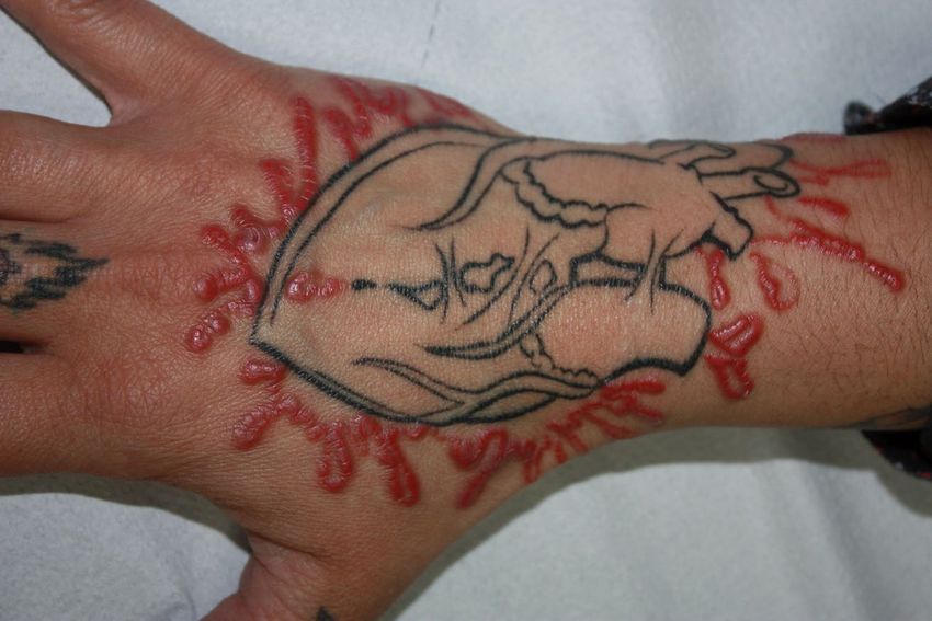
7

pseudolymphoma, we must demonstrate that the inflammatory infiltrate is polyclonal by
means of molecular biological studies [1,7,11,17].

Patch Testing
       Patch testing is used for the study of allergic contact dermatitis, which is caused
by haptens that come into contact with the epidermis and are capable of eliciting a
delayed immune response mediated by lymphocytes. It is not clear whether this test is
useful for diagnosing delayed hypersensitivity reactions provoked by antigens that are
inoculated directly into the dermis. However, it is true that many cases of
(noneczematous) lichenoid reactions to metals have been described, as well as drug
eruptions with positive patch tests, in which the allergen has not penetrated through the
epidermis but was instead administered systemically [18]. Theoretically, intradermal
tests would be the most specific for the study of these reactions; however, they are not
risk-free. Permanent pigment may remain in the dermis following the tests, or a chronic
reaction may be triggered. Thus, performing these tests is not considered appropriate or
ethical [19,20].
       Patch testing in the presence of a suspected tattoo allergy has often been
undertaken without knowledge of the ingredients in the ink used. Indeed, in many
countries, ink manufacture and composition are still unregulated, with no labelling
obligations. This is not the case in Spain, where the Spanish Agency of Medicines and
Medical Devices approves tattoo inks for marketing and mandates labelling of their
ingredients [21]. Thus, faced with a suspected tattoo allergy, clinicians can and should
request the safety data sheets and batch numbers for the products used by the tattooist
(Figure 12). In clinical practice, these are not always easy to obtain; securing this
information depends on the collaboration of both the patient and the tattooist. The
patient often fails to obtain the ink, or the tattooist might provide an unlabeled sample
that may or may not be the one used for the tattoo. The patient may even provide safety
sheets detailing identical compositions for the entire array of pigments sold under a
particular brand name.
       It is not clear which allergens should be studied through patch tests for suspected
tattoo allergy; patch tests are mainly performed for potential allergens identified from
inks on other occasions, albeit with no certainty that they are present in the problem ink

J Investig Allergol Clin Immunol 2019; Vol. 29(6)            © 2019 Esmon Publicidad
doi: 10.18176/jiaci.0383
8

[22]. We usually apply the Spanish baseline series, sometimes adding a specific series
of metals and/or textile dyes. If possible, we also patch test the problem ink and the
potential allergens listed on the safety sheet (including auxiliary ingredients and
excipients) [18].
       The metals series is used because inks may contain pigments from inorganic
metal salts; the most frequent of these are titanium (white ink) along with barium and
iron (red), although other salts that may be present include those from mercury (red),
cadmium (yellow, red), zinc (white), nickel and chrome (green), cobalt (blue),
aluminum and manganese (purple), and gold [1,11]. The metal that has been most
frequently identified as an allergen is mercury, which was a component of red ink.
Today, metallic salt components, which used to be very common, are giving way to
organic molecules such as azo pigments, which could now be present in up to 60% of
color inks [23]. The textile dyes series includes a number of azo pigments, which is why
this series tends to be used in patients with cutaneous reactions to tattoos, even though
we are not sure that the series contains the azo colors used in the tattoo [24].
     Few studies reflect the utility of patch testing in these situations. Most published
articles correspond to isolated clinical cases with positive results and an adequate
clinical correlation. Serup et al [25] undertook patch testing in 90 patients with chronic
reactions to tattoos, although the results were discouraging. The baseline series showed
positive findings for metal allergens that the authors determined were not relevant
(Figure 13), and the specific series for dyes and the problem ink showed positive results
for only 5% and 8% of the cases, respectively (Figure 14) [25].
     This lack of sensitivity and specificity to patch testing in the detection of allergic
reactions to tattoo inks has prompted several hypotheses. Some authors propose that the
scant penetration of the test ink into the skin is the reason for the negative results. More
recently, investigators speculated that the real allergen was not present in its native form
in the ink itself, but rather was generated in the dermis from pigments or other ink
components through a haptenization process that could take weeks, months, or even
years. The culprit allergen could even be a metabolite. The concentration of azo
pigments in the skin has been shown to decrease over time, as the pigments are
transported in part by the lymphatic system and in part degraded in the dermis into
products such as amino-naphthol-AS and naphthol-AS. These metabolic products could

J Investig Allergol Clin Immunol 2019; Vol. 29(6)              © 2019 Esmon Publicidad
doi: 10.18176/jiaci.0383
9

potentially be the ones responsible for the allergic reactions [26]. However, they have
been tested in 3 patients, with negative results [23]. Some researchers have postulated
that sunlight could induce a process of photochemical cleavage of the tattoo pigments,
particularly the azo pigments, which would enable chemical substances to act as haptens
[27,28]. This observation could explain numerous phenomena of solar intolerance that
have been described in tattooed areas and opens up new possibilities in how to approach
these patients, whether through photopatch testing or patch testing with substances
processed with ultraviolet light. Two studies have explored these additional tests, both
with negative results [29,30]. A further consideration is that tattoo inks also contain
other auxiliary ingredients and excipients, such as propylene glycol and shellac, which
could also be the culprit allergens and should be considered in patch testing [31].

Diagnostic Algorithm
       Based on our experience, we propose an algorithm for managing patients with
reactions to a tattoo (Figure 15). Our approach depends on initially whether the lesion
presents early or late [6].
       If an early reaction in the area of the tattoo shows clinical signs of infection, it is
essential to perform a culture of the purulent discharge as well as a biopsy for a
histological and microbiological study (bacterial and microbial culture). Empirical
antibiotics can also be started. If a sample of the ink used is available, it would be
desirable to perform a culture on it to rule out contamination. For early reactions
without clinical signs of infection, we recommend initiating treatment with topical
corticosteroids. If the reaction is refractory to treatment, a biopsy is warranted.
       In cases of late reactions, we recommend undertaking a skin biopsy to rule out
infection, systemic granulomatous diseases, and lymphatic neoplasms. If the biopsy
shows a lichenoid or spongiotic pattern and the patient shows clinical signs that are
compatible with an allergic reaction, we advise performing patch tests with at least the
Spanish baseline series, and if possible, also with the problem ink and all of the
ingredients listed on the safety data sheet. If the biopsy reveals an abscess, a sample
should be sent for culture, whereas a granulomatous pattern would require us to rule out
systemic disease and send a sample for fungal and microbial culture. In patients with a
histological pattern of B- or T-cell pseudolymphoma, a molecular study is advised.

J Investig Allergol Clin Immunol 2019; Vol. 29(6)               © 2019 Esmon Publicidad
doi: 10.18176/jiaci.0383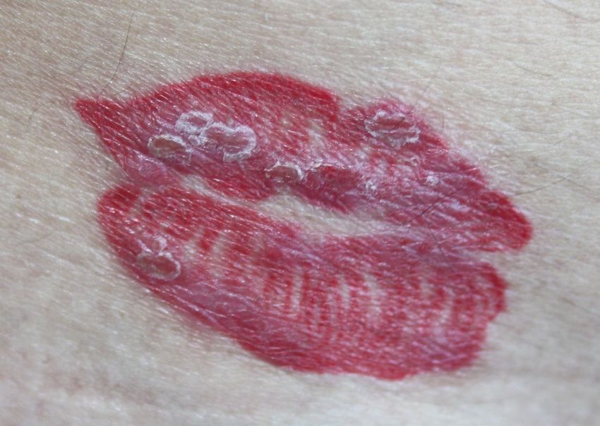
10

CONCLUSIONS
       Diagnosis of allergic reactions to ink in permanent tattoos is very challenging.
While clinical and histological data can help clinicians recognize the reactions, these
indications are not specific. We consider that biopsy is essential in late reactions, and
while we also recommend performing patch testing, this approach currently has a low
yield. We hope that more information on the composition of tattoo inks will be available
in the future, so that clinicians can choose allergens for patch tests more accurately and
thus identify the likely cause of allergic reactions to tattoos.

Funding
The authors declare that no funding was received for the present study.
Conflicts of Interest
The authors declare that they have no conflicts of interest.

J Investig Allergol Clin Immunol 2019; Vol. 29(6)                  © 2019 Esmon Publicidad
doi: 10.18176/jiaci.0383
11
References
1. Mataix J, Silvestre JF. Reacciones cutáneas adversas por tatuajes y piercings. Actas
Dermosifiliogr. 2009;100:643-56.
2. Kluger N. Cutaneous complications related to tattoos: 31 cases from Finland.
Dermatology. 2017;233:100-9.
3. Hogsber T, Hutton K, Serup J. High prevalence of minor symptoms in tattoos
among a young population tattooed with carbon black and organic pigments. J Eur
Acad Dermatol Venereol. 2013;27:846-52.
4. Kluger N. Self-reported tattoo reactions in a cohort of 448 French tattooists. Int J
Dermatol. 2016;55:764-8.
5. Brady BG, Gold H, Leger EA, Leger MC. Self-reported adverse tattoo reactions: a
New York City Central Park study. Contact Dermatitis. 2015;73:91-9.
6. González-Villanueva I, Silvestre Salvador JF. Diagnostic Tools to Use When We
Suspect an Allergic Reaction to a Tattoo: A Proposal Based on Cases at Our
Hospital. Actas Dermosifiliogr. 2018;109:162-72
7. Serup J. Diagnostic tools for doctors’ evaluation of tattoo complications. Curr Probl
Dermatol 2017;52:42-57.
8. González I, Silvestre JF. Claves clínicas ante una sospecha de reacción alérgica a un
tatuaje. Piel. 2018;33:575-80.
9. Serup J, Carlsen KH, Sepehri M. Tattoo complaints and complications: diagnosis
and clinical spectrum. Curr Probl Dermatol. 2015;48:48-60.
10. Serup J. How to diagnose and classify tattoo complications in the clinic: a system of
distinctive patterns. Curr Probl Dermatol. 2017;52:58-73.
11. González I, Silvestre JF. Reacciones alérgicas frente a las tintas de los tatuajes. In:
Muñoz D, Ed. Tratado sobre los tatuajes. Claves para su eliminación con láser.
Buenos Aires: Ediciones Journal, 2017; p. 69-76.
12. Morte PD, Magee M. Hyperalgesia after volar wrist tattoo: a case of complex
regional pain syndrome? J Clin Neuromuscul. 2011;12:118-21.
13. Zemtsov A, Wilson L. CO2 laser treatment causes local tattoo allergic reaction to
become generalized. Acta Derm Venereol. 1997;77:497.
14. Simunovic C, Shinohara MM. Complications of decorative tattoos: Recognition and
management. Am J Clin Dermatol. 2014;15:525-36.
J Investig Allergol Clin Immunol 2019; Vol. 29(6) © 2019 Esmon Publicidad
doi: 10.18176/jiaci.038312 15. Romani N, Clausen B, Stoitzner P. Langerhans cells and more: langerin-expressing dendritic cell subsets in the skin. Immunol Rev. 2010;234:120-41. 16. Kimber I, Dearman RJ, Basketter DA. Dendritic cells and the assessment in vitro of skin sensitizing potential. Cutan Ocul Toxicol. 2013;32:54-9. 17. Høgsberg T, Thomsen BM, Serup J. Histopathology and immune histochemistry of red tattoo reactions. Skin Res Technol. 2015;21:449-58. 18. Silvestre JF, González I. Utilidad de las pruebas epicutáneas en el estudio de las reacciones cutáneas a tatuajes. Piel. 2018;33:549-51. 19. McFadden N, Lyberg T, Hensten-Pettersen A. Aluminum-induced granulomas in a tattoo. J Am Acad Dermatol. 1989;20:903-8. 20. Gutermuth J, Hein R, Fend F, Ring J, Jakob T. Cutaneous pseudolymphoma arising after tattoo placement. J Eur Acad Dermatol Venereol. 2007;21:566-7. 21. Michel R. Manufacturing of tattoo ink products today and in future: Europe. Curr Probl Dermatol. 2015;48:103-11. 22. Serup J, Kluger N, Bäumler W. Tattoo inks: legislation, pigments, metals and chemical analysis. Curr Probl Dermatol. 2015;48:152-7. 23. Gaudron S, Ferrier-Le Bouëdec MC, Franck F, D'Incan M. Azo pigments and quinacridones induce delayed hypersensitivity in red tattoos. Contact Dermatitis. 2015;72:97-105. 24. González-Villanueva I, Álvarez-Chinchilla P, Silvestre JF. Allergic reaction to 3 tattoo inks containing Pigment Yellow 65. Contact Dermatitis. 2018;79:107-8 25. Serup J, Hutton Carlsen K. Patch test study of 90 patients with tattoo reactions: negative outcome of allergy patch test to baseline batteries and culprit inks suggests allergens are generated in the skin through haptenization. Contact Dermatitis. 2014;71:255-63. 26. Bäumler W. Absorption, distribution, metabolism and excretion of tattoo colorants and ingredients in mouse and man: the known and the unknown. Curr Probl Dermatol. 2015;48:176-84. 27. Engel E, Spannberger A, Vasold R, König Landthaler M, Bäumler W. Photochemical cleavage of a tattoo pigment by UVB radiation or natural sunlight. J Dtsch Dermatol Ges. 2007;5:583-9. J Investig Allergol Clin Immunol 2019; Vol. 29(6) © 2019 Esmon Publicidad doi: 10.18176/jiaci.0383
13

28. Hutton Carlsen K, Serup J. Photosensitivity and photodynamic events in black, red
   and blue tattoos are common: a ‘Beach study’. J Eur Acad Dermatol Venereol.
   2014;28:231-7.
29. Tresukosol P, Ophaswongse S, Kullavanijaya P. Cutaneous reaction to cosmetic lip
   tattooing. Contact Dermatitis. 1997;36:176-7.
30. Steinbrecher I, Hemmer W, Jarisch R. Adverse reaction to the azo dye Pigment Red
   170 in a tattoo. J Dtsch Dermatol Ges. 2004;(2):1007-8.
31. González-Villanueva I, Hispan P, Silvestre JF. Allergic contact dermatitis caused by
   a black tattoo ink in a patient allergic to shellac. Contact Dermatitis. 2016;75:247.

Juan Francisco Silvestre Salvador

Pintor Baeza 12

03010 Alicante, Spain

E-mail: silvestre_jfr@gva.es

J Investig Allergol Clin Immunol 2019; Vol. 29(6)             © 2019 Esmon Publicidad
doi: 10.18176/jiaci.0383
14

LEGENDS TO THE FIGURES

Figure 1. The presence of crusted honey-colored lesions distributed randomly in a
recently tattooed area should lead us to suspect a bacterial infection.

Figure 2. The appearance of sarcoidosis on previous cutaneous scars is a well-known
phenomenon (scar sarcoidosis). It can also occur in tattooed areas and mimic a local
reaction to the ink used in the tattoo.

J Investig Allergol Clin Immunol 2019; Vol. 29(6)          © 2019 Esmon Publicidad
doi: 10.18176/jiaci.0383
15

Figure 3. The late appearance of dry eczematous lesions with exclusive involvement of
the tattooed area increases the suspicion of an allergic reaction.

Figure 4. The appearance of flattened papular lesions (lichenoid pattern) that affect
the entire tattooed surface of the same color is characteristic of allergic reactions to
the inks used in tattoos.

J Investig Allergol Clin Immunol 2019; Vol. 29(6)             © 2019 Esmon Publicidad
doi: 10.18176/jiaci.0383
16

Figure 5. The presence of scar areas is a final stage to which most of these clinical
patterns could progress.

J Investig Allergol Clin Immunol 2019; Vol. 29(6)             © 2019 Esmon Publicidad
doi: 10.18176/jiaci.0383
17

Figure 6. In this case, eczema-like lesions are observed in all red tattooed areas. The
remaining colors do not present complications. Regardless of the clinical pattern, an
essential finding to confirm a suspected allergic reaction is a uniform, monomorphic
appearance throughout the area tattooed with the problem ink. Red ink is more
commonly associated with complications than other colors.

Figure 7. Deposition of the pigment of the ink around the sensory nerves of the dermis
seems to be the origin of the discomfort that many patients experience in tattoos,
apparently without lesions (neuropathic pattern). Allergy should not be suspected in
these cases.

J Investig Allergol Clin Immunol 2019; Vol. 29(6)             © 2019 Esmon Publicidad
doi: 10.18176/jiaci.0383
18

Figure 8. A, Eczematous plaque reaction in the red color of a tattoo placed a month
and a half before the photo was taken. B, Similar simultaneous reaction in a red tattoo
placed 2 years previously. This phenomenon is very suggestive of allergic reaction to
tattoo ink.

J Investig Allergol Clin Immunol 2019; Vol. 29(6)           © 2019 Esmon Publicidad
doi: 10.18176/jiaci.0383
19

Figure 9. Psoriasis presents as an isomorphic phenomenon and may appear on
previously injured skin such as scars, surgical wounds, or tattoos.

Figure 10. The presence of interface dermatitis with predominantly lymphocytic band-
like inflammatory infiltrate in the dermoepidermal junction and in the superficial
dermis, whether associated or not with spongiotic dermatitis, is a frequent histological
finding in allergic reactions to tattoo ink.

J Investig Allergol Clin Immunol 2019; Vol. 29(6)           © 2019 Esmon Publicidad
doi: 10.18176/jiaci.0383
20

Figure 11. Pseudoepitheliomatous hyperplasia with transepidermal pigment
elimination is a histological finding suggestive of allergic reaction to tattoo ink.

Figure 12. Prior to performing patch testing, it is advisable to obtain the problem ink
and its safety data sheet.

J Investig Allergol Clin Immunol 2019; Vol. 29(6)               © 2019 Esmon Publicidad
doi: 10.18176/jiaci.0383
21

Figure 13. A, Patient with eczematous skin reaction to “apparently” red tattoo ink. B,
Results of patch testing with positivity to cobalt chloride. The patient told us that she
had previously been tattooed with a blue ink, but that she was retattooed with a red
ink in order to improve the appearance of the tattoo. No relevance could be
established, given that it was unknown whether the blue ink contained cobalt chloride.

J Investig Allergol Clin Immunol 2019; Vol. 29(6)            © 2019 Esmon Publicidad
doi: 10.18176/jiaci.0383
22

Figure 14. Patch testing with the problem ink (as is) is advisable, although it is generally
negative. In this case, it was positive, and we discovered that the patient was allergic
to shellac, which is used to bind the pigment particles in black inks.

J Investig Allergol Clin Immunol 2019; Vol. 29(6)              © 2019 Esmon Publicidad
doi: 10.18176/jiaci.0383
23

Figure 15. Diagnostic algorithm for managing patients with cutaneous reactions to a
tattoo.

                                                                NO        TOPICAL
                                                                      CORTICOSTEROIDS

     EARLY REACTION                    CLINICAL SUSPICION OF            LACK OF
                                                                                             BIOPSY
       (? 1 month)                          I N F E CTI O N ?          RESPONSE

                                                                        CULTIVE +
                                                                YES     EMPIRICAL
                                                                       ANTIBIOTICS

      LATE REACTION                               ABSCESS
       (> 1 month)
                                                                          RULE OUT SYSTEMIC DISEASE/
                                              GRANULOMATOUS               MYCOBACTERIAL OR FUN GAL
                                                                                  INFECTION

                                                                            MOLECULAR BIOLOGICAL
                                          PSEUDOLIMPHOMATOUS
                                                                                  STUDY
               BIOPSY

                                           INTERFACE DERMATITIS                   PATCH TESTING

          ¿ C L I N I C A L S I GN S
             SUGGESTING                             YES
               ALLERGY?

J Investig Allergol Clin Immunol 2019; Vol. 29(6)                                 © 2019 Esmon Publicidad
doi: 10.18176/jiaci.0383
24
Table 1. Recommended History for a Skin Reaction in a Tattooed Area
Medical history
- History of contact allergies (especially to metallic salts or textile dyes)
- Previous relevant dermatoses (eg, psoriasis, eczema, urticaria, lichen planus,
cutaneous lupus erythematous, and pyoderma gangrenosum)
- Previous infections (hepatitis, HIV)
- Systemic diseases (eg, sarcoidosis)
- Neoplastic diseases
Symptoms present
- How much time passed between completion of the tattoo and the appearance of
the lesions?
- Is there pruritus, pain, or dysesthesia in the tattooed area?
- Does exposure to sunlight trigger or exacerbate the complaint?
- Is fever, arthralgia, lymph node involvement or another systemic manifestation
present?
- What treatments have been used for this problem, and what were the results?
- Have any lesions appeared on previous tattoos?
Tattoo process
- When and where was the tattoo performed, and who did it (professional tattooist
or amateur)?
- Were different colors used in the tattoo? To obtain the color(s), were the inks
mixed?
- Do you have other tattoos? Is this the first time you were tattooed with this color
ink?
- Can you get a sample of the problem ink and/or its safety data sheet?
J Investig Allergol Clin Immunol 2019; Vol. 29(6) © 2019 Esmon Publicidad
doi: 10.18176/jiaci.038325 Table 2. Clinical Signs of Allergic Contact Dermatitis to Tattoo Ink LATE reaction (weeks, months, or years after being tattooed) PERSISTENT reaction HOMOGENEOUS involvement of ALL AREAS where the ink of the same COLOR was injected Clinical pattern: PLAQUE-LIKE, LICHENOID, HYPERKERATOTIC, or ULCERO- NECROTIC DISTANT REACTION in OTHER PREVIOUS TATTOOS of the SAME COLOR POOR RESPONSE to topical corticosteroids J Investig Allergol Clin Immunol 2019; Vol. 29(6) © 2019 Esmon Publicidad doi: 10.18176/jiaci.0383
26

J Investig Allergol Clin Immunol 2019; Vol. 29(6)   © 2019 Esmon Publicidad
doi: 10.18176/jiaci.0383
You can also read