Prevalence, Types and Clinical Presentation of Heart Failure among Hypertensive Patients Seen at a Tertiary Hospital in Dar Es Salaam, Tanzania
←
→
Page content transcription
If your browser does not render page correctly, please read the page content below
ISSN: 2378-2951
                                                                                           Nyaisonga and Chillo. Int J Clin Cardiol 2021, 8:230
                                                                                                         DOI: 10.23937/2378-2951/1410230
                                                                                                                             Volume 8 | Issue 3
                   International Journal of                                                                                       Open Access

                   Clinical Cardiology
Original Research Article

Prevalence, Types and Clinical Presentation of Heart Failure
among Hypertensive Patients Seen at a Tertiary Hospital in
Dar Es Salaam, Tanzania
Gervas George Nyaisonga, MD, MMED and Pilly Chillo, MD, MMED, PhD*                                        iD
                                                                                                                                  Check for
Department of Internal Medicine, Muhimbili University of Health and Allied Sciences, Tanzania                                     updates

*Corresponding author: Pilly Chillo, MD, MMED, PhD, Section of Cardiovascular Medicine, Department of Internal
Medicine, Muhimbili University of Health and Allied Sciences, PO BOX 65001, Dar es Salaam, Tanzania, Tel: +255-22-215-
0603; +255-713-779781

 Abstract                                                                  Conclusion: The prevalence of HF among hypertensive
                                                                           patients seen at a tertiary hospital in Tanzania is 29.5%,
 Background: Heart failure (HF) is a common complication                   majority of them having HFpEF. HFpEF differs from HFrEF
 in patients with hypertension who may present as HF with                  in terms of BP levels, obesity status and some echocar-
 preserved ejection fraction (HFpEF) or HF with reduced                    diographic parameters. These factors need to be carefully
 ejection fraction (HFrEF). These categories have different                examined when HF is suspected in otherwise less sympto-
 clinical presentations and may require special attention to
                                                                           matic patients.
 diagnose, especially when the presentation is HFpEF. The
 aim of this study was to assess the prevalence, types and                 Keywords
 clinical presentation of HF among hypertensive patients
 being followed-up at a tertiary hospital in Tanzania.                     Heart failure, Hypertension, Heart failure with preserved
                                                                           ejection fraction, Heart failure with reduced ejection fraction,
 Methods: We included all known and newly diagnosed                        Diastology, Echocardiogram, Sub Saharan Africa, Tanzania
 hypertensive adults (≥ 18 years) referred for echocardiogram
 examination at the Muhimbili National Hospital - Mloganzila,
 between June and December 2019. A detailed cardiovascu-                  Introduction
 lar history, physical, laboratory and echocardiogram exa-                    Heart failure (HF) is a global pandemic that affects
 mination was performed in all patients. HF was diagnosed
 according to the Framingham criteria and was further cate-               approximately 64.3 million people worldwide [1], rep-
 gorized as HFpEF (EF ≥ 50%) or HFrEF (EF < 50%), accor-                  resenting an important cause of morbidity and mortal-
 ding to the echocardiography findings. Patients from these               ity [2]. The age-standardized prevalence rates of HF is
 two groups were then compared in terms of demographic,                   increasing, and is accompanied with an increase in mor-
 clinical, laboratory and echocardiographic characteristics.
                                                                          tality and years lived with disability, especially in low
 The chi-square and Student's t test was used to compare
 categorical and continuous data respectively. A p-value of <             and middle income countries (LMIC) [1]. The increas-
 0.05 indicated a statistically significant difference.                   ing HF burden is especially significant in sub Saharan
 Results: Out of 633 hypertensive patients seen during                    Africa (SSA), including Tanzania, which is experiencing
 the study period, 346 (54.7%) fulfilled the inclusion criteria           a change in epidemiology of diseases from communica-
 and were enrolled. Mean ± SD age was 58.3 ± 12.4 years,                  ble to non-communicable diseases [3]. Hypertension is
 and 60.4% were women. Mean ± SD systolic and diastolic                   by far the most common underlying cause contributing
 BP was 152 ± 23 and 91 ± 15, respectively. A total of 102
 (29.5%) patients were found to have HF. Three quarters of
                                                                          to the increase of HF burden in SSA [4], being present
 HF patients (74.5%) had HFpEF and the remaining (25.5%)                  in 14.9% to 29.8% of the adult population [5,6], and in
 had HFrEF. In comparison, patients with HFpEF were more                  up to 50% of those aged ≥ 55 years [5]. Furthermore,
 likely to be outpatients, older, obese, and with higher mean             hypertension in SSA is more severe and results in early
 BP and more concentric left ventricular hypertrophy when                 end-organ damage, including HF, chronic kidney dis-
 compared to those with HFrEF, all p < 0.05.

                                       Citation: Nyaisonga GG, Chillo P (2021) Prevalence, Types and Clinical Presentation of Heart Failure
                                       among Hypertensive Patients Seen at a Tertiary Hospital in Dar Es Salaam, Tanzania. Int J Clin Cardiol
                                       8:230. doi.org/10.23937/2378-2951/1410230
                                       Accepted: May 29, 2021: Published: May 31, 2021
                                       Copyright: © 2021 Nyaisonga GG, et al. This is an open-access article distributed under the terms of the
                                       Creative Commons Attribution License, which permits unrestricted use, distribution, and reproduction
                                       in any medium, provided the original author and source are credited.

Nyaisonga and Chillo. Int J Clin Cardiol 2021, 8:230                                                                         • Page 1 of 8 •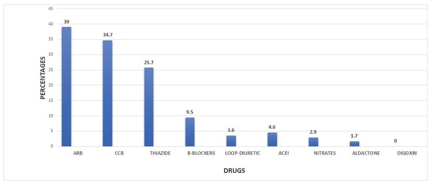
DOI: 10.23937/2378-2951/1410230                                                                           ISSN: 2378-2951

ease and stroke [7].                                         when serum total cholesterol was > 5.2 mmol/l and
                                                             low HDL-C was defined when serum HDL-C was < 1.04
    Regardless of the type, a diagnosis of HF carries a
                                                             mmol/l, according to the Muhimbili National Hospital’s
significant morbidity and mortality risk [8,9], and ef-
                                                             laboratory reference values. Estimated glomerular fil-
forts should be made to diagnose HF earlier than later.
                                                             tration rate (eGFR) was calculated from CKD-EPI equa-
With the aid of echocardiogram, HF has been classified
                                                             tions [18] and renal dysfunction was considered to be
into two major categories: HF with preserved ejection
                                                             present when a patient had eGFR of less than 60 ml/
fraction (HFpEF) and HF with reduced ejection fraction
                                                             min/1.73 m2. Anemia was defined as hemoglobin of less
(HFrEF) [10]. Among patients with hypertension, studies
                                                             than 13 g/dl in men and 12 g/dl in women according to
have found HFpEF to be more common, mainly due to
                                                             the World Health Organization [19].
diastolic dysfunction of the hypertrophied left ventricle
[11,12]. HFpEF needs careful attention to diagnose, as            Echocardiograms were performed using a General
traditionally HF was defined as a presence of reduced        Electric (GE) Vivid S3 echocardiogram machine equipped
ejection fraction on echocardiogram. Ascertaining the        with a 3.5 MHZ transducer, and the protocol followed
proportion of hypertensive patients with HFpEF is im-        the American Society of Echocardiology recommenda-
portant as it will increase awareness among clinicians       tions [20]. Left ventricular (LV) hypertrophy (LVH) was
of this otherwise obscured disease [13]. However, only       considered present when LV mass (LVM) indexed to
few studies have reported on the types of HF among           body surface area (LVMI) was > 95 g/m2 in women and
hypertensive patients in SSA [8,14,15], and most of pre-     > 115 g/m2 in men. LVEF was determined using M-mode
vious studies did not systematically look for HF symp-       guided parasternal long-axis images of the left ventricle
toms therefore may have missed those with mild to mo-        and was taken as a measure of LV systolic function. EF
derate symptoms. Furthermore, no previous study in           of < 50% was considered as systolic dysfunction [20]. LV
Tanzania has reported on the types of HF among exclu-        filling was obtained by determining peak early velocity
sively hypertensive patients. This study was therefore       (E) at the level of the mitral leaflets’ tips, and the medial
set out to determine the prevalence, types and clinical      early diastolic mitral annular velocity (E′) was measured
characteristics of HF in a population of hypertensives at-   by spectral tissue Doppler imaging in apical four-cham-
tending a tertiary hospital in Tanzania.                     ber views. The ratio of E to E′ velocity (E/E′ ratio) was
                                                             taken as an estimation of LV filling pressure and diastol-
Methodology                                                  ic dysfunction was considered present when the E/E’
Data collection process and definition of terms              was ≥ 15 [21].
    A structured questionnaire was used to collect pa-           Patients with HF as per Framingham criteria were
tients’ socio-demographic and clinical data. Information     further categorized as HFrEF (when EF < 50%) or HFpEF
collected included gender, age of the patient, area of       (EF ≥ 50%) [10]. The diagnosis of HFpEF required the fol-
residence, cardiovascular risk factors, symptoms of HF,      lowing conditions to be satisfied: (i) Positive diagnosis
etc. A thorough physical examination was done and            of HF as per Framingham criterion; (ii) LV EF ≥ 50%; (iii)
cardiovascular signs like ankle edema, upper quadrant        LV diastolic dysfunction, i.e. E/E’ ≥ 15. The diagnosis of
abdominal tenderness, chest rales, S3 gallop, and jugu-      HFrEF was reached when the following conditions were
lar venous pulse were looked for and recorded whether        satisfied (i) Positive diagnosis of HF as per Framingham
present or not. The Framingham criteria was used to as-      criterion and (ii) Reduced LV systolic function on echo-
sess for the presence of HF among patients [16].             cardiogram (i.e. LV ejection < 50%) [10].
    Blood pressure (BP) was measured according to gui-       Data handling and analysis
delines [17], using a standard automated BP machine
                                                                 All questionnaires were scanned for completeness
(Heuer Company, from USA). Hypertension was defined
                                                             and coded before being entered into the dataset. Sta-
as systolic BP of ≥ 140 mmHg and/or diastolic BP of ≥ 90
                                                             tistical package of Social Science for Windows (SPSS)
mmHg, or known hypertensive on medications, and was
                                                             version 21 was used for statistical analysis. Continuous
categorized as grade 1 (140-159/90-99 mmHg), grade
                                                             variables were expressed as the mean ± SD, and catego-
2 (160-179/100-109 mmHg) and grade 3 (≥ 180/≥ 110
                                                             rical variables as n (%). The χ2 or Fisher's exact test was
mmHg) according to European Society of Cardiology
                                                             used to compare categorical variables, as appropriate.
guidelines [17]. Height, weight, waist and hip circumfe-
                                                             Student's t test was used to compare the mean values.
rences were measured following standard guidelines.
                                                             For statistical tests a two-tailed p-value < 0.05 was con-
   For each patient, a 10 ml of venous blood was col-        sidered significant.
lected and analyzed for cholesterol, glucose, creatinine,
urea nitrogen and hemoglobin levels. High triglyceride
                                                             Ethical considerations
levels were defined when serum triglyceride was > 1.69          This study was conducted in accordance with the
mmol/l, raised LDL-C was defined when serum LDL-C            Helsinki Declaration of studies on human subjects. Ethi-
was > 3.34 mmol/l, high total cholesterol was defined        cal approval to conduct the study was obtained from

Nyaisonga and Chillo. Int J Clin Cardiol 2021, 8:230                                                      • Page 2 of 8 •
DOI: 10.23937/2378-2951/1410230                                                                                  ISSN: 2378-2951

                        Table 1: Demographic, clinical and laboratory characteristics of the study population.

Variable                                                                               n (%) or mean ± SD
Mean ± SD Age (years)                                                                  58.3 ± 12.4
Age Categories, n (%)
 18-40                                                                                 28 (8.1)
 41-54                                                                                 100 (28.9)
 ≥ 55                                                                                  218 (63)
Female Sex, n (%)                                                                      209 (60.4)
Place Of Referral, n (%)
 Outpatient                                                                            305 (88.1)
 Inpatient                                                                             41 (11.9)
Cardiovascular Risk Factors, n (%)
 Diabetes                                                                              44 (12.7)
 Smoking                                                                               34 (9.8)
 Alcohol                                                                               146 (42.2)
 Family History of Cardiovascular Disease                                              152 (43.9)
Anthropometric Variables
 Mean ± SD Height (cm)                                                                 165.2 ± 9.2
 Mean ± SD Weight (kg)                                                                 75.9 ± 16.2
 Mean ± SD BMI (kg/m2)                                                                 27.8 ± 5.3
Obesity Status, n (%)
 Normal Weight                                                                         127 (36.7)
 Overweight                                                                            113 (32.7)
 Obese                                                                                 106 (30.6)
Mean ± SD Duration of hypertension                                                     6.8 ± 9.1
Mean ± SD Systolic BP (mmhg)(mmhg)                                                     152 ± 23
Mean ± SD Diastolic BP (mmhg)                                                          91 ± 15
Hypertension Stage, n (%)
Normal (Controlled) BP                                                                 100 (28.9)
Stage 1                                                                                98 (28.3)
Stage 2                                                                                85 (24.6)
Stage 3                                                                                63 (18.2)
Mean ± SD Pulse Rate (beats/min)                                                       80 ± 15
Tachycardia (≥ 100b/min), n (%)                                                        47 (13.6)
Laboratory Findings
 Mean ± SD Triglyceride (mmol/L)                                                       1.5 ± 0.7
Raised Triglyceride, n (%)                                                             120 (34.7)
 Mean ± SD Cholesterol (mmol/L)                                                        5.3 ± 3.4
Raised Cholesterol, n (%)                                                              165 (47.7)
 Mean ± SD LdL-C (mmol/L)                                                              3.6 ± 1.2
Raised LDL-C, n (%)                                                                    202 (58.4)
 Mean ± SD HdL-C (mmol/L)                                                              1.1 ± 0.2
Low HDL-C, N (%)                                                                       154 (44.5)
 Mean ± Hemoglobin (g/dl)                                                              12.6 ± 2
Anemia, n (%)                                                                          110 (31.8)
Mean ± SD eGFR (ml/min/1.73 m2)                                                        72.7 ± 22
Mean ± SD Urea Nitrogen (mmol/L)                                                       10.9 ± 7.7
Proportion With Renal Dysfunction, n (%)                                               85 (24.5)

BMI: Body Mass Index; BP: Blood Pressure; LDL: Low Density Lipoprotein; HDL: High Density Lipoprotein; eGFR: Estimated
Glomerular Filtration Rate

Nyaisonga and Chillo. Int J Clin Cardiol 2021, 8:230                                                             • Page 3 of 8 •
DOI: 10.23937/2378-2951/1410230 ISSN: 2378-2951
the Directorate of Research and Publications of the Prevalence of HF
Muhimbili University of Health and Allied Sciences. All
One hundred and eight out of the 346 studied popu-
patients had to sign an informed consent form before
lation fulfilled the Framingham criteria for HF. Among
any data was collected.
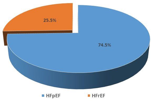
the 108 patients who met clinical criteria for HF, 26 had
Results EF < 50%, while 82 had EF ≥ 50% in echocardiogram. Of
the 82 participants with clinical HF and EF ≥ 50%, 6 did
Socio-demographic and clinical findings not meet the echocardiographic definition of diastolic
Three hundred and forty six (54.7%) out of 633 dysfunction and therefore they did not have HF, leaving
hypertensive patients that were screened fulfilled the 102/346 (29.5%) as the true prevalence of HF in this po-
inclusion and exclusion criteria and were enrolled. Of pulation. Among 102 patients with HF, 76 (74.5%) had
those excluded, 88 did not give consent, 12 were pre- HFpEF and the remaining 26 (25.5%) had HFrEF (Figure
gnant, 16 were admitted in the Intensive Care Unit and 2).
171 were referred more than once. Table 1 shows the
socio-demographic and clinical characteristics of the Table 2: Symptoms and signs of HF among study patients.
study participants. The mean age of the total studied Variable n (%)
population was 58.3 years (range 28-89 years). Majori-
Symptoms
ty (88.1%) of participants were from outpatient clinics.
Obesity was present in 30.6% of the participants. The Shortness of breath 185 53.5
mean ± SD systolic and diastolic blood pressure in the Palpitation 182 52.6
total population was 152 ± 23 mmHg and 91 ± 15 mmHg Lower limb swelling 132 38.2
respectively, and those with stage 3 hypertension were Nocturnal cough 65 18.8
18.2%. Anemia was present in 31.3% of the total popu- Orthopnea 47 13.6
lation, and 24.5% had renal dysfunction. Paroxysmal nocturnal dyspnea 34 9.8
Figure 1 shows the drugs used by study patients, Right upper quadrant pain 7 2
while the symptoms and signs of HF experienced by Signs
study patients are shown in Table 2. Antihypertensives Lower limb edema 69 19.9
receptor blockers were the most used antihypertensive
Gallop 24 6.9
(39%), followed by calcium channel blockers (34.7%),
Shift of apex beat 16 4.6
thiazide diuretics (25.7%) and beta blockers (9.5%).
Raised jugular venous pressure 8 2.3
Echocardiogram findings of the study patients are
Heaving apex beat 19 5.5
shown in Table 3. As seen in the table, more than half
Basal crepitation 10 2.9
(52.9%) of the total studied had LVH, 9.5% had LV systo-
lic dysfunction, and 20.5% had LV diastolic dysfunction. Tenderness of right upper quadrant 4 1.2
Figure 1: Types of drugs used by hypertensive patients.
ARB: Angiotensin Receptor Blocker; CCB: Calcium Channel Blocker; ACEI: Angiotensin Converting Enzyme Inhibitor
Nyaisonga and Chillo. Int J Clin Cardiol 2021, 8:230 • Page 4 of 8 •DOI: 10.23937/2378-2951/1410230 ISSN: 2378-2951
Comparison between hypertensive patients with significantly more concentric LV geometry, while those
HFpEF and HFrEF found to have HFrEF had significantly higher left atrial
volume index as well as left ventricle internal diameter,
Patients with HFpEF were more likely to be outpa- indicating larger LV dimensions all p < 0.05 (Table 4).
tients and showed a trend of being older than patients
with HFrEF. In terms of symptoms and signs, patients Discussion
with HFpEF were less likely to have paroxysmal noctur- HF is a common complication of hypertension, and
nal dyspnoea, shifted apex beat, gallop rhythm and ba- its burden may be higher in SSA due to the increased
sal crepitations, all p < 0.05 (Table 4). In terms of echo- hypertension prevalence [5,6], late hospital presenta-
cardiographic findings, patients with HFpEF were having tion [3] and limited ability to diagnose the early disease
Table 3: Echocardiographic findings of the study population. [1]. In this cross-sectional study of hypertensive patien-
ts attending a referral hospital in Tanzania, we highlight
Variable n (%) or mean 3 important findings that add to the current knowledge
± SD
of hypertensive heart disease in the region. First, the
Interventricular Septum in Diastole (cm) 1.18 ± 0.2 prevalence of HF among hypertensive patients seen at
LV Posterior Wall in Diastole (cm) 1.12 ± 0.3 a referral hospital in Tanzania is 29.5%; second, majo-
LV Internal Diameter in Diastole (cm) 4.6 ± 0.7 rity of hypertensive patients with HF have HFpEF; and
LV Mass Index (g/m ) 2
111.1 ± 37.3 third, hypertensive patients with HFpEF differ from tho-
Proportion with LV Hypertrophy 183 (52.9) se with HFrEF in a number of demographic and clinical
characteristics.
Fractional Shortening (%) 37.7 ± 8.6
EF (%) 66.8 ± 12.4 The prevalence of HF found in this study is in keeping
Proportion with Reduced EF 33 (9.5) with the findings from a meta-analysis of 23 blood pres-
sure-lowering clinical trials involving 193,424 hyperten-
Left Atrial Diameter (cm) 3.8 ± 0.6
sives, in whom HF occurred in 28.9% [22]. The present
Left Atrial Volume Index (ml/m ) 2
30 ± 12 findings also echo our understanding that hypertension
Proportion with Left Atrial Enlargement 97 (28) is the most common underlying risk factor for HF in
E (m/s) 0.6 ± 0.2
SSA accounting up to 45% of HF cases [4]. In popula-
tion studies, hypertension confers a 2-3 folds increased
A (m/s) 0.75 ± 0.1
hazard to HF development [23], indicating very strong
E/A Ratio 0.9 ± 0.5
links between hypertension and HF. The mechanism of
Deceleration Time (ms) 155 ± 62.1 HF in hypertension has been termed to involve chronic
Isovolumic Relaxation Time (ms) 101 ± 22 pressure overload that leads to the development of left
E’ (m/s)) 0.08 ± 0.2 ventricular hypertrophy and fibrotic changes that lead
E/E’ Ratio 12 ± 22 to progressive diastolic dysfunction and failure, while
Proportion with E/E’ ≥ 15 71 (20.5)
another subset of patients progresses to systolic dy-
sfunction in the presence of chronic volume and pres-
LV: Left Ventricular; EF: Ejection Fraction
Figure 2: Distribution of HFpEF and HFrEF among hypertensive patients with HF.
Nyaisonga and Chillo. Int J Clin Cardiol 2021, 8:230 • Page 5 of 8 •DOI: 10.23937/2378-2951/1410230 ISSN: 2378-2951
Table 4: Comparison between hypertensive patients with HFpEF and HFrEF.
Variables HFpEF HFrEF p value
n = 76 n = 26
Mean ± SD age (years) 61 ± 11 57 ± 14 0.113
Age ≥ 55 years, n (%) 57 (75) 14 (53.8) 0.023
Women, n (%) 49 (64.5) 15 (58) 0.64
Inpatients, n (%) 14 (18.4) 11 (42.3) 0.02
Diabetes, n (%) 10 (13.2) 6 (23.1) 0.348
Symptoms, n (%)
Shortness of breath 68 (89.5) 25 (96.2) 0.442
Palpitation 48 (63.2) 16 (61.5) 0.912
Orthopnea 51 (67.1) 18 (69.2) 0.987
PND 17 (22.4) 15 (57.7) 0.001
Lower limb swelling 48 (63.2) 15 (57.7) 0.367
Right upper quadrant pain 3 (4) 3 (11.4) 0.334
Nocturnal cough 42 (55.3) 20 (76.9) 0.051
Physical findings
Mean ± SD Pulse rate (beats/min) 79 ± 18 86 ± 17 0.063
Mean ± SD SBP (mmHg) 157 ± 23 142 ± 28 0.011
Mean ± SD DBP (mmHg) 92 ± 23 89 ± 17 0.258
Mean ± SD BMI (kg/m2) 29 ± 6.4 26.2 ± 6.6 0.058
Obesity, n (%) 33 (43.4) 6 (23.1) 0.001
Signs, n (%)
Raised JVP 6 (7.9) 3 (11.5) 0.690
Shifted Apex beat 13 (17.1) 12 (46.2) 0.005
Gallop rhythm 11 (14.5) 20 (76.9) 0.000
Basal crepitation 3 (3.9) 7 (26.9) 0.002
Lower limb edema 30 (39.5) 13 (50) 0.367
Laboratory findings
Mean ± SD Hemoglobin (g/dl) 12.4 ± 1.6 11.6 ± 2 0.001
Mean ± SD Creatinine (µmol/l) 121.5 ± 87 130.7 ± 117 0.11
Mean ± SD BUN (mmol/l) 12 ± 12.7 12.2 ± 8.8 0.15
Mean ± SD eGFR (ml/min/1.73 m ) 2
68.6 ± 24 70.6 ± 30 0.204
Renal dysfunction, n (%) 22 (28.9) 10 (38.5) 0.463
Echocardiographic findings
Interventricular septum in diastole (cm) 1.35 ± 0.2 1.1 ± 0.3 0.001
LV internal diameter in diastole (cm) 4.6 ± 0.6 5.6 ± 0.8 0.000
Left atrial volume index (ml/m2) 32.5 ± 12 43.4 ± 16 0.001
Proportion with LVH, n (%)s 57 (75) 22 (84.6) 0.419
Proportion with enlarged LA n (%) 32 (42.1) 16 (61.5) 0.112
Mean ± SD E/E’ 15.8 ± 5 20.9 ± 11 0.002
BMI: Body Mass Index; PND: Paroxysmal Nocturnal Dyspnea; JVP: Jugular Venous Pressure; n: Number; SD: Standard Deviation,
HFpEF: Heart Failure with Preserved Ejection Fraction; HFrEF: Heart Failure with Reduced Ejection Fraction
sure overload [24]. to the current findings [15]. The difference between
ours and the study by Ogah is likely due to the diffe-
Our finding that majority of patients with hyperten-
rences in the study population, as HF registry tend to
sive HF have HFpEF is similar to many previous studies
include patients with end stage hypertensive heart di-
that studied hypertensive-only HF cohorts [25-27], and
sease, where those with diastolic dysfunction progress
underscores the importance of diastolic HF in this popu-
to have LV dilatation and eventual systolic dysfunction
lation. However, Ogah, et al. found a 35% proportion of
at the end of the hypertensive heart disease spectrum
HFpEF in a hypertensive HF registry in Nigeria contrary
Nyaisonga and Chillo. Int J Clin Cardiol 2021, 8:230 • Page 6 of 8 •DOI: 10.23937/2378-2951/1410230 ISSN: 2378-2951
[24]. Nevertheless, active search for HF among hyper- obstructive pulmonary disease. However, the use of
tensive patients is recommended [21] as most of the the Framingham criteria together with echocardiogram
patients with HFpEF could have been missed if only must have offset most of these biases.
ejection fraction was used to categorize HF. Of note,
the diagnosis of HFpEF is tricky and it requires thorough
Conclusion
assessment of diastolic function to determine presen- In conclusion, the prevalence of HF among hyper-
ce of increased LV filling pressures [13]. Our definition tensive patients being followed-up at a tertiary hospi-
of diastolic dysfunction as E/E’ of ≥ 15 indicate marked tal in Tanzania is high, and the majority of patients with
raise in LV filling pressures, and therefore true diastolic HF present as HFpEF. We recommend active screening
dysfunction. Of note, these patients had similar propor- for HF especially in the obese, elderly and uncontrolled
tion of dyspnea which is the hallmark of HF, similar to hypertensive patients, as they may present with HFpEF
patients with HFrEF (Table 4). As suggested by guideli- which can pass unnoticed.
nes, patients with diastolic HF require similar medica-
tions and follow-up as for those with HFrEF [25].
Disclosure Statement
The authors report no conflicts of interest.
In this study, hypertensive patients found to have
HFpEF were more likely to be older adults (≥ 55 years), Funding Details
obese and with higher mean systolic BP when compared
Not applicable.
to those with HFrEF. These findings are similar to cur-
rent knowledge of this subset of HF patients [8,12,27], Competing Interests
and our findings confirms this observation also among
The authors declare that they have no competing in-
native Tanzanian hypertensives. However, other risk
terests.
factors including diabetes mellitus and female gender
did not show significant associations as previously re- Availability of Data and Materials
ported [8,28-30], most likely due to the fact that the
The datasets used and/or analyzed during the cur-
current study was not powered to detect these associa-
rent study are available from the corresponding author
tions, and only trends could be seen. Not surprisingly,
on reasonable request.
clinical signs of shifted apex beat, gallop rhythm, basal
crepitations which signify more volume overload were Authors’ Contributions
more frequently seen in HFrEF when compared to the
GN and PC conceived the research idea. GN recrui-
group found to have HFpEF, in keeping with previous
ted patients and did data entry. GN and PC performed
studies in literature [26].
echocardiogram, analyzed and interpret data. Both au-
As expected, patients with HFpEF had more concen- thors drafted the manuscript, have read and approved
tric LV geometry on echocardiogram, which is the un- the final manuscript.
derlying cause of diastolic dysfunction in hypertensive
patients [31]. Of note, while the mean LA volume was Acknowledgements
high in both groups, those with HFrEF had higher volu- We extend our gratitude to all the study participants
mes, indicating that patients with HFrEF are at the end for their cooperation offered during this study. Special
spectrum of hypertensive HF, and they are likely to have thanks to Doctor Emmanuel Matulo, Sr. Rose and Sr.
passed the LV diastolic dysfunction before progressing Sophia whose work on data collection, recruitment and
to HFrEF [32]. This is also confirmed by the higher mean disposal of patients cannot go unmentioned.
E/E’ in the HFrEF group. In short, patients with HFrEF also
have diastolic dysfunction while patients with HFpEF References
have diastolic dysfunction without systolic dysfunction, 1. Bragazzi NL, Zhong W, Shu J, Abu Much A, Lotan D, et
confirming the notion that diastolic and systolic HF are al. (2021) Burden of heart failure and underlying causes in
195 countries and territories from 1990 to 2017. European
not independent or separate entities, rather HF is a sin- Journal of Preventive Cardiology 12.
gle continuous disease spectrum and systolic and dia-
2. Jones NR, Roalfe AK, Adoki I, Hobbs FDR, Taylor CJ
stolic HF are phenotypes at two extremes; as advocated
(2019) Survival of patients with chronic heart failure in the
by the single syndrome hypothesis of HF [32]. community: A systematic review and meta-analysis. Eur J
Heart Fail 21: 1306-1325.
The strength of this study include its prospective
nature which allowed for objective and thorough as- 3. Agbor VN, Essouma M, Ntusi NAB, Nyaga UF, Bigna JJ, et
sessment of clinical and echocardiographic parameters, al. (2018) Heart failure in sub-Saharan Africa: A contempo-
raneous systematic review and meta-analysis. Int J Cardiol
therefore likely to have captured most of hypertensive 257: 207-215.
patients with HF. We did not systematically determine
4. Damasceno A, Mayosi BM, Sani M, Ogah OS, Mondo C,
biomarkers of HF like NT-Pro BNP levels in this study,
et al. (2012) The causes, treatment, and outcome of acute
therefore it is possible that some of the HF symptoms heart failure in 1006 Africans from 9 countries. Arch Intern
could have been due to other conditions like chronic Med 172: 1386-1394.
Nyaisonga and Chillo. Int J Clin Cardiol 2021, 8:230 • Page 7 of 8 •DOI: 10.23937/2378-2951/1410230 ISSN: 2378-2951
5. Hendriks ME, Wit FW, Roos MT, Brewster LM, Akan- 20. Lang RM, Badano LP, Mor-Avi V, Afilalo J, Armstrong A, et
de TM, et al. (2012) Hypertension in sub-Saharan Africa: al. (2015) Recommendations for cardiac chamber quantifi-
Cross-sectional surveys in four rural and urban communi- cation by echocardiography in adults: An update from the
ties. PLoS One 7: e32638. American Society of Echocardiography and the European
Association of Cardiovascular Imaging. European heart
6. Guwatudde D, Nankya-Mutyoba J, Kalyesubula R, Lauren-
journal Cardiovascular Imaging 16: 233-271.
ce C, Adebamowo C, et al. (2015) The burden of hyperten-
sion in sub-Saharan Africa: A four-country cross sectional 21. Nagueh SF, Smiseth OA, Appleton CP, Byrd BF, Dokaini-
study. BMC Public Health 15: 1211. sh H, et al. (2016) Recommendations for the evaluation of
left ventricular diastolic function by echocardiography: An
7. Yoruk A, Boulos PK, Bisognano JD (2018) The state of
update from the american society of echocardiography and
hypertension in Sub-Saharan Africa: Review and Commen-
the european association of cardiovascular imaging. Eur
tary. Am J Hypertens 31: 387-388.
Heart J Cardiovasc Imaging 17: 1321-1360.
8. Abebe TB, Gebreyohannes EA, Tefera YG, Abegaz TM
22. Tocci G, Sciarretta S, Volpe M (2008) Development of heart
(2016) Patients with HFpEF and HFrEF have different cli-
failure in recent hypertension trials. J Hypertens 26: 1477-
nical characteristics but similar prognosis: A retrospective
1486.
cohort study. BMC Cardiovasc Disord 16: 232.
23. Ho KK, Pinsky JL, Kannel WB, Levy D (1993) The epide-
9. Magnussen C, Niiranen TJ, Ojeda FM, Gianfagna F, Blan-
miology of heart failure: The framingham study. J Am Coll
kenberg S, et al. (2019) Sex-specific epidemiology of heart
Cardiol 22: 6A-13A.
failure risk and mortality in Europe: Results From the Bio-
marCaRE Consortium. JACC Heart Fail 7: 204-213. 24. Levy D, Larson MG, Vasan RS, Kannel WB, Ho KK (1996)
The progression from hypertension to congestive heart fai-
10. Paulus WJ, Tschope C, Sanderson JE, Rusconi C, Fla-
lure. JAMA 275: 1557-1562.
chskampf FA, et al. (2007) How to diagnose diastolic heart
failure: A consensus statement on the diagnosis of heart 25. Pfeffer MA, Shah AM, Borlaug BA (2019) Heart failure with
failure with normal left ventricular ejection fraction by the preserved ejection fraction in perspective. Circ Res 124:
Heart Failure and Echocardiography Associations of the 1598-1617.
European Society of Cardiology. Eur Heart J 28: 2539-
26. Vasan RS, Larson MG, Benjamin EJ, Evans JC, Reiss CK,
2550.
et al. (1999) Congestive heart failure in subjects with nor-
11. Sun JP, Xu TY, Lee AP, Yang XS, Liu M, et al. (2015) Early mal versus reduced left ventricular ejection fraction: Preva-
diastolic dyssynchrony in relation to left ventricular remo- lence and mortality in a population-based cohort. J Am Coll
deling and function in hypertension. Int J Cardiol 179: 195- Cardiol 33: 1948-1955.
200.
27. Devereux RB, Roman MJ, Liu JE, Welty TK, Lee ET, et al.
12. Gradman AH, Wilson JT (2009) Hypertension and diastolic (2000) Congestive heart failure despite normal left ventri-
heart failure. Curr Cardiol Rep 11: 422-429. cular systolic function in a population-based sample: The
strong heart study. Am J Cardiol 86:1090-1096.
13. Gutierrez C, Blanchard DG (2004) Diastolic heart failure:
Challenges of diagnosis and treatment. Am Fam Physician 28. Shah RU, Klein L, Lloyd Jones DM (2009) Heart failure in
69: 2609-2616. women: Epidemiology, biology and treatment. Womens he-
alth 5: 517-527.
14. Adebayo AK, Adebiyi AA, Oladapo OO, Ogah OS, Aje A,
et al. (2009) Characterisation of heart failure with normal 29. Lawson CA, Zaccardi F, Squire I, Ling S, Davies MJ, et al.
ejection fraction in a tertiary hospital in Nigeria. BMC Car- (2019) 20-year trends in cause-specific heart failure outco-
diovasc Disord 9: 52. mes by sex, socioeconomic status, and place of diagno-
sis: A population-based study. The Lancet Public health 4:
15. Ogah OS, Sliwa K, Akinyemi JO, Falase AO, Stewart S
e406-e420.
(2015) Hypertensive heart failure in Nigerian Africans: In-
sights from the Abeokuta Heart Failure Registry. J Clin 30. Meagher P, Adam M, Civitarese R, Bugyei Twum A,
Hypertens 17: 263-272. Connelly KA (2018) Heart failure with preserved ejection
fraction in diabetes: Mechanisms and management. Can J
16. McKee PA, Castelli WP, McNamara PM, Kannel WB (1971)
Cardiol 34:632-643.
The natural history of congestive heart failure: The Framin-
gham study. N Engl J Med 285:1441-1446. 31. Komamura K (2013) Similarities and differences between
the pathogenesis and pathophysiology of diastolic and sy-
17. Williams B, Mancia G, Spiering W, Agabiti Rosei E, Azizi
stolic heart failure. Cardiol Res Pract 2013: 824135.
M, et al. (2018) 2018 ESC/ESH Guidelines for the manage-
ment of arterial hypertension. Eur Heart J 39: 3021-3104. 32. Ouzounian M, Lee DS, Liu PP (2008) Diastolic heart failu-
re: Mechanisms and controversies. Nature Clinical Practice
18. Burballa C, Crespo M, Redondo-Pachon D, Perez-Saez
Cardiovascular Medicine 5: 375-386.
MJ, Mir M, et al. (2018) MDRD or CKD-EPI for glomerular
filtration rate estimation in living kidney donors. Nefrologia
38: 207-212.
19. https://www.who.int/nutrition/publications/en/ida_asses-
sment_prevention_control.pdf
Nyaisonga and Chillo. Int J Clin Cardiol 2021, 8:230 • Page 8 of 8 •You can also read