The Journal of Rheumatology
←
→
Page content transcription
If your browser does not render page correctly, please read the page content below
The Journal of Rheumatology

Risk of Cancer in 767 Patients with Giant Cell Arteritis in Western Norway: A
Retrospective Cohort with Matched Controls
Lene Kristin Brekke, Bjørg-Tilde Svanes Fevang, Andreas P. Diamantopoulos, Jörg
Assmus, Elisabet Esperø and Clara Gram Gjesdal

DOI: 10.3899/jrheum.190147
http://www.jrheum.org/content/early/2020/01/10/jrheum.190147

               1. Sign up for TOCs and other alerts
                 http://www.jrheum.org/alerts

               2. Information on Subscriptions
                 http://jrheum.com/faq

               3. Information on permissions/orders of reprints
                 http://jrheum.com/reprints_permissions

The Journal of Rheumatology is a monthly international serial edited by Earl D.
Silverman featuring research articles on clinical subjects from scientists working
in rheumatology and related fields.

      Downloaded from www.jrheum.org on January 29, 2020 - Published by The
      Journal of Rheumatology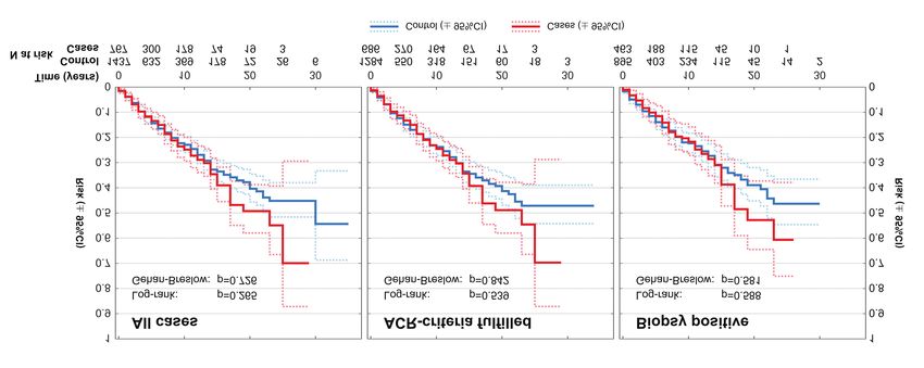
Risk of Cancer in 767 Patients with Giant Cell Arteritis
in Western Norway: A Retrospective Cohort with
Matched Controls
Lene Kristin Brekke, Bjørg-Tilde Svanes Fevang, Andreas P. Diamantopoulos,
Jörg Assmus, Elisabet Esperø, and Clara Gram Gjesdal
ABSTRACT. Objective. To determine the risk of cancer in a large Norwegian cohort of patients with giant cell
arteritis (GCA).
Methods. This is a hospital-based, retrospective, observational cohort study including patients
diagnosed with GCA in the Bergen Health Area during 1972–2012. Patients were identified through
computerized hospital records using the International Classification of Diseases coding system.
Medical records were reviewed. Each patient was randomly assigned population controls matched on
age, sex, and geography from the Central Population Registry of Norway. Data on the occurrence of
cancer were obtained from the Cancer Registry of Norway. The cumulative risk of malignancy was
estimated using Kaplan-Meier methods and potential differences were analyzed using the
Gehan-Breslow and log-rank tests.
Results. We identified 881 cases with a clinical diagnosis of GCA, of which 792 fulfilled the American
College of Rheumatology (ACR) 1990 classification criteria and 528 were biopsy-verified. Cases
with no registered cancer prior to GCA diagnosis were included in a time-to-event analysis, with first
cancer as the event (n = 767 with clinical GCA diagnosis, 686 fulfilling ACR criteria for GCA, 463
biopsy-verified). These cases were matched with previously cancer-free population controls (n = 1437,
1284, 895, respectively). We found no significant difference in the risk of malignancy after time of
diagnosis/matching for GCA patients compared to population controls (p > 0.05).
Conclusion. In this study of a large and well-characterized cohort of patients with GCA, there was
no difference in the risk of malignancy in patients with GCA compared to matched population controls.
(J Rheumatol First Release January 15 2020; doi:10.3899/jrheum.190147)
Key Indexing Terms:
VASCULITIS GIANT CELL ARTERITIS TEMPORAL ARTERITIS
MALIGNANCY CANCER EPIDEMIOLOGY
Giant cell arteritis (GCA) is the most common systemic its complexity and still many unknown factors, has been
vasculitis in adults. The immunopathogenesis of GCA, with outlined in a review by Weyand, et al and more recently by
Hid Cadena, et al1,2. These reviews describe several lineages
From the Hospital for Rheumatic Diseases, Haugesund; Department of of dysfunctional immune cells as well as age-related remod-
Clinical Science, University of Bergen, Bergen; Bergen Group of
Epidemiology and Biomarkers in Rheumatic Disease (BEaBIRD), eling of the immune system in GCA. The balance between
Department of Rheumatology, and Centre for Clinical Research, the expression of coinhibitory and costimulatory immune
Haukeland University Hospital, Bergen; Martina Hansens Hospital, checkpoint molecules appears to be crucial in fine-tuning the
Bærum, Norway.
This work was supported by unrestricted research grants from the
immune response and preventing autoimmunity. However,
Norwegian Association of Heart and Lung Patients; The Norwegian activation of specific inhibitory signals also allows cancer
Rheumatism Association; Marit Hansens Memorial Fund; Merck Sharp & cells to avoid recognition and destruction3,4. Immune check-
Dohme; Odd Fellow Medical Research Fund; and The Raagholt
Foundation.
point inhibitors, used in cancer treatment, have been followed
L.K. Brekke, MD, Hospital for Rheumatic Diseases, and Department of by a range of immune-related adverse events, including
Clinical Science, University of Bergen; B.T. Fevang, MD, PhD, polymyalgia rheumatica (PMR) and large-vessel vasculitis
Department of Clinical Science, University of Bergen, and BEaBIRD, (LVV)5,6,7,8,9. The increasing use of these drugs, which
generate antitumor activity but also enhance autoimmunity,
Department of Rheumatology, Haukeland University Hospital; A.P.
Diamantopoulos, MD, PhD, MPH, Martina Hansens Hospital; J. Assmus,
PhD, Centre for Clinical Research, Haukeland University Hospital; E. draws new attention to the “old” question of whether there is
Esperø, MD, Hospital for Rheumatic Diseases; C. Gram Gjesdal, MD, an association between GCA and cancer. Several investi-
gators have addressed this question, but published reports
PhD, Department of Clinical Science, University of Bergen, and
BEaBIRD, Department of Rheumatology, Haukeland University Hospital.
Address correspondence to Dr. L.K. Brekke, HSR AS, PB 2175, 5504 have shown conflicting results10–18. A metaanalysis from
Haugesund, Norway. E-mail: lene.kristin.brekke@hsr.as 2014 reported a low but statistically significant increased
Accepted for publication June 25, 2019. malignancy risk for patients with GCA19. However, most
Personal non-commercial use only. The Journal of Rheumatology Copyright © 2020. All rights reserved.
Brekke, et al: Cancer risk in GCA 1
Downloaded from www.jrheum.org on January 29, 2020 - Published by The
Journal of Rheumatologystudies on the risk of cancer in GCA have been limited by ously cancer-free cases, we dropped from 3:1 to 2:1 matching prior to the
small sample sizes, possible misclassification bias, and/or cancer-specific analyses. This was performed by random selection/inclusion
of 2 of the original 3 controls when none of the controls had previous cancer.
short periods of followup. We report a large cohort study in When only 1 of the 3 original controls had previous cancer, this control was
which GCA diagnoses have been thoroughly verified and excluded but the 2 cancer-free controls were kept in the analysis along with
cancer diagnoses were obtained from a national registry with the cancer-free matched case. If 2 or all 3 original controls had previous
mandatory reporting, providing virtually no loss to followup. cancer, we excluded all controls and the corresponding case from the
This study may contribute to clarifying the association time-to-event analysis.
Extensive demographic and clinical data were collected for the cases but
between GCA and the risk of malignancy. for the population controls, we had no available information on potential
risk factors, comorbid conditions, or other clinical data. Information on the
MATERIALS AND METHODS occurrence of cancer was obtained from the CRN. The CRN registration is
This is a retrospective, observational cohort study including patients based on mandatory reporting by physicians involved in the investigation,
diagnosed with GCA in the Bergen Health Area during 1972–2012. Our treatment, or followup of cancer patients in Norway. CRN also receives
material represents a predominantly white referral cohort from mixed rural information about all cancer deaths registered by the Norwegian Cause of
and urban areas. The study setting was the Bergen Health Area, consisting Death Registry. This ensures near-complete ascertainment of incident
of 3 somatic hospitals: Haukeland University Hospital, Haraldsplass cancers in the Norwegian population and the CRN is among the oldest and
Deaconess Hospital, and Voss Hospital. Together these hospitals provide most complete cancer registries in the world21. Every cancer diagnosis is
specialist healthcare services to the inhabitants of 22 municipalities in required to be reported, except basal cell carcinoma (BCC) in adults.
Hordaland County in western Norway. By population, Hordaland is the third Precancerous lesions, benign tumors, and tumors with uncertain/unknown
largest county in Norway, and home to 10% of Norway’s total population potential for malignancy are also reported. When the study was performed,
(Statistics Norway 2016). In this region there is only 1 laboratory for the registration of cancers in CRN was complete for the entire study period.
pathology, and no private hospitals that care for rheumatology patients. The Variables received from the CRN include date and ICD code of the cancer
majority (> 90%) of rheumatologists and internal medicine specialists in the diagnosis, and the diagnostic basis/accuracy of the malignancy. Cancer
area are hospital-based, with only 3 private rheumatologists in the region, diagnoses were grouped according to the European Shortlist for Causes of
all publicly funded and collaborating closely with the hospital departments. Death, 2012 version (COD-SL-2012). The interpretation and reporting of
Patients were identified through hospital records using the International these data are the sole responsibility of the authors, and no endorsement by
Classification of Diseases (ICD) coding system: ICD-8 (446.4) for 1972– the CRN is intended nor should be inferred. The study was approved by the
1987, ICD-9 (446.5) for 1987–1998, and ICD-10 (M31.5-6) for 1999–2012. REK sør-øst B regional ethics committee (REC), which approved the study
Electronic medical records of hospital diagnoses were available from 1972. for all the hospitals involved (study reference number 2012/643/REK sør-
Medical records that were completely electronic were available from 2001. øst B). REC granted permission to access records without obtaining consent
For patients diagnosed in 1972 through 2001, we also obtained access to from patients or their relatives owing to the long duration of the study and
archived paper-based medical records to complete the extraction of clinical late onset of the disease. REC also granted permission to obtain a control
data. We note that cancer data were obtained separately, because this infor- cohort (3:1) matched on age, sex, and geography from the Central Population
mation was provided by the Cancer Registry of Norway (CRN), in which Registry of Norway.
all new cases of cancer in Norway have been registered since 1952. We Statistical analysis. Descriptive statistics were used to characterize the
collected other data by reviewing medical records of every patient registered sample. The cumulative risk of cancer in cases and controls was estimated
with the diagnosis of GCA following an outpatient visit or admission to any using Kaplan-Meier plots with the diagnosis of cancer as the event. P values
ward in one of the 3 study hospitals between January 1, 1972, and December comparing the Kaplan-Meier curves were calculated using the
31, 2012 (41-yr period). We excluded patients if their GCA diagnosis origi- Gehan-Breslow and log-rank tests. Followup time was estimated using the
nated prior to the beginning of our study, if data were unavailable, or if the reverse Kaplan-Meier method. HR were estimated using Cox regression with
review of records from the time of diagnosis concluded that GCA was an adjustment for time of diagnosis/matching (both linearly and categorized by
implausible diagnosis. We subclassified patients according to the American decades). The significance level was set to 0.05. The computing was done
College of Rheumatology (ACR) 1990 criteria for GCA and according to using the Statistical Package for the Social Sciences (SPSS) software version
temporal artery biopsy (TAB) result. Further details about the selection 24 (IBM Corp.) and R software version 3.4 (R Foundation for Statistical
process including characteristics of the excluded cases have been published Computing).
previously20. Every Norwegian is given a unique 11-digit identification
number at birth or time of immigration. Each patient was initially matched RESULTS
for age (date of birth ± 1 month), sex, and county of residence to 3 control
We identified 881 patients with a clinical diagnosis of GCA,
subjects randomly selected from the Central Population Registry of Norway.
The controls were required to be alive at the time of GCA diagnosis for their of whom 792 fulfilled the ACR 1990 classification criteria
matched case and this date was defined as the start of the observation period for GCA and 528 had biopsy-verified GCA based on TAB.
for the control. The observation period ended with the diagnosis of cancer, Incidence numbers for GCA in the cohort were comparable
death, or end of study (December 31, 2012), whichever came first. We to previous reports, with a mean annual cumulative incidence
excluded duplicate control subjects, control subjects who were also among
of 18.4 per 100,000 persons aged 50 years or more for clini-
the cases, and all subjects with precancerous lesions or unverified malig-
nancy (e.g., tumor with uncertain topography and/or unverified histo- cally diagnosed GCA, 16.7 for cases fulfilling the ACR 1990
pathology). For persons with ≥ 2 registered cancers, we included only the criteria, and 11.2 for biopsy-verified cases alone20. After
first cancer. Only individuals with no registered cancer before the time of excluding persons with registered cancer prior to GCA
GCA diagnosis or corresponding date among matched controls (i.e., diagnosis, persons with registered cancer with uncertain
outcome-/event-free on entry to the cohort) were included in the
diagnostic accuracy, controls that were also among the cases,
time-to-event analysis. The original target of 3:1 matching was designed
with the intent to analyze also cause-specific mortality, including rare causes and duplicate control subjects, we included the following
of death. However, the high prevalence of cancer in the community created numbers of cases and controls: 767 cases with a clinical
an imbalance in this matching, and to avoid unnecessary exclusion of previ- diagnosis of GCA, of which 686 cases fulfilled the ACR 1990
Personal non-commercial use only. The Journal of Rheumatology Copyright © 2020. All rights reserved.
2 The Journal of Rheumatology 2020; 47:doi:10.3899/jrheum.190147
Downloaded from www.jrheum.org on January 29, 2020 - Published by The
Journal of Rheumatologycriteria for GCA and 463 were biopsy-verified. These were well with epidemiological data showing little or no difference
matched (2:1) with 1437/1284/895 previously cancer-free in overall mortality rates of patients with GCA compared to
population controls, respectively. Core characteristics of the the general population22,23,24. However, our result differs from
included cases and controls are presented in Table 1. that of some previous reports, in which an increased risk of
At the end of study (December 31, 2012), a total of cancer was seen in patients with GCA (Table 3).
120/107/69 patients with GCA and 227/204/141 population Ungprasert, et al, in a metaanalysis from 2014, concluded
controls had been registered with a verified first malignancy with a low but statistically significant increased cancer risk,
in CRN after the time of GCA diagnosis/matching (Table 1). reporting a pooled risk ratio (RR) of 1.14 overall, 2.16 in the
We found no significant difference in the risk of malignancy first 6–12 months after GCA diagnosis, and 1.35 for
after time of diagnosis/matching for any subgroup of patients biopsy-proven GCA19. However, excluding a study with
with GCA compared to population controls (Figure 1, Table potential selection bias reduced the pooled RR to a nonsignifi-
2). Followup times ranged from 0 to 35 years, with a median cant level of 1.08. The excluded study was a large retro-
followup time of 3 years for cases and 5 years for controls. spective cohort study from Sweden reporting on 35,918
We did not compare the risk of cancer prior to GCA patients registered with a diagnostic code of GCA or PMR in
diagnosis/matching because of missing data on cancers a nationwide hospital discharge database16. The inclusion of
diagnosed before 1972. Key features of our study compared only hospitalized patients may have led to a selection bias,
to previous reports evaluating the risk of cancer in GCA favoring patients with severe disease who seem to have a
cohorts from various populations are presented in Table 3. higher cancer risk. A study by Michet, et al from the Mayo
The overall distribution of various cancer types, grouped Clinic examined hospitalizations in the years 1996–2012 for
by codes in COD-SL-2012, showed no difference between patients with known GCA to evaluate whether hospital-
cases and controls (p = 0.768; Figure 2). The numbers for ization-related diagnoses accurately identified patients with
each cancer diagnosis are small and our study lacks sufficient this disease25. They found that the GCA diagnosis was
power to analyze the risk of different cancer diagnoses mentioned in only 31% of 502 hospitalizations among
separately. However, the combined lymphoid and hemato- patients with GCA. This illustrates the potential failure of
poietic malignancies (COD-SL-2012 codes 2.1.19–2.1.21) identification that may bias any study based on hospital-
accounted for almost one-quarter of all registered cancers in administrative diagnostic coding, ours included.
both cases and controls in our study, afflicting 3.8% of all The highest risk of cancer in the Swedish study was noted
GCA cases and 3.5% of their corresponding controls. during the first year after hospitalization for GCA or PMR16.
Malignant neoplasms of the colon, rectum, and anus This study was based solely on ICD coding without verifi-
(COD-SL-2012 code 2.1.4) accounted for another quarter of cation of the diagnosis through journal review. Consequently,
the registered cancers in cases and about 20% of the incident cancers may have been misdiagnosed as GCA or
registered cancers in controls. Cancers of the breast PMR initially and thus contributed to the excess rate of
(COD-SL-2012 code 2.1.10) followed, accounting for 10.8% cancer reported in the study. Symptoms similar to PMR and
of the registered malignancies in the patients with GCA and the sometimes-nonspecific presentation of GCA may mimic
12.8% in the controls. the presenting features of a variety of cancer diagnoses26–32.
In the selection process for our study, 35% of the initially
DISCUSSION selected patients coded as GCA were excluded to ensure a
This 41-year study of 881 Norwegian patients with GCA cohort of correctly diagnosed GCA20. We verified GCA
showed no difference in the risk of cancer compared to age-, diagnoses by reviewing patient records from the time of
sex-, and geographically matched population controls. This fits diagnosis and we excluded patients if the reviewing rheuma-
Table 1. Core characteristics of cases and controls.
Characteristics Clinical Diagnosis ACR 1990 Criteria Biopsy-verified
Cases, n = 767 Controls, n = 1437 Cases, n = 686 Controls, n = 1284 Cases, n = 463 Controls, n = 895
Mean age at time of GCA diagnosis/
matching, yrs (SD) 72.5 (8.5) 72.4 (8.5) 72.5 (8.4) 72.4 (8.4) 73.1 (7.8) 73.1 (7.8)
Female sex, n (%) 551 (71.8) 1030 (71.7) 497 (72.4) 928 (72.3) 339 (73.2) 656 (73.3)
Median observation
time*, yrs (95% CI) 3 (2–3) 5 (4–5) 3 (2–3) 4 (3–4) 3 (1–3) 5 (4–5)
Events (first cancer), n (%) 120 (15.6) 227 (15.8) 107 (15.6) 204 (15.9) 69 (14.9) 141 (15.8)
Deaths during observation,
n (%) 352 (45.9) 680 (47.3) 309 (45.0) 589 (45.9) 216 (46.7) 432 (48.3)
* Observation time according to reverse Kaplan-Meier. ACR: American College of Rheumatology; GCA: giant cell arteritis.
Personal non-commercial use only. The Journal of Rheumatology Copyright © 2020. All rights reserved.
Brekke, et al: Cancer risk in GCA 3
Downloaded from www.jrheum.org on January 29, 2020 - Published by The
Journal of RheumatologyFigure 1. Cumulative risk of first malignancy after time of GCA diagnosis or matching. GCA: giant cell arteritis; ACR: American College of Rheumatology.
Figure 2. The distribution of different cancers in patients with GCA and matched controls in the Bergen Health Area (1972–2012). All values represent the
number (%) of persons with registered cancer. A. Incident cancers for 767 patients with clinically diagnosed GCA and 1437 matched controls. B. Incident
cancers for 686 patients with GCA diagnosis fulfilling the ACR 1990 classification criteria for GCA and their 1284 matched controls. C. Incident cancers for
463 biopsy-proven patients with GCA and 895 matched controls. Cancers are grouped according to COD-SL-2012: lip, oropharynx, esophagus, stomach (codes
2.1.1-3); colon, rectum, anus (code 2.1.4); liver, pancreas (codes 2.1.5–6); larynx, trachea, bronchus, lung (codes 2.1.7–8); malignant melanoma of skin (code
2.1.9); breast (code 2.1.10); cervix uteri, uterus, ovary (codes 2.1.11–13); prostate (code 2.1.14); kidney (code 2.1.15); hematological, lymphoid (codes 2.1.19–
21); and other (codes 2.1.16–18, 2.1.22). ACR: American College of Rheumatology; GCA: giant cell arteritis; COD-SL-2012: European Shortlist for Causes
of Death (2012 version).
tologist concluded that GCA was an implausible diagnosis coding in the Swedish hospital discharge study would
based on clinical information available at that time. represent a significant limitation of their results.
Characteristics of the excluded cases have been published The excellent completeness of the CRN ensures that
previously20. A similar proportion of erroneous diagnostic nearly all incident cancers are registered21. A study conducted
Personal non-commercial use only. The Journal of Rheumatology Copyright © 2020. All rights reserved.
4 The Journal of Rheumatology 2020; 47:doi:10.3899/jrheum.190147
Downloaded from www.jrheum.org on January 29, 2020 - Published by The
Journal of RheumatologyTable 2. HR for the risk of cancer in patients with GCA versus population controls.
HR Lower CI Upper CI
All Cases, n = 2204
Unadjusted 1.13 0.91 1.42
Adjusted for time of diagnosis/matching 1.16 0.93 1.45
Adjusted for time period (decades*) 1.16 0.93 1.45
ACR 1990 criteria fulfilled, n = 1970
Unadjusted 1.12 0.85 1.36
Adjusted for time of diagnosis/matching 1.12 0.87 1.39
Adjusted for time period (decades*) 1.12 0.87 1.39
Biopsy-positive, n = 1358
Unadjusted 1.09 0.81 1.45
Adjusted for time of diagnosis/matching 1.12 0.84 1.49
Adjusted for time period (decades*) 1.11 0.83 1.49
* Decades defined as the time periods 1972–1982, 1983–1992, 1993–2002, and 2003–2012. ACR: American
College of Rheumatology; GCA: giant cell arteritis.
in a population where registration of cancers is not mandatory tigation, in which concomitant cancers are detected earlier
might be biased by an increased registration of cancers in than they otherwise would have been, thus shifting the timing
patients followed within specialist care, compared to previ- of cancer diagnosis from post-GCA to pre-GCA. However, a
ously healthy individuals. Being monitored for a chronic large population-based study from the UK found no
disease might thus increase the probability of a subsequent difference in the frequency of prior cancer between patients
cancer being registered. If so, this could partly explain the with and without GCA17, and Kermani, et al found that
increased risk of cancer noted in patients with GCA in some patients with GCA in a cohort from Olmsted County (USA)
studies. had significantly fewer malignancies prior to diagnosis
To date no definite association between GCA/PMR and compared to controls33.
cancer has been agreed on because of conflicting epidemio- The 2 previously published reports on cancer risk in
logical evidence, as illustrated in Table 3. The majority of Norwegian patients with GCA concluded differently10,11.
previous studies have been retrospective cohort studies. Myklebust, et al concluded that there was no difference in
However, 2 of the studies had a prospective design although risk in their cohort of 398 patients with GCA and 1592
differently executed11,18. Prospective design is often con- controls11. Haga, et al published a case-control study based
sidered superior as it allows an evaluation of the temporal on prospectively included cases of GCA or PMR during
sequence between exposure and outcome, and possibly 1978–1983 with subsequent retrospective analysis of cancer
reduces the risk of loss to followup. However, prospective risk based on cancer (registered in the CRN) from the
studies are cost- and time-consuming, and often difficult to inception of the registry through 198810. The inclusion
maintain for a long observation period, making them less criteria were biopsy-proven GCA or fulfillment of
suitable for the study of seldom and potentially late outcomes study-specific clinical criteria for either GCA or PMR, and
such as malignant diseases. Also, patients included in a 185 cases and 925 controls were included (Table 3). While
prospective study are regularly evaluated and more likely to Haga, et al found no difference in overall cancer risk, they
undergo extensive medical investigations, possibly leading found an increased risk in biopsy-proven GCA with HR of
to earlier detection and even an increased risk of cancer. As 2.35 for cases versus controls. They also reported an
such, prospective studies run the risk of overestimating the increased HR of 7.25 for biopsy-positive compared to
incidence of cancer in the patient population compared to the biopsy-negative cases. The Haga study included patients
controls. In the smaller prospective study, they found a from hospitals within our catchment area and within our
standardized incidence ratio of malignancy of 4.6118. study period. However, they also included isolated PMR (91
However, the number of patients was rather low (n = 107) of the 185 cases) and the study-specific inclusion criteria
and the number of recent and successive cancers was low were somewhat different from ours. Further, the number of
(n = 7), rendering these numbers uncertain. Also, there were biopsy-proven GCA was low (n = 65), and the finding of 16
no age- and sex-matched controls, only the use of crude cancers among these might have been coincidental because
incidence rates. we found no such difference in our 528 biopsy-proven cases.
It may be argued that a surveillance bias applies to any Two other previously published reports limited to
patient with GCA, often presenting with elevated inflam- biopsy-proven GCA cases have not confirmed the finding of
matory markers and nonspecific symptoms and signs. This increased risk associated with positive biopsy. In disagree-
clinical presentation may start an extensive diagnostic inves- ment with Haga, et al10, they found no overall difference in
Personal non-commercial use only. The Journal of Rheumatology Copyright © 2020. All rights reserved.
Brekke, et al: Cancer risk in GCA 5
Downloaded from www.jrheum.org on January 29, 2020 - Published by The
Journal of Rheumatology6
Table 3. Key features of previous studies on cancer risk in patients with giant cell arteritis (GCA).
Ungprasert, Brekke, Jese, Li, Ji, Kermani, Hill, Gonzalez-Gay, Liozon, Myklebust, Haga,
201419 2018 (PS) 201818 201717 201016 201015 201014 200713 200612 200211 199310
Country and NA Norway, Slovenia, UK, Sweden, USA, Australia, Spain, France, Norway, Norway,
time period 1972–2012 2011–2015 1990–2014 1965–2006 1950–2006 1991–2006 1981–2005 1976–2005 1987–1998 1978–1988
Study design Metaanal. Retro Prosp Retro Retro Retro Retro Retro Retro Prosp Retro
Inclusion criteria NA GCA defined ACR 1990 Diagnostic code Diagnostic code ACR 1990 Biopsy-proven Biopsy-proven ACR 1990 Biopsy-proven Biopsy-proven
by clinical criteria for GCA in GCA or PMR in criteria for GCA GCA criteria for GCA GCA, Bird GCA or study-
diagnosis, cranial GCA, nationwide nationwide GCA or Hamrin specific clinical
ACR1990 criteria, imaging general practice hospital discharge criteria for PMR criteria for
or positive biopsy criteria for database database GCA or PMR
purely extracranial
GCA
Controls NA Age-, sex-, and General Age-, sex-, and General Age-, sex-, and General NR General Age-, sex-, and Age-, sex-, and
Journal of Rheumatology
geographically population geographically population geographically population population geographically geographically
matched controls > 50 yrs (by practice) (database matched (age- and sex- (same age group) matched matched
matched controls subjects without controls specific) controls controls
without GCA the disease) without GCA
No. cases / Pooled cases: 767 / 1437 107 / NR 9778 / 92,268 35,918 / NR 204 / 407 226 / NR 255 / NR 271 /NR 398 / 1592 185 / 925
controls 39,808
Method of cancer NA Norwegian Prosp data + Diagnostic Swedish cancer Review of South Australia Review of Review of Norwegian Norwegian
identification Cancer Registry Slovenian code in general registry complete cancer medical medical Cancer Cancer
cancer registry practice database medical records registry records records + regional Registry Registry
cancer registry
Main conclusion Increased risk No difference Increased risk No difference Increased risk No difference No difference No difference Increased risk No difference Increased risk
(if TAB-positive)
ACR:American College of Rheumatology; GCA: giant cell arteritis; NA: not applicable (metaanalysis reporting pooled data from several cohort studies10,11,14,15,16); Metaanal.: metaanalysis; Retro: retrospective;
Prosp: prospective; NR: not reported; PMR: polymyalgia rheumatica; PS: present study; TAB: temporal artery biopsy.
Downloaded from www.jrheum.org on January 29, 2020 - Published by The
Personal non-commercial use only. The Journal of Rheumatology Copyright © 2020. All rights reserved.
The Journal of Rheumatology 2020; 47:doi:10.3899/jrheum.190147risk of malignancy in biopsy-proven GCA compared to the yrs) is a strength that reduces the risk of missing
general population13,14. late-occurring cancers as well as diminishes the risk of
Published reports have varied regarding the distribution erroneous conclusion based on variations through time.
of different types of cancers that patients with GCA However, the long duration also poses a challenge because
experience. Kermani, et al found that nonmelanoma skin the diagnostic criteria for both cancer and GCA might have
cancers were the most common cancers in both cases and changed substantially during such a long period. Even so,
controls15. This finding has not been confirmed by other time-dependent changes are controlled for by including the
studies, although Ji, et al also found an excess of skin control population on which such factors presumably would
cancers16. However, several studies have not included BCC have a similar effect.
of the skin as a malignancy10,11,12,14. BCC in adults are not Based on our findings, patients with GCA are not at
reported to the CRN and were thus not included among increased risk of cancer following the diagnosis of their
cancers observed in our study or in the previous Norwegian vasculitis. From a clinical point of view, our results indicate
studies. that patients with an unequivocal diagnosis of GCA do not
Liozon, et al found a strong temporal association between need specific screening for malignancy.
GCA and myelodysplastic syndromes and reported a
concurrent (occurring 1 yr before or after GCA diagnosis) ACKNOWLEDGMENT
hematological malignancy in 3.6% of their cases12. The The authors thank Dr. Rabab Adam, MD, for contributions in the early phase
combined malignancies of lymphoid and hematopoietic of the study. The authors also thank Alf Aksland (IT consultant, Haukeland
University Hospital), Anita Mellingen (Department of Rheumatology,
tissues accounted for almost one-quarter of the registered Haukeland University Hospital), Bjørg Sandvik (Department of Pathology,
cancers in our study, afflicting 3.8% of all GCA cases and Haukeland University Hospital), Robinson Lazcano (Central archive,
3.5% of the matched controls. Overall, both frequency and Haukeland University Hospital), Lisbeth Thorsen (Haraldsplass Deaconess
types of cancers found in patients with GCA and population Hospital), Laila Kvåle (Voss Hospital), and their coworkers, who aided with
controls in our study were similar. This is in agreement with the identification of patients or the extensive retrieval of patient records.
the findings reported by Myklebust, et al and largely with the
sizeable Swedish study, except for their finding of more skin REFERENCES
1. Weyand CM, Liao YJ, Goronzy JJ. The immunopathology of giant
cancers and leukemia11,16. The numbers for each cancer cell arteritis: diagnostic and therapeutic implications.
diagnosis are small and most studies lack sufficient power to J Neuroophthalmol 2012;32:259-65.
analyze the risk of different cancer diagnoses separately. 2. Hid Cadena R, Abdulahad WH, Hospers GA, Wind TT, Boots AM,
We have discussed factors that may have contributed to Heeringa P, et al. Checks and balances in autoimmune vasculitis.
our findings being different from those of some other studies. Front Immunol 2018;9:315.
3. Calabrese LH. Sorting out the complexities of autoimmunity and
Our data are limited by the retrospective design and have checkpoint inhibitors: not so easy. Ann Intern Med 2018;
incomplete (for cases) and missing (for controls) data on 168:149-50.
important cancer risk factors such as smoking, use of 4. Calabrese L, Velcheti V. Checkpoint immunotherapy: good for
cytotoxic medications or hormones, and other potential cancer therapy, bad for rheumatic diseases. Ann Rheum Dis
confounders such as body mass index and family history of 2017;76:1-3.
5. Goldstein BL, Gedmintas L, Todd DJ. Drug-associated polymyalgia
malignancy. For example, if the prevalence of smokers is rheumatica/giant cell arteritis occurring in two patients after
reduced in patients with GCA compared to the controls, our treatment with ipilimumab, an antagonist of CTLA-4. Arthritis
study could underestimate the true cancer risk associated with Rheumatol 2014;66:768-9.
GCA. We also note that our cohort consists of cases with 6. Calabrese C, Kirchner E, Kontzias K, Velcheti V, Calabrese LH.
predominantly cranial GCA (65% with positive TAB). Thus, Rheumatic immune-related adverse events of checkpoint therapy for
our results may not be representative for cases with purely cancer: case series of a new nosological entity. RMD Open
2017;3:e000412.
extracranial LVV. One strength of our study is the 7. Hodi FS, Lawrence D, Lezcano C, Wu X, Zhou J, Sasada T, et al.
completeness and high quality of the CRN. It has been Bevacizumab plus ipilimumab in patients with metastatic
mandatory to register every new case of cancer in Norway in melanoma. Cancer Immunol Res 2014;2:632-42.
this registry since 1952. Another strength is the large and 8. Micaily I, Chernoff M. An unknown reaction to pembrolizumab:
well-defined cohort of GCA cases resulting from a thorough giant cell arteritis. Ann Oncol 2017;28:2621-2.
9. Kuswanto WF, MacFarlane LA, Gedmintas L, Mulloy A, Choueiri
review of clinical data, excluding misclassified cases, and TK, Bermas BL. Rheumatologic symptoms in oncologic patients on
including hospitalized patients, as well as those only treated PD-1 inhibitors. Semin Arthritis Rheum 2018;47:907-10.
in outpatient clinics. The study also included a large cohort 10. Haga HJ, Eide GE, Brun J, Johansen A, Langmark F. Cancer in
of population controls that were tightly matched regarding association with polymyalgia rheumatica and temporal arteritis.
the most significant of all cancer risk factors — age. The J Rheumatol 1993;20:1335-9.
11. Myklebust G, Wilsgaard T, Jacobsen BK, Gran JT. No increased
large sample size of both cases and controls rendered a frequency of malignant neoplasms in polymyalgia rheumatica and
well-powered analysis, allowing us to detect relevant differ- temporal arteritis. A prospective longitudinal study of 398 cases and
ences between the groups. The long duration of our study (41 matched population controls. J Rheumatol 2002;29:2143-7.
Personal non-commercial use only. The Journal of Rheumatology Copyright © 2020. All rights reserved.
Brekke, et al: Cancer risk in GCA 7
Downloaded from www.jrheum.org on January 29, 2020 - Published by The
Journal of Rheumatology12. Liozon E, Loustaud V, Fauchais AL, Soria P, Ly K, Ouattara B, et al. 22. Lee YH, Song GG. Overall and cause-specific mortality in giant cell
Concurrent temporal (giant cell) arteritis and malignancy: report of arteritis: a meta-analysis. Z Rheumatol 2018;77:946-51.
20 patients with review of the literature. J Rheumatol 2006; 23. Catanoso M, Macchioni P, Boiardi L, Muratore F, Restuccia G,
33:1606-14. Cavazza A, et al. Incidence, prevalence, and survival of
13. Gonzalez-Gay MA, Lopez-Diaz MJ, Martinez-Lado L, biopsy-proven giant cell arteritis in Northern Italy during a 26-year
Pena-Sagredo JL, Lopez-Agreda H, Miranda-Filloy JA, et al. period. Arthritis Care Res 2017;69:430-8.
Cancer in biopsy-proven giant cell arteritis. A population-based 24. Mohammad AJ, Nilsson JA, Jacobsson LT, Merkel PA, Turesson C.
study. Semin Arthritis Rheum 2007;37:156-63. Incidence and mortality rates of biopsy-proven giant cell arteritis in
14. Hill CL, Cole A, Rischmueller M, Dodd T, Coleman M, Tucker G, southern Sweden. Ann Rheum Dis 2015;74:993-7.
et al. Risk of cancer in patients with biopsy-proven giant cell 25. Michet CJ 3rd, Crowson CS, Achenbach SJ, Matteson EL. The
arteritis. Rheumatology 2010;49:756-9. detection of rheumatic disease through hospital diagnoses with
15. Kermani TA, Schafer VS, Crowson CS, Hunder GG, Gabriel SE, examples of rheumatoid arthritis and giant cell arteritis: what are we
Ytterberg SR, et al. Malignancy risk in patients with giant cell missing? J Rheumatol 2015;42:2071-4.
arteritis: a population-based cohort study. Arthritis Care Res 26. Aguiar T, Vincent MB. Giant cell arteritis and polymyalgia
2010;62:149-54. rheumatica as first manifestation of typical pulmonary carcinoid
16. Ji J, Liu X, Sundquist K, Sundquist J, Hemminki K. Cancer risk in tumor. Reumatismo 2015;67:165-8.
patients hospitalized with polymyalgia rheumatica and giant cell 27. Bhatti MT, Furman J, Gupta S, Tabandeh H, Monshizadeh R.
arteritis: a follow-up study in Sweden. Rheumatology Superficial temporal artery biopsy diagnostic for lung carcinoma.
2010;49:1158-63. Am J Ophthalmol 2001;132:135-8.
17. Li L, Neogi T, Jick S. Giant cell arteritis and vascular disease-risk 28. Andersen SA, Kiss K. Primary temporal region squamous cell
factors and outcomes: a cohort study using UK Clinical Practice carcinoma diagnosed by a superficial temporal artery biopsy. Eur
Research Datalink. Rheumatology 2017;56:753-62. Ann Otorhinolaryngol Head Neck Dis 2015;132:91-2.
18. Ješe R, Rotar Ž, Tomšič M, Hočevar A. Giant cell arteritis and 29. Masood I, While B, Mudhar HS. Perivascular mantle cell lymphoma
malignancy-more than just a coincidence? J Clin Rheumatol affecting a temporal artery—a highly unusual cause of temporal
2018;24:85-6. headache. Cardiovasc Pathol 2011;20:244-6.
19. Ungprasert P, Sanguankeo A, Upala S, Knight EL. Risk of 30. Linxweiler M, Hasenfus A, Wolf G, Schick B. Perivascular marginal
malignancy in patients with giant cell arteritis and polymyalgia zone lymphoma mimicking temporal arteritis. Otolaryngol Head
rheumatica: a systematic review and meta-analysis. Semin Arthritis Neck Surg 2015;152:187-8.
Rheum 2014;44:366-70. 31. Naschitz JE, Slobodin G, Yeshurun D, Rozenbaum M, Rosner I.
20. Brekke LK, Diamantopoulos AP, Fevang BT, Assmus J, Espero E, Atypical polymyalgia rheumatica as a presentation of metastatic
Gjesdal CG. Incidence of giant cell arteritis in Western Norway cancer. Arch Intern Med 1997;157:2381.
1972-2012: a retrospective cohort study. Arthritis Res Ther 32. Jalava-Karvinen P, Kemppainen J, Saario R, Kotilainen P.
2017;19:278. Metastasis in the temporal bone mimicking temporal arteritis. J Clin
21. Larsen IK, Smastuen M, Johannesen TB, Langmark F, Parkin DM, Rheumatol 2010;16:19-21.
Bray F, et al. Data quality at the Cancer Registry of Norway: an 33. Kermani TA, Schafer VS, Crowson CS, Hunder GG, Ytterberg SR,
overview of comparability, completeness, validity and timeliness. Matteson EL, et al. Cancer preceding giant cell arteritis: a
Eur J Cancer 2009;45:1218-31. case-control study. Arthritis Rheum 2010;62:1763-9.
Personal non-commercial use only. The Journal of Rheumatology Copyright © 2020. All rights reserved.
8 The Journal of Rheumatology 2020; 47:doi:10.3899/jrheum.190147
Downloaded from www.jrheum.org on January 29, 2020 - Published by The
Journal of RheumatologyYou can also read