A twisting story: how a single gene twists a snail? Mechanogenetics - Cambridge University Press
←
→
Page content transcription
If your browser does not render page correctly, please read the page content below
PERSPECTIVE

                  A twisting story: how a single gene
                  twists a snail? Mechanogenetics
                  Reiko Kuroda*

                  Institute for Science and Technology, Tokyo University of Science, Noda, Chiba 278-8510, Japan

                  Quarterly Reviews of Biophysics (2015), 48(4), pages 445–452   doi:10.1017/S0033583515000098

                  Abstract. Left–right (l–r) symmetry breaking and the establishment of asymmetric animal body plan during embryonic development
                  are fundamental questions in nature. The molecular basis of l–r symmetry breaking of snails is a fascinating topic as it is determined
                  by a maternal single handedness-determining locus at a very early developmental stage. This perspective describes the current state of the
                  art of the chiromorphogenesis, mainly based on our own work, i.e. the first step of l–r symmetry breaking, as proven by our
                  “Mechanogenetics”, before the start of zygotic gene expression, transfer of chirality information to the cell-fate determining stage, and the
                  expression of nodal at the blastula stage. The Nodal signalling pathway is a common mechanism in vertebrates’ chiromorphogenesis in
                  later development. Studies on snails, especially Lymnaea (L.) stagnalis, shall give important insights into the molecular basis of chiromorpho-
                  genesis not only in Lophotrochozoa but in vertebrates as well.

                  Key words: Chirality chiromorphology, left-right symmetry, breaking developmental biology, snail handedness.

                  Introduction
                  Chirality is expressed throughout nature, whether micro-                         I have been interested in the molecular basis of left–right
                  scopic or macroscopic, and whether animate or inanimate.                         (l–r) symmetry breaking of animal body plan that starts from
                  Some molecules are either chiral or non-chiral, and billions                     a single fertilized egg, as it is determined genetically by intri-
                  of them self-assemble to form either chiral or non-chiral                        cate molecular interactions among genes and proteins. The
                  crystals by way of chiral discrimination at the molecular                        directions of anterior–posterior and dorsal–ventral axes
                  level. In the biological world, chirality is also observed at                    during the embryonic morphogenesis are determined pri-
                  all levels of hierarchy, from the macroscopic individual                         marily, and they have been well characterized. In contrast,
                  organisms down to molecules. At the molecular level, bio-                        the l–r direction emerges secondarily to the other two
                  logical world is totally homochiral; i.e. all living organisms                   axes, and unlike the two axes which define dichotomies in
                  on Earth use molecules of a unique invariant handedness:                         biological and physical attributes such as head and tail,
                  only D-(deoxy) ribose in nucleic acids and only L-amino                          and dorsal and ventral, no functional difference is inherent
                  acids in proteins. At the macroscopic level, most animals ap-                    to the left and right distinction. Embryonic morphogenesis
                  pear symmetric externally, but exhibit chirality within the                      along the l–r axis has only recently begun to be elucidated.
                  body cavity, i.e. in terms of asymmetric organ position, di-                     Several mechanisms for l–r asymmetry determination have
                  rectional organ looping and lateralized organ function in the                    been proposed for vertebrates and invertebrates. In verte-
                  larva and adult. Thus, both in animate and inanimate                             brates, there is a general agreement that l–r asymmetry is
                  domains, chirality is an excellent approach for studying                         determined by directional nodal flow across the embryonic
                  the link between macroscopic and microscopic phenomena.                          midline at a late development stage. Several unified models
                                                                                                   have been proposed, however, still little is known about the
                                                                                                   initial step of handedness determination for both in verte-
                                                                                                   brates and invertebrates (see recent Review articles by
                    * Authors for correspondence: R. Kuroda, Research Institute for Science
                  and Technology, Tokyo University of Science, Noda, Chiba 278-8510, Japan.        Blum et al. 2014; Coutelis et al. 2014; Vandenberg &
                  Tel: +81-4-7124-1501; Email: rkuroda@rs.tus.acp                                  Levin, 2013, and references therein).

                  © Cambridge University Press 2015. This is an Open Access article, distributed under the terms of the Creative Commons Attribution licence (http://
Downloaded from https://www.cambridge.org/core.   IP address:
                    creativecommons.org/licenses/by/3.0/),    176.9.8.24,
                                                           which  permitsonunrestricted
                                                                            28 Apr 2020re-use,
                                                                                        at 02:37:29, subject to
                                                                                               distribution,    thereproduction
                                                                                                              and   Cambridge Core  terms
                                                                                                                                in any    of use, available
                                                                                                                                        medium,   providedatthe
                                                                                                                                                             https://www.cambridge.org/core/terms.
                                                                                                                                                                original work is
https://doi.org/10.1017/S0033583515000098
                    properly cited.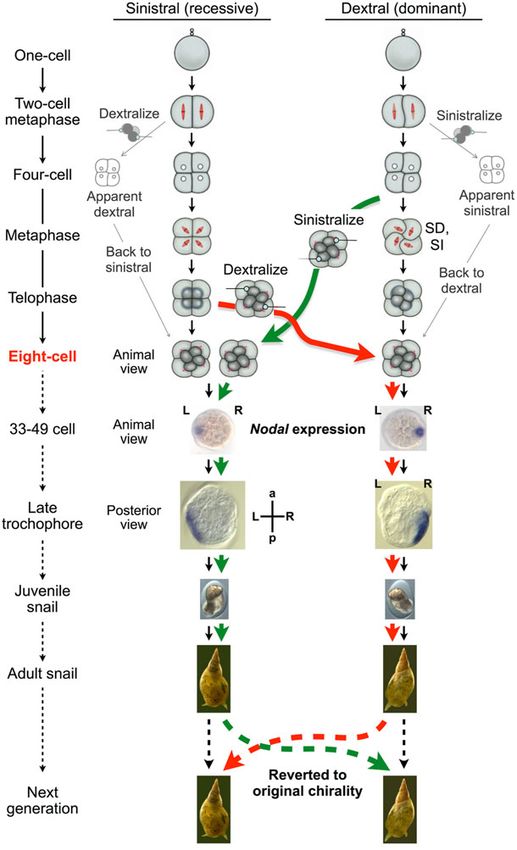
I conceived the study on snails thinking that snails may be-                      known since 1894 (Crampton, 1894). We closely analysed
                  come an ideal model system to study the initial step of chir-                     the temporal and spatial behaviour of cytoskeletons of the
                  ality determination. There are several unique features in                         dextral and the sinistral L. stagnalis during the early clea-
                  snails. First, the chirality is determined by maternal genetic                    vages by staining filamentous actin with fluorescently
                  inheritance. Namely, it is not the genotype of the individual                     labelled phalloidin and by visualizing spindles with indirect
                  but that of the mother that determines the chirality. The                         immunofluorescence with anti-β-tubulin antibody.
                  maternal inheritance is a crucial advantage as the chirality
                                                                                                    Contrary to what had been believed and illustrated in many
                  is determined at the early developmental stage. Recently,
                                                                                                    standard textbooks, we revealed non-mirror-image cytoske-
                  we proved that chirality is determined by a single locus
                                                                                                    letal dynamics for the sinistral and dextral embryos at the
                  (Hosoiri et al. 2003; Kuroda, 2014; Kuroda et al. 2009;
                                                                                                    most critical chiromorphogenetic step (Shibazaki et al.
                  Shibazaki et al. 2004), although it had been postulated
                                                                                                    2004). A helical deformation of blastomere shape was
                  based on the early 20th century work (Boycott et al. 1930;
                                                                                                    observed for the dextral embryos at the animal hemisphere
                  Sturtevant, 1923). Secondly, snails are hermaphrodite, re-
                                                                                                    in metaphase to anaphase (Fig. 1a I), as described ‘spiral de-
                  producing offspring by self- and cross-fertilization, which
                                                                                                    formation (SD)’ (Meshcheryakov, 1978). Consequently, a
                  is advantageous in tracing genetic traits. Thirdly, the geneti-
                                                                                                    quartet of micromeres protruded and budded off from the
                  cally determined chirality depends on species, ca. 90% dex-
                                                                                                    sister macromeres dextrotropically and rotated in a clockwise
                  tral only, 10% sinistral only, and rare dimorphic with both
                                                                                                    sense when viewed from the animal pole. In metaphase–ana-
                  chirality within a species. Fourthly, most snails display chir-
                                                                                                    phase, spindles were oriented with helicity along the A–V
                  ality not only internally but externally as well at the macro-
                                                                                                    axis. We named this spindle inclination (SI) (Fig. 1a I). In
                  scopic level, which makes experiments easier.
                                                                                                    marked contrast, neither SD nor SI was observed in meta-
                  We embarked on chiromorphology study using dimorphic                              phase to anaphase of sinistral embryos and each blastomere
                  fresh water snail, Lymnaea stagnalis. As the species had                          presented a symmetrical shape. Thus, the new micromeres
                  not been well studied compared with Lymnaea peregra                               emerged almost directly above the paired macromeres
                  (Freeman & Lundelius, 1982) except for the detailed obser-                        along the A–V axis. Chirality was not obvious until furrow
                  vation of development (Meshcheryakov, 1990), we studied                           ingression, when new micromeres rotated levotropically.
                  the breeding behaviour (Hosoiri et al. 2003), correlation of                      The spindles in metaphase–anaphase did not exhibit
                  cleavage pattern and organismal morphology (Shibazaki                             ‘reversed’ helicity but were positioned radially, devoid of
                  et al. 2004), cytoplasm injections to the 1-cell embryos of                       chirality, and almost parallel to the A–V axis (Fig. 1a II).
                  opposite chirality (Kuroda, 2014), cytoskeletal dynamics                          The blastomere shape and the spindle orientation as well
                  during spiral cleavages (Shibazaki et al. 2004), expression                       as the timing of chiromorphogenesis are different for the
                  of nodal-Pitx genes (Kuroda et al. 2009), creation of mirror-                     dextral and sinistral embryos within a species, and are far
                  image healthy animals by twisting blastomeres at the third                        from enantiomorphs (Shibazaki et al. 2004).
                  cleavage (Kuroda et al. 2009), etc. We also constructed back-
                  crossed congenic animals (Hosoiri et al. 2003; Kuroda, 2014;
                  Kuroda et al. 2009; Shibazaki et al. 2004) and carried out                        SD depends on actin polymerization
                  positional cloning in order to identify the handedness deter-
                                                                                                    When the dextral embryos were treated with actin depoly-
                  mining gene(s).
                                                                                                    merization agents, latrunculin A or B, they showed a con-
                                                                                                    comitant absence of SD and SI (Fig. 1b I left), resembling
                                                                                                    sinistral embryos (Fig. 1a II). The treatment of sinistral em-
                  Cytoskeletal dynamics during the third                                            bryos showed no change (Fig. 1b II left). When the dextral
                  cleavage                                                                          embryos in prophase were treated with 200 nM nocodazole,
                                                                                                    microtubule depolymerization agent, they did not form spin-
                  Non-mirror image relationship
                                                                                                    dles as expected, but exhibited SD slightly more prominent
                  The Spiralia (Lophotrochozoa) is one of the three major                           (Fig. 1b I right) than that of untreated embryos (Fig. 1a I).
                  clades of bilaterian metazoan which includes molluscs and                         Nocodazole treated sinistral embryos are devoid of spindles
                  annelids. The Spiralian clade of animals are morphologically                      and the symmetrical shape of the blastomeres remained
                  diverse but exhibit canonical spiral cleavage. Spiral cleavage                    (Fig. 1b II right). These results suggest that actin polymeriza-
                  where the cleavage planes are at oblique angles to the                            tion is involved in the dextral-specific SD. The cause and ef-
                  animal–vegetal (A–V) axis of the egg is characterized by                          fect of spindle orientation and blastomere shape has become
                  the alternating clockwise and anticlockwise rotation particu-                     clear: spindles do not deform blastomeres but the blastomere
                  larly during the 3rd–5th cleavages (Verdonk & van der                             deformation orients spindles. It is interesting to note that
                  Biggelaar, 1983). The correlation of the shell-coiling direc-                     there seem to be some cross-talk between the actins and
                  tion with the handedness of the third blastomere cleavage                         microtubules as the extent of SD enhances when the forma-
                  (i.e. from the four-cell to the eight-cell stage) has been                        tion of spindles is inhibited (Shibazaki et al. 2004).
                                                                                              446
Downloaded from https://www.cambridge.org/core. IP address: 176.9.8.24, on 28 Apr 2020 at 02:37:29, subject to the Cambridge Core terms of use, available at https://www.cambridge.org/core/terms.
https://doi.org/10.1017/S0033583515000098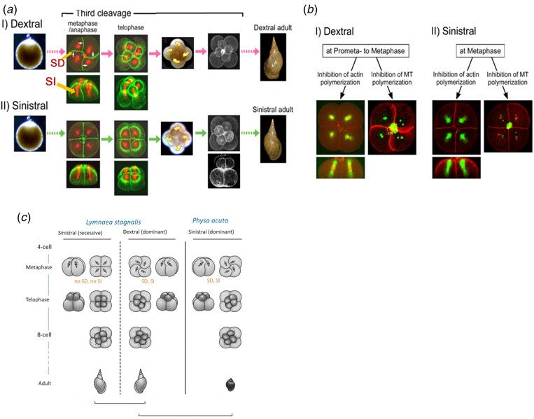
Fig. 1. The third cell cleavage of L. stagnalis for the dextral (I) and sinistral (II) embryos, and the schematic representation including
                  P. acuta. (a) SD and SI occur only in the dextral embryo. Filamentous actin: green; microtubules: red. (b) Dextral-specific SD and SI are
                  inhibited by treatment with an actin depolymerizing agent. F-actin and DNA: red; microtubules: green. (c) Schematic representation of
                  non-mirror image relationship of the mirror-image snails within a species but mirror image relationship across species.

                  Levotropical SD and SI of Physa (P.) acuta                                        frequent incident of insufficient and heterogeneous rotation
                                                                                                    of micromeres at the third cleavage of the sinistral embryos,
                  We found that P. acuta, a monomorphic sinistral gastropod,
                                                                                                    resulting in death before hatching. This clearly shows the
                  exhibits substantial SD and SI levotropically (Fig. 1c).
                                                                                                    role of SD to assure correct and sufficient rotation of blasto-
                  Nocodazole and latrunculin A treatments on P. acuta em-
                                                                                                    meres and the tilting of spindles, and hence the correct
                  bryos induced similar results to those observed for the dex-
                                                                                                    micromere–macromere contacts at the eight-cell stage
                  tral L. stagnalis, i.e. SD without formation of spindles for the
                                                                                                    (Kuroda, 2014).
                  nocodazole treatment, and loss of SD and SI for latrunculin
                  A treatment. Although the recessive sinistral embryos of
                  dextral-dominant L. stagnalis and L. peregra do not exhibit
                  a mirror-image cleavage pattern of their respective dextral                       The body handedness is determined by
                  embryos at the third cleavage, cleavage pattern of the em-                        a single locus that control cytoskeletal
                  bryos of sinistral only P. acuta displays a mirror image pat-                     dynamics
                  tern of dominant dextral Lymnaes. Figures in standard
                  textbooks illustrate a mirror-image relationship during the                       To prove that the handedness is determined by a single gene
                  embryogenesis for the enantiomorphic pair of snails within                        or genes at closely linked loci, we carried out backcross
                  a species; however, they must be amended to the enantio-                          breeding between inbred dextral and sinistral strains of
                  morphic pair across species as in Fig. 1c.                                        L. stagnalis cultivated in our laboratory. We used dextral
                                                                                                    animals as donor ‘fathers’ and sinistral animals as recurrent
                                                                                                    ‘mothers’, and the chirality-determining part of the genome
                  Functions of SD and SI
                                                                                                    from dextral inbred strain is introgressed on the genetic
                  SD and SI are unique features of dominantly handed snails                         background of the sinistral inbred strain. Each of the back-
                  at the eight-cell stage. We have observed that only half of the                   cross progeny obtained was typed for the chirality of the
                  embryos grew to normal snails in the case of recessive sin-                       next generation oviposited by it, because the genotype for
                  istral L. stagnalis, which is in sharp contrast to more than                      the handedness locus emerges as filial chirality. We have
                  90% in the dominant dextral embryos. This is due to the                           carried out serial backcrossing experiments (Kuroda et al.
                                                                                              447
Downloaded from https://www.cambridge.org/core. IP address: 176.9.8.24, on 28 Apr 2020 at 02:37:29, subject to the Cambridge Core terms of use, available at https://www.cambridge.org/core/terms.
https://doi.org/10.1017/S0033583515000098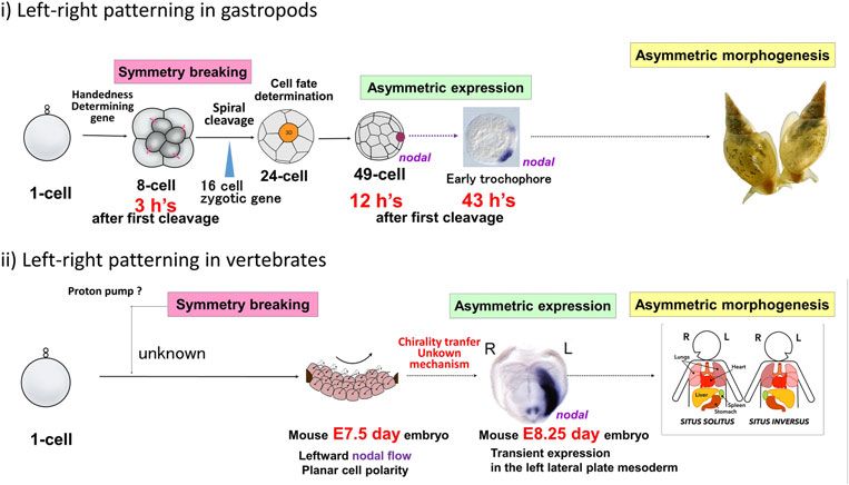
2009). We eventually succeeded in constructing the con- well (Abe et al. 2014). Thus, the creation of mirror-image
genic strain F10 that on average inherit 99·9% of the individuals is not restricted to dimorphic species. Similar
sinistral-derived genome and only 0·1% of the dextral strain- handedness-determining mechanisms must operate for
derived genome. All the dextral embryos oviposited by F10 L. stagnalis and P. acuta.
animals that inherited the dextrality gene(s) within the
0·1% of the dextral-derived genome exhibited dextrotropi- Eight-cell stage is the key determinant step
cal rotation with SD/SI at the third cleavage, and were
By similar mechanical manipulation, we altered the direc-
grown to possess dextral body shape without an excep-
tions of blastomere rotations of both the sinistral and dextral
tion. Equally, all the sinistral embryos oviposited by F10
embryos at the second cleavage to produce reversed blasto-
animals that do not inherit the dextrality-determining
mere configuration at the four-cell stage. However, the
gene(s) showed levotropical rotation without SD/SI, and
manipulated embryos all reverted to the original-type con-
were grown to possess sinistral body shape, again without
figuration at the third cleavage (Kuroda et al. 2009). We
an exception (Kuroda, 2014; Kuroda et al. 2009). This is a
also observed that sinistral embryos occasionally showed
strong indication that dextrality is determined by a single
dextral-type blastomere arrangement at the four-cell stage
gene or by genes at closely linked loci. This was recently
even inside the egg capsules, but they showed normal
confirmed by DNA mapping using RAD-Seq and
counterclockwise cleavage at the third division (Kuroda
Fibre-FISH techniques (Liu et al. 2013). Furthermore,
et al. 2009). It was reported that chirality is distinguishable
we could show that the gene dictates the cytoskeletal
as early as the second or even the first cleavage
dynamics at the third cleavage.
(Meshcheryakov & Beloussov, 1975); however, our micro-
manipulation experiments have proven that the macro-
mere–micromere cell contacts at the eight-cell stage
Creation of mirror-image animals by embryo is the first determining step for asymmetric
twisting blastomeres at the third development.
cleavage
Not epigenetics
Cause and effect
The reversed-coiled snails were fertile, and produced sinis-
To prove directly the role of blastomere arrangements on the
tral or dextral progenies dictated by their genotype and
organismal handedness, we carried out “Mechanogenetics”
not by the reversed body handedness (Kuroda et al. 2009)
and reversed the genetically specified third-cleavage directions
(Fig. 2). This is a direct proof that the birth of mirror-image
by micromanipulation both in sinistral and dextral embryos
animals is not due to the epigenetic consequence but to the
of L. stagnalis. At metaphase–anaphase (for dextral snails)
chiral positioning of blastomeres at the eight-cell stage. In
or telophase (for sinistral snails) of the third cleavage when
other words, the twisting of the blastomeres is the very func-
embryonic chirality is about to be established, we pushed,
tion of the handedness determining gene, and we played the
by thin glass rods, the animal surface of each blastomere
function by our hands though in opposite directions to ob-
gently in the directions opposite to the normal third cleavage
serve the results of manipulation (“Mechanogenetics”).
until contacts between newly formed adjacent pairs of a
micromere and a macromere were established. We incubated
the manipulated embryos in a glass capillary tube, and after
about 17 days 1/3 to 1/2 of the inverted embryos developed
Nodal pathway common to vertebrates
into juvenile snails, and then to adults. Remarkably, all of operates
them are situs inversus, and neither situs solitus nor situs am- Continued l–r asymmetric expression of nodal-Pitx
biguous were observed (Fig. 2). We could show that the effect genes in chiromorphogenesis
of the blastomere chirality at the third cleavage continues
Asymmetric activation of the Nodal pathway is a conserved
throughout the whole subsequent developmental programme
feature of deuterostomes for the determination of the asym-
(Kuroda et al. 2009).
metric body plan (Duboc & Lepage, 2008). In vertebrate,
asymmetric expression of nodal occurs in the lateral plate
Twisting of embryos of P. acuta, a sinistral-only mesoderm transiently at the late developmental stage. The
gastropod
Nodal pathway was found in gastropods as well first in
One may argue that the success in reversing genetically de- the sinistral snail Biomphalaria glabrata and the dextral
termined handedness by mechanical manipulation is due to snail Lottia gigantea (Grande & Patel, 2009). We cloned
the fact that L. stagnalis is dimorphic, which may somehow the orthologues of nodal and its downstream Pitx genes
have a potential to accommodate the reversing physical and investigated their expression patterns in L. stagnalis
force. By similar twisting of the embryos at the third cleav- (Abe & Kuroda, 2013; Kuroda et al. 2009) and P. acuta
age, we obtained situs inversus for sinistral-only P. acuta as (Abe et al. 2014) by whole mount in situ hybridization. In
448
Downloaded from https://www.cambridge.org/core. IP address: 176.9.8.24, on 28 Apr 2020 at 02:37:29, subject to the Cambridge Core terms of use, available at https://www.cambridge.org/core/terms.
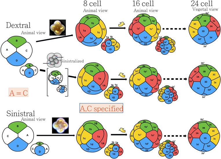
https://doi.org/10.1017/S0033583515000098Fig. 2. Chirality-reversed embryos by manipulation at the third cleavage developed to snails with an oppositely-coiled shell and visceral
situs inversus. Nodal expression site was inverted by the micromanipulation, throughout the development from the starting 33–49 cell
towards the late veliger stage. The chirality of the progenies was dictated by the mother’s genotype.
L. stagnalis, nodal and Pitx gene expressions were detected Twisting of blastomeres results in mirror-image
as early as cell stages 33–49 and 49–64, respectively, in a Nodal expression
specific blastomere destined to develop into ectoderm.
Nodal expression continued up to veliger stage, and then Embryos of progenies of sinistral and dextral F7 congenic
decreased gradually and eventually disappeared when the snails exhibited asymmetric nodal and Pitx patterns exactly
asymmetrical morphology was noticeable and the shell the same as wild type, indicating that the Nodal pathway
started to develop. The expression sites for the dextral and acts downstream of the handedness-determining gene prod-
sinistral L. stagnalis embryos were invariably mirror images uct(s) (Kuroda et al. 2009). Remarkably, when chirality at the
of each other following the third cleavage chirality (Abe & eight-cell stage was reversed by micromanipulation, the
Kuroda, 2013; Kuroda et al. 2009). nodal and Pitx expression patterns were completely reversed
449
Downloaded from https://www.cambridge.org/core. IP address: 176.9.8.24, on 28 Apr 2020 at 02:37:29, subject to the Cambridge Core terms of use, available at https://www.cambridge.org/core/terms.
https://doi.org/10.1017/S0033583515000098Fig. 3. Blastomere arrangements during the spiral cleavages for the dextral and sinistral embryos and the consequence of micromanipula-
tion at the third cleavage. A–C distinction is established at the eight-cell stage.
(Fig. 2) (Abe & Kuroda, 2013; Kuroda et al. 2009). Thus, the tissues interact with one another to orchestrate developmen-
maternally-determined blastomere arrangement at the eight- tal processes? Spemann’s classical experiments on salaman-
cell stage dictates the zygotic Nodal signalling pathway. der (Spemann & Mangold, 1924) have shown the existence
of an organizer, now called Spemann organizer, that instructs
both neutralization and dorsalization, and that cells can
Spiral cleavage conveys chirality
adopt their developmental fate according to their position
information when instructed by other cells. In the gastropods, the macro-
We followed and compared the cytoskeletal dynamics at the mere 3D is known to act as an organizer. How and when is the
fourth cleavage in detail for the mechanically reversed and non- D quadrant specification made from which 3D is derived?
manipulated embryos of L. stagnalis and P. acuta. After the There are two types of spiral cleavage patterns, i.e. equal
recurrent blastomere compaction during post-mitotic phase and unequal cell divisions in the first and second cleavages.
in which the morphological chirality of embryos was seemingly In the unequal cleaving gastropods such as Ilyanassa, D
lost, macromeres (1Q, Q = A, B, C and D) and then micromeres blastomere is much larger than the other A–C cells at the
(1q, q = a, b, c and d) underwent spiral cleavages in the four-cell stage and is easily recognized. In the case of equal
opposite sense to the third cleavage, regardless they were non- cleaving Lymnaea, however, all four blastomeres are of
manipulated or manipulated, keeping the features of alternat- equal size. Blastomeres A/C and B/D can be identified as A
ing rotation direction in the successive cleavages (Fig. 3) (Abe and C are in contact at the animal pole along the cross-
et al. 2014; Kuroda et al. 2009). The direction of the fourth furrow, whereas B and D are in contact at the vegetal pole.
cleavage was not influenced by intra-cellular event caused by But, A or C, and similarly B or D cannot be distinguished
cell pushing, but was determined by micromere (1q)– from each other from the appearance (Fig. 3). At the 24-cell
macromere (1Q) contacts established by the third cleavage stage, the macromere 3D moves to the central location of veg-
(Abe et al. 2014). We believe that spiral cleavage is the way to etal side, fills almost entire cleavage cavity where it contacts
transfer chirality information from the chirality-determining with micromeres including animal-most 1st quartet 1q1
third cleavage to the later developmental stage. and 1q2 (q = a, b, c, d). These cell arrangements take place
during the long interval between the fifth and sixth cleavages
(Meshcheryakov, 1990). Whether the contacts trigger a cell to
Cell-fate become 3D or already specified macromere 3D moves to the
Multicellular organisms are derived from a single fertilized position is still a matter of debate. This series of events
egg. Are cell fates somehow predetermined or do cells and appears to trigger MAPK activation within the 3D
450
Downloaded from https://www.cambridge.org/core. IP address: 176.9.8.24, on 28 Apr 2020 at 02:37:29, subject to the Cambridge Core terms of use, available at https://www.cambridge.org/core/terms.
https://doi.org/10.1017/S0033583515000098Fig. 4. Snails provide excellent model systems for the study of chiromorphogenesis. Symmetry breaking occurs at the eight-cell stage,
and asymmetric expression of nodal gene starts at 33–49 cell, only 12 h after the first cell cleavage, which can be compared with 8·25
days in the case of mouse.
macromere (Henry & Perry, 2008), and then MAPK acts up- this stage, the organizer 3D macromere activates, via still un-
stream of Nodal gene (Lambert & Nagy, 2003). We are cur- known mechanisms, MAPK signal leading to the asymmetric
rently analysing the molecular processes of these events. zygotic nodal gene expression at the 33–49 cell stage, only
12 h after the first cleavage. Nodal pathway is directly
Blastomere which expresses Nodal gene is conventionally
involved in asymmetric morphogenesis both in vertebrate
regarded as derived from the macromere C. As described ear-
and invertebrate. The continued expression of nodal gene
lier, our micromanipulation at the third cleavage of L. stagna-
in snails is clearly different from the transient expression
lis shifted the nodal expression to the mirror-image site
in vertebrates at the late developmental stage. In the case
(Fig. 2), however, this is not because the macromere C was
of mouse, nodal flow and asymmetric nodal expression
moved to the other side of midline by twisting (Fig. 3). Our
were observed at E7·5 and E8·25 days, respectively, a long
experiments have clearly shown that the cell fate A or C is
step from the first symmetry breaking (Fig. 4). There are
not yet specified at the four-cell stage. In other words, l–r sym-
still many questions to be answered. Why and how the spiral
metry is not yet established before the third cleavage.
cleavages occur in alternating sense? The same mechanisms
seem to operate for dominant dextral L. stagnalis and sinis-
tral only P. acuta. Then, what determines the direction of ro-
Outlook tation? What drives levotropic rotation of micromeres in
sinistral L. stagnalis after the non-chiral budding of micro-
The feature of maternal inheritance of chirality, and the cor-
meres at the third cleavage? How and when is the organizer
relation of shell coiling and the spiral blastomere cleavage in
3D macromere’s fate determined? How is MAPK activated
gastropods were long observed, but, the nature of the link be-
and how does it lead to Nodal expression?
tween them had remained obscure. So far, we have clarified
(Fig. 4) that the initial step in the l–r symmetry breaking It is not clear how the asymmetry of animal body plan is asso-
occurs as early as the third (i.e. the first spiral) cleavage, ciated with homochiral biomolecules. The situation is differ-
only 3 h after the first cleavage of a fertilized egg (before ent from chemical chiromophology where enantiomorphic
the start of zygotic gene expression), when newly formed crystals are made up of enantiomorphic molecules. Left-
micromeres are rotated levotropically and dextrotropically hand and right-hand shell coiling snails are of course
with respected to the macromeres for the sinistral and dex- made up of biomolecules of the same unique handedness.
tral snails, respectively. Only the dominant chirality shows Twenty-five years ago, Brown & Wolpert (1990) proposed a
SD of blastomeres which assures correct micromere–macro- model for handedness development in bilateral animals. Out
mere contacts. The single handedness-determining locus dic- of their three components, conversion from a handedness at
tates the cytoskeletal dynamics at the third cleavage, as the molecular level to handedness at the cellular and multicel-
proven by our micromanipulation experiments. Chirality lular level (F-molecule) is intriguing, but their model is just an
thus established is transferred to the cell-fate determination abstract one, devoid of real molecular systems. We have been
step by spiral cleavages, which end at the 24-cell stage. At trying to identify the single handedness-determining gene in
451
Downloaded from https://www.cambridge.org/core. IP address: 176.9.8.24, on 28 Apr 2020 at 02:37:29, subject to the Cambridge Core terms of use, available at https://www.cambridge.org/core/terms.
https://doi.org/10.1017/S0033583515000098L. stagnalis by positional cloning. If the gene and its product HOSOIRI, Y., HARADA, Y. & KURODA, R. (2003). Construction of a
protein are identified, understanding of chirality determi- backcross progeny collection of dextral and sinistral individuals
nation at the molecular level will be greatly advanced. of a freshwater gastropod, Lymnaea stagnalis. Development
Genes and Evolution 213, 193–198.
KURODA, R. (2014). How a single gene twists a snail. Integrative and
Comparative Biology e54, 677–687.
References KURODA, R., ENDO, B., ABE, M. & SHIMIZU, M. (2009). Chiral blasto-
ABE, M. & KURODA, R. (2013). Continued left-fight asymmetric mere arrangement dictates zygotic l–r asymmetry pathway in
expression of nodal-Pitx genes throughout the development is re- snails. Nature 462, 790–794.
quired for the gastropod Lymnaea stagnalis’s chiromophogenesis, LAMBERT, J. D & NAGY, L. M. (2003). The MAPK cascade on equally
P2-0603. In The 36th Annual Meeting of the Molecular Biology cleaving spiralian embryos. Developmental Biology 264, 231–241.
Society of Japan, Kobe, Japan. LIU, M. M., DAVEY, J. W., BANERJEE, R., HAN, J., YANG, F., ABOOBAKER,
ABE, M., TAKAHASHI, H. & KURODA, R. (2014). Spiral cleavages deter- A., BLAXTER, M. & DAVISON, A. (2013). Fine mapping of the pond
mine the left–right body plan by regulating Nodal pathway in snail l–r asymmetry (Chirality) locus using RAD-Seq and
monomorphic gastropods, Physa acuta. International Journal of Fibre-FISH. PLoS ONE 8, e71067.
Developmental Biology 58, 513–520. Special issue: “Spiralian MESHCHERYAKOV, V. N. (1978). Orientation of the cleavage spindles
Model Systems.” in pulmonate molluscs. II. The role of the architecture of the in-
BLUM, M., FIESTED, K., THUMBERGER, T. & SCHWEICKERT, A. (2014). tercellular contacts in III and IV cleavage spindle orientation.
The evolution and conservation of left-right patterning mechan- Ontogenez 9, 567–575.
isms. Development 141, 1603–1613. MESHCHERYAKOV, V. N. (1990). The common pond snail Lymnaea
BOYCOTT, A. E., DIVER, C. & GARSTANG, S. L., HARDY, A. C. & TURNER, stagnalis. In Animal Species for Developmental Studies, Volume
F. M. (1930). The inheritance of sinistrality in Lymnaea peregra. 1. Invertebrates (eds. T. A. DETTLAFF & S. G. VASSETZKY), pp.
Philosophical Transactions of Royal Society of London B 219, 69–132. New York: Consultants Bureau.
51–131. MESHCHERYAKOV, V. N. & BELOUSSOV, L. V. (1975). Asymmetrical
BROWN, N. A. & WOLPERT, L. (1990). The development of handed- rotations of blastomeres in early cleavage of gastropoda.
ness in l–r asymmetry. Development 109, 1–9. Wilhelm Roux’s Archives 177, 193–203.
COUTELIS, J-B., GONZALEZ-MORALES, N., GEMINARD, C. & NOSELLI, S. SHIBAZAKI, Y., SHIMIZU, M. & KURODA, R. (2004). Body handedness is
(2014). Diversity and convergence in the mechanisms establish- directed by genetically determined cytoskeletal dynamics in the
ing L/R asymmetry in metazoan. EMBO 15, 926–937. early embryo. Current Biology 14, 1462–1467.
CRAMPTON, H. E. (1894). Reversal of cleavage in a sinistral gastropod. SPEMANN, H. & MANGOLD, H. (1924). Über Induktion von
Annals of the New York Academy of Sciences 8, 167–170. Embryonalanlagen durch Implantation artfremder Organisa-
DUBOC, V. & LEPAGE, T. (2008). A conserved role for the Nodal sig- toren (Induction of embryonic primordia by implantation
naling pathway in the establishment of dorso–ventral and left– of organizers from a different species). Roux’s Archiv für
right axes in deuterostomes. Journal of Experimental Zoology Entwicklungsmechanik 100, 599–638.
(Mol Dev Evol) 310B, 41–53. STURTEVANT, A. H. (1923). Inheritance of direction of coiling in
FREEMAN, G. & LUNDELIUS, J. W. (1982). The developmental Lymnaea. Science 58, 269–270.
genetics of dextrality and sinistrality in the gastropod Lymnaea VANDENVERG, L. N. & LEVIN, M. (2013). A unified model for left–
peregra. Wilhelm Roux Archive Developmental Biology 191, right asymmetry? Comparison and synthesis of molecular
69–83. models of embryonic laterality. Developmental Biology 379, 1–15.
GRANDE, C. & PATEL, N. H. (2009). Nodal signalling is involved in l–r VERDONK, N. H. & VAN DEN BIGGELAAR, J. A. M. (1983). Early devel-
asymmetry in snails. Nature 457, 1007–1011. opment and the formation of the germ layers. In The Mollusca,
HENRY, J. J. & PERRY, K. J. (2008). MAPK activation and the specifi- Volume 3. Development (eds. N. H. VERDONK, J. A. M. VAN DEN
cation of the D quadrant in the gastropod mollusc, Crepidula BIGGELAAR & A. S. TOMPA), pp. 91–122. New York: Academic
fornicata. Developmental Biology 313, 181–195. Press.
452
Downloaded from https://www.cambridge.org/core. IP address: 176.9.8.24, on 28 Apr 2020 at 02:37:29, subject to the Cambridge Core terms of use, available at https://www.cambridge.org/core/terms.
https://doi.org/10.1017/S0033583515000098You can also read