Expanded human NK cells from lung cancer patients sensitize patients' PDL1 negative tumors to PD1- blockade therapy
←
→
Page content transcription
If your browser does not render page correctly, please read the page content below
Open access Short report
Expanded human NK cells from lung
J Immunother Cancer: first published as 10.1136/jitc-2020-001933 on 21 January 2021. Downloaded from http://jitc.bmj.com/ on July 25, 2021 by guest. Protected by copyright.
cancer patients sensitize patients’
PDL1−negative tumors to PD1-
blockade therapy
Sophie M Poznanski,1 Tyrah M Ritchie,1 Isabella Y Fan,1 Abdullah El-Sayes,1
Ana L Portillo,1 Ronny Ben-Avi,2 Eduardo A Rojas,1 Marianne V Chew,1
Yaron Shargall,2 Ali A Ashkar 1
To cite: Poznanski SM, ABSTRACT made by immune checkpoint inhibitors over
Ritchie TM, Fan IY, et al. Lung cancer remains the leading cause of cancer death the past decade has been revolutionary, with
Expanded human NK cells from worldwide despite the significant progress made by
lung cancer patients sensitize
antibody blockade of programmed death
immune checkpoint inhibitors, including programmed receptor-1 (PD1) achieving unprecedented
patients’ PDL1−negative tumors
death receptor-1 (PD1)/PD ligand 1 (PDL1)-blockade durable tumor regression in some patients
to PD1-blockade therapy.
Journal for ImmunoTherapy therapy. PD1/PDL1−blockade has achieved unprecedented
with advanced lung tumors, melanoma and a
of Cancer 2021;9:e001933. tumor regression in some patients with advanced
lung cancer. However, the majority of patients fail growing list of other cancers. However, only
doi:10.1136/jitc-2020-001933
to respond to PD1/PDL1 inhibitors. The high rate of ~10% of patients benefit from the therapy.2
►► Additional material is therapy non-response results from insufficient PDL1 Non- response to PD1- blockade therapy
published online only. To view, expression on most patients’ tumors and the presence is associated with insufficient PD ligand
please visit the journal online of further immunosuppressive mechanisms in the tumor 1 (PDL1) expression (Tumor Proportion
(http://dx.doi.org/10.1136/jitc- microenvironment. Here, we sensitize non-responding Score, TPS) on patient tumors and addi-
2020-001933). tumors from patients with lung cancer to PD1-blockade tional mechanisms of immunosuppression
therapy using highly cytotoxic expanded natural killer (NK) in the tumor microenvironment.3–5 Indeed,
Accepted 11 December 2020 cells. We uncover that NK cells expanded from patients
a number of landmark trials have shown
with lung cancer dismantle the immunosuppressive
that an important component for sustained
tumor microenvironment by maintaining strong antitumor
activity against both PDL1+ and PDL1− patient tumors. In immunotherapeutic efficacy is the ability to
the process, through a contact-independent mechanism shift the immunosuppressive tumor microen-
involving interferon γ, expanded NK cells rescued tumor vironment to a proinflammatory milieu and
killing by exhausted endogenous TILs and upregulated the restore the antitumor functions of exhausted
tumor proportion score of PDL1 across patient tumors. endogenous immune cells.2 3 6–9 Thus, new
In contrast, unexpanded NK cells, which are susceptible immunotherapies that can both sustain
to tumor-induced immunosuppression, had no effect on strong inflammatory antitumor activity in
tumor PDL1. As a result, combined treatment of expanded the tumor microenvironment and increase
NK cells and PD1-blockade resulted in robust synergistic
PDL1 TPS would hold promise to extend
tumor destruction of initially non-responding patient
tumors. Thus, expanded NK cells may overcome the critical
the remarkable therapeutic benefits of PD1-
roadblocks to extending the prodigious benefits of PD1- blockade therapy to more patients.
blockade therapy to more patients with lung cancer and The antitumor cytokine interferon-gamma
© Author(s) (or their (IFNγ), released by cytotoxic natural killer
other tumor types.
employer(s)) 2021. Re-use
permitted under CC BY-NC. No (NK) cells and T cells, is a critical driver of
commercial re-use. See rights PDL1 expression on tumors and a positive
and permissions. Published by BACKGROUND predictor of clinical response to immuno-
BMJ.
1
Lung cancer is the leading cause of cancer therapies.8 10–12 However, the antitumor
McMaster Immunology functions of NK cells and T cells are signifi-
Research Centre, Department of
death worldwide. In 2018 alone, there were
Medicine, McMaster University, over 1.7 million lung cancer-related deaths, cantly hindered by the tumor microenvi-
Hamilton, Ontario, Canada reflecting a dismal 5-year survival rate of ronment.13 14 Our previous work uncovered
2
Surgery, McMaster University, less than 18%. At the time of diagnosis, the that NK cells from the peripheral blood and
Hamilton, Ontario, Canada majority (80%) of lung cancer patients (LCP) tumors of breast and patients with ovarian
Correspondence to already have locally advanced or metastatic cancer could be expanded ex vivo for cell
Dr Ali A Ashkar; disease, which continues to progress despite therapy. On adoptive transfer to mice, these
ashkara@mcmaster.c a chemotherapy.1 As a result, the progress expanded NK cells (exNK) were capable of
Poznanski SM, et al. J Immunother Cancer 2021;9:e001933. doi:10.1136/jitc-2020-001933 1
Open access
sustaining antitumor activity against tumors and elimi- stained with extracellular antibodies for 30 min. Cells not
J Immunother Cancer: first published as 10.1136/jitc-2020-001933 on 21 January 2021. Downloaded from http://jitc.bmj.com/ on July 25, 2021 by guest. Protected by copyright.
nated macroscopic ovarian tumors.15–17 Another study undergoing intracellular staining were then fixed in 1%
has also demonstrated that NK cells expanded using paraformaldehyde. For intracellular staining, cells were
membrane particles were capable of upregulating PDL1 fixed with Cytofix/Cytoperm from BD Biosciences for 20
on tumor cell lines.18 In the current study, we sought to min and then stained with intracellular antibodies in a 1X
assess (1) the therapeutic potential of exNK cells against Perm/Wash solution (BD Bioscience) for 30 min. Sample
tumors from advanced-stage LCP and (2) whether exNK acquisition was carried out on a BD LSRFortessa and
cells can additionally sensitize patients’ non-responding analyzed on FlowJo software. See (online supplemental
tumors to PD1-blockade therapy. methods) for a complete list of the antibodies used.
Functional assays and PD1/PDL1 expression
METHODS Flow cytometry- based killing and degranulation assays
Peripheral blood, pleural effusions, and tumor pieces were conducted in complete RPMI media as described
were obtained with written informed consent from LCP previously.15 16 Specifically, for killing assays against A549
at St. Joseph’s Healthcare in Hamilton, Ontario. Pleural cells, NK cells were coincubated with CFSE-labeled A549s
effusions were collected from patients via thoracentesis at 1:1, 5:1, and 10:1 effector-to-target ratios for 5 hours,
and tumor pieces were collected via surgical resection, following which cells were stained with fixable viability
both of which were conducted as part of the patients’ stain. For killing assays against PDL1+ A549 cells, A549s
standard care. Peripheral blood was collected from were pretreated with rhIFNγ (20 ng/mL) for 48 hours to
healthy donors (HD) with written informed consent at induce PDL1 expression and washed three times prior to
McMaster University. NOD-Rag1null IL2rgnull (NRG) mice seeding for the assay.
were originally obtained from Jackson Laboratory and Killing assays against patient tumors were conducted
bred and housed in specific pathogen-free conditions at using a transwell model ex vivo. Patient tumors were
McMaster’s Central Animal Facility. seeded on both apical and basolateral surfaces of the
transwell. NK cells were added to the apical chamber
Processing of lung tumors, pleural effusions and peripheral at a 10:1 effector-to-target ratio and coincubated for 48
blood hours unless indicated otherwise. Tumors from the same
Lung tumors were minced into ~1 mm3 pieces in αMEM patients were used across NK cell groups (HD periph-
medium containing collagenase IV and DNase I. Tumor eral blood (pb)NK, and LCP pbNK and tumor-associated
pieces were then incubated in the media on a plate shaker NK). Nivolumab, a PD1 blocking antibody used clinically
at 37°C for 2 x, 1-hour intervals and pipetted vigorously for the treatment of PDL1+ cancer,11 was added to wells at
in between incubations to break up the pieces. Cells were 1 µg/mL where indicated. Low dose IL-15 (1 ng/mL) was
filtered through a 70 µm filter, pelleted via centrifugation, added as an NK cell survival factor for this extended incu-
then washed with phosphate-buffered saline (PBS). Cells bation. Following incubation, cells in the apical chamber
from pleural effusions and peripheral blood were isolated were stained to assess direct NK cell killing and cells in
via density-gradient centrifugation with Lymphoprep the basolateral chamber were stained to assess killing by
(Stemcell Technologies, Vancouver, British Columbia, endogenous tumor-infiltrating lymphocytes (TILs).
Canada), then washed with PBS. For degranulation assays and IFNγ expression against
patient tumors, NK cells were coincubated with target
NK cell expansion and isolation cells at a 1:1 ratio for 5 hours. Golgi Stop (BD Biosci-
NK cells were cultured in RPMI medium supplemented ences) was added following the first hour of incubation.
with 10% FBS, 1% hepes, 1% penicillin- streptomycin, PD1 expression on NK cells and PDL1 expression
and 1% L-glutamine. NK cells were expanded from the on tumor cells were assessed by co-incubating NK cells
peripheral blood mononuclear cells (PBMCs) of HD, with tumor cells for 48 hours using the transwell model
or PBMCs, pleural effusion cells, or tumors of LCP. NK described above. PD1 expression was assessed by staining
cells were expanded by coculture with irradiated K562- cells in the apical chamber for NK cell markers and PD1.
membrane- bound--interleukin (IL)-21 feeder cells and PDL1 expression was assessed on live tumor cells in the
recombinant human (rh) IL-2 as described previously.16 basolateral chamber.
Expansion was conducted for at least 3 weeks prior to
functional assays. For experiments using freshly isolated Statistics
NK cells, NK cells were isolated via positive selection Statistical analysis was conducted using GraphPad Prism
from PBMCs using a CD56+ selection kit from Stemcell, software. Graphs with two groups were analyzed using
according to the manufacturer’s instructions. a two-tailed unpaired t-test. Graphs with three or more
conditions with one independent variable were analyzed
Flow cytometric staining by one- way analysis of variance (ANOVA) with Tukey
To discriminate live/dead cells, cells were first stained for correction for multiple comparisons. Graphs with two
30 min with fixable viability stain (eBioscience). Cells were independent variables were analyzed by two-way ANOVA
then washed with 0.2% bovine serum albumin in PBS and with Tukey correction. D’Agostino and Pearson normality
2 Poznanski SM, et al. J Immunother Cancer 2021;9:e001933. doi:10.1136/jitc-2020-001933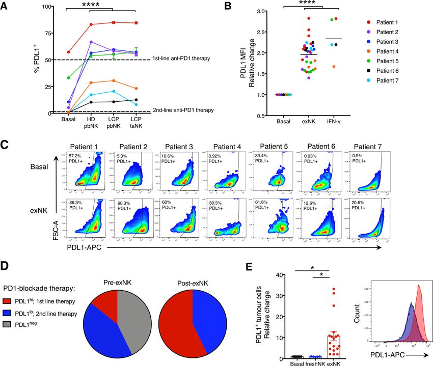
Open access
J Immunother Cancer: first published as 10.1136/jitc-2020-001933 on 21 January 2021. Downloaded from http://jitc.bmj.com/ on July 25, 2021 by guest. Protected by copyright.
Table 1 Study population and tumor characteristics
Patient # Age Sex Tumor classification PDL1 Status ALK EGFR
1 87 F NSCLC-adeno P (HI, >50%) N N
2 74 F NSCLC-squamous P (LOW, 1%–49%) N N
3 62 F NSCLC-squamous P (LOW, 1%–49%) NA NA
4 65 M NSCLC-adeno N (50%) N N
10 63 F NSCLC-adeno P (LOW, 1%–49%) N P
11 78 M NSCLC-adeno P (HI, >50%) N N
12 79 F NSCLC-adeno N (50%) N N
16 63 M NSCLC-squamous P (LOW, 1%–49%) NA NA
High-positive (HI); TPS ≥50%. Low-positive (LOW); TPS ≥1% and≤49%. Negative; TPSOpen access
J Immunother Cancer: first published as 10.1136/jitc-2020-001933 on 21 January 2021. Downloaded from http://jitc.bmj.com/ on July 25, 2021 by guest. Protected by copyright.
Figure 1 NK cells expanded from lung cancer patients (LCPs) exert strong antitumor activity against patient tumors and
rescue tumor killing by endogenous TILs. NK cells were expanded from the peripheral blood (pbNK), pleural effusions (peNK)
or tumors (taNK) of LCPs or peripheral blood of healthy donors (HD). (A) Fold expansion of NK cells from PDL1+ versus PDL1−
tumors and representative flow plots showing NK cell (CD56+CD3-) purity pre-expansion and postexpansion. Expression of
activation (B) and inhibitory (C) receptors on exNK cells from PDL1+ and PDL1− tumors at 28-day expansion. (D) Five-hour
killing of human A549 lung cancer cells by exNK cells. (E) Schematic: exNK cells were adoptively transferred to NRG mice 2
and 5 days after intravenous infusion of Luciferase-expressing A549s (A549-Luc). Graph: quantification of tumor burden via
bioluminescence at the indicated days. (F–I) Expanded LCP taNK and pbNK cells were coincubated with LCP tumors. Five-
hour (F) killing and relative increase in (G) degranulation (CD107a) and (H) IFNγ expression against patient tumors compared
with expanded HD pbNK cells against these same patient tumors. (I) Representative flow plots of NK cell CD107a and IFNγ
expression. (J) Schematic: patient tumors were seeded in transwell on apical and basolateral surfaces. exNK cells or rhIFNγ
(20 ng/mL) or neither (basal) were added to the apical chamber. Graph shows killing of tumors in the basolateral chamber by
endogenous TILs after 48 hours. (K) Schematic summarizing results. Data show means±SEM of three to eight replicates per
condition. Results analyzed by two-way ANOVA (A, D, E, G, H), unpaired t-test (B, C) or one-way ANOVA (D, F, J). **POpen access
J Immunother Cancer: first published as 10.1136/jitc-2020-001933 on 21 January 2021. Downloaded from http://jitc.bmj.com/ on July 25, 2021 by guest. Protected by copyright.
Figure 2 exNK cells convert lung cancer patient PDL1− tumors to PDL1+/hi. Lung cancer patient tumors were seeded
in transwell on apical and basolateral surfaces. exNK cells or rhIFNγ (20 ng/mL) or neither (basal) were added to the apical
chamber and incubated for 48 hours. (A) PDL1 TPS and (B) PDL1 mean fluorescence intensity (MFI) on basolateral tumor cells.
(C) Representative flow plots of PDL1 on exNK-treated versus untreated patient tumors. (D) Proportion of patient tumors that
were PDL1neg, PDL1lo or PDL1hi pre-exNK and post-exNK treatment. (E) Quantification and representative histogram of PDL1
expression on lung cancer patient tumors treated with expanded or unexpanded freshly isolated (fresh) pbNK cells. Data show
means±SEM of 5–32 replicates per condition. A, B, E analyzed via one-way ANOVA. *POpen access
J Immunother Cancer: first published as 10.1136/jitc-2020-001933 on 21 January 2021. Downloaded from http://jitc.bmj.com/ on July 25, 2021 by guest. Protected by copyright.
Figure 3 exNK cells sensitize patients’ non-responding tumors to PD1-blockade therapy. (A) exNK cells were incubated with
or without lung cancer patient tumors. Per cent PD1+ exNK cells and representative histogram of PD1 expression after 48-
hour incubation. (B, C) A549s were left untreated or treated with rhIFNγ (20 ng/mL) for 48 hours to induce PDL1 expression,
then washed three times and incubated with exNK cells with or without nivolumab (1 µg/mL) for 5 hours. Relative exNK cell
killing of (B) IFNγ-treated (PDL1+) A549s or (C) untreated (PDL1−) A549s. (D–F) Schematic shows experimental design: patient
tumors were seeded on apical and basolateral transwell surfaces. exNK cells and/or nivolumab (Nivo; 1 µg/mL) were added to
the apical chamber for 48 hours. (D) Tumor killing in the apical chamber. (E) Tumor killing by endogenous TILs in the basolateral
chamber. (F) Correlation between change in tumor PDL1 expression induced by exNK cells and apical tumor killing. (G)
Graphical summary of the study’s main findings. Data show means±SEM of 4–36 replicates per condition. Results analyzed
via unpaired t-test (A) two-way ANOVA (B, C, E), one-way ANOVA (D) or pearson correlation (F). *POpen access
predictor for tumors that will be most responsive to exNK Nevertheless, the proven safety profile shown in this
J Immunother Cancer: first published as 10.1136/jitc-2020-001933 on 21 January 2021. Downloaded from http://jitc.bmj.com/ on July 25, 2021 by guest. Protected by copyright.
cell therapy. previous trial, together with the robust synergistic tumor
killing we observed on combination treatment of exNK
cells and nivolumab against initially PDL1− tumors,
DISCUSSION suggests that such combination treatment should be
Patients with advanced tumors that do not respond to investigated in patients with advanced PDL1− tumors.
checkpoint blockade immunotherapy face a dearth
of effective treatment options. Although studies have Acknowledgements We thank Rebecca Long for administrative assistance and all
lung cancer patient and healthy donors who donated samples.
predominantly focused on the role of T cells to mediate
the antitumor effects of immune checkpoint blockade Contributors SMP and AAA conceived the project and designed the experiments;
YS provided intellectual input, contributed to experimental design and guided
therapy, there is a growing understanding for the clinical sample acquisition; SMP, TR and IYF performed experiments. AE-S, ALP,
important role of other immune cell types, including NK RB-A, ER and MC contributed to performing experiments. YS and RB-A obtained
cells and B cells, in mediating this response.24 25 Notably, the clinical samples. SMP curated and formally analyzed the data. SMP and AAA
NK cell depletion was shown to reduce response to PD1/ wrote the manuscript; TR and YS edited the manuscript; AAA secured funding and
supervised the project.
L1-blockade therapy in a syngeneic murine model of
colon carcinoma.24 A previous study also showed that Funding This work was supported by the Canadian Institutes for Health Research
(CIHR) (20 009 360 to AAA). AAA holds a tier 1 Canada Research Chair in Natural
NK cells expanded from induced-pluripotent stem cells Immunity and NK Cell Function. SMP is supported by a CIHR Vanier Canada
increased PDL1 TPS on tumor cell lines.18 Our study iden- Graduate Scholarship.
tifies highly cytotoxic exNK cells as a promising therapy Competing interests None declared.
for lung cancer that may also further extend the benefits Patient consent for publication Not required.
of PD1-blockade therapy to patients with non-responding
Ethics approval All research involving human samples was approved by the
tumors (figure 3G). A significant barrier to developing Hamilton Integrated Research Ethics Board at McMaster University. All research
broadly effective immunotherapies against solid tumors involving animals was approved by the Animal Research Ethics Board at McMaster
has been the inability to sustain cytotoxic and proinflam- university.
matory immune cell functions in the tumor microenviron- Provenance and peer review Not commissioned; externally peer reviewed.
ment.3–5 Our findings that NK cells expanded from LCP Data availability statement All data relevant to the study are included in the
maintain strong tumor killing and IFNγ production, both article or uploaded as online supplemental information.
in xenograft models in vivo and over prolonged exposure Supplemental material This content has been supplied by the author(s). It has
to patient tumors ex vivo, suggest that exNK cells over- not been vetted by BMJ Publishing Group Limited (BMJ) and may not have been
come the critical hurdle of tumor-induced suppression. peer-reviewed. Any opinions or recommendations discussed are solely those
of the author(s) and are not endorsed by BMJ. BMJ disclaims all liability and
Further, our results that exNK cells restore the antitumor responsibility arising from any reliance placed on the content. Where the content
activity of endogenous TILs indicates a striking ability to includes any translated material, BMJ does not warrant the accuracy and reliability
not only resist, but dismantle, the immunosuppressive of the translations (including but not limited to local regulations, clinical guidelines,
tumor microenvironment and unleash the patient’s own terminology, drug names and drug dosages), and is not responsible for any error
and/or omissions arising from translation and adaptation or otherwise.
antitumor immunity.
Open access This is an open access article distributed in accordance with the
Previous work by our group showed that NK cells
Creative Commons Attribution Non Commercial (CC BY-NC 4.0) license, which
expanded from patients with breast and ovarian cancer permits others to distribute, remix, adapt, build upon this work non-commercially,
have comparable antitumor activity against the patients’ and license their derivative works on different terms, provided the original work is
own autologous tumors as NK cells expanded from properly cited, appropriate credit is given, any changes made indicated, and the use
is non-commercial. See http://creativecommons.org/licenses/by-nc/4.0/.
HD.15–17 The present study extends these findings to NK
cells from LCP, identifying exNK cells as a promising ORCID iD
autologous cell therapy for lung cancer. Ali A Ashkar http://orcid.org/0000-0003-1139-1468
A seminal study recently found that anti-PDL1−mAbs
can directly activate the cytotoxic effector functions of
PDL1+ NK cells, irrespective of tumor PDL1 status.26 REFERENCES
These findings together with the results in the present 1 Howlader N, Noone AM, Krapcho M. SEER cancer statistic review.
Bethesda, MD: National Cancer Institute, 2019.
study, identify NK cells as critical effectors for inducing 2 Herbst RS, Soria J-C, Kowanetz M, et al. Predictive correlates of
responses to PD1/L1- blockade therapy in initially response to the anti-PD-L1 antibody MPDL3280A in cancer patients.
PDL1− tumors. Nature 2014;515:563–7.
3 Yu Y, Zeng D, Ou Q, et al. Association of survival and immune-related
A recent randomized control trial in patients with biomarkers with immunotherapy in patients with non-small cell lung
PDL1+ non-small-cell lung carcinoma found that combi- cancer: a meta-analysis and individual patient-level analysis. JAMA
Netw Open 2019;2:e196879.
nation treatment of NK cells with the PD1 inhibitor 4 Topalian SL, Hodi FS, Brahmer JR, et al. Safety, activity, and
pembrolizumab was well-tolerated and improved overall immune correlates of anti-PD-1 antibody in cancer. N Engl J Med
and progression-free survival in patients compared with 2012;366:2443–54.
5 Benci JL, Xu B, Qiu Y, et al. Tumor interferon signaling regulates a
pembrolizumab treatment alone.27 Importantly, the multigenic resistance program to immune checkpoint blockade. Cell
trial found that there were no adverse events associated 2016;167:1540–54.
6 Amaria RN, Reddy SM, Tawbi HA, et al. Neoadjuvant immune
with the addition of NK cell therapy. A limitation of our checkpoint blockade in high-risk resectable melanoma. Nat Med
current study is the relatively small study population. 2018;24:1649–54.
Poznanski SM, et al. J Immunother Cancer 2021;9:e001933. doi:10.1136/jitc-2020-001933 7Open access
7 Fehrenbacher L, Spira A, Ballinger M, et al. Atezolizumab versus 17 Shenouda MM, Gillgrass A, Nham T, et al. Ex vivo expanded natural
J Immunother Cancer: first published as 10.1136/jitc-2020-001933 on 21 January 2021. Downloaded from http://jitc.bmj.com/ on July 25, 2021 by guest. Protected by copyright.
docetaxel for patients with previously treated non-small-cell lung killer cells from breast cancer patients and healthy donors are
cancer (poplar): a multicentre, open-label, phase 2 randomised highly cytotoxic against breast cancer cell lines and patient-derived
controlled trial. Lancet 2016;387:1837–46. tumours. Breast Cancer Res 2017;19:76.
8 Ayers M, Lunceford J, Nebozhyn M, et al. IFN-γ-related mRNA 18 Oyer JL, Gitto SB, Altomare DA, et al. PD-L1 blockade
profile predicts clinical response to PD-1 blockade. J Clin Invest enhances anti-tumor efficacy of NK cells. Oncoimmunology
2017;127:2930–40. 2018;7:e1509819.
9 McDermott DF, Huseni MA, Atkins MB, et al. Clinical activity 19 Ciurea SO, Schafer JR, Bassett R, et al. Phase 1 clinical trial using
and molecular correlates of response to atezolizumab alone or mbIL21 ex vivo-expanded donor-derived NK cells after haploidentical
in combination with bevacizumab versus sunitinib in renal cell transplantation. Blood 2017;130:1857–68.
carcinoma. Nat Med 2018;24:749–57. 20 Garon EB, Rizvi NA, Hui R, et al. Pembrolizumab for the treatment of
10 Taube JM, Anders RA, Young GD, et al. Colocalization of non-small-cell lung cancer. N Engl J Med 2015;372:2018–28.
inflammatory response with B7-H1 expression in human melanocytic 21 Pai-Scherf L, Blumenthal GM, Li H, et al. Fda approval summary:
lesions supports an adaptive resistance mechanism of immune pembrolizumab for treatment of metastatic non-small cell lung
escape. Sci Transl Med 2012;4:127ra37. cancer: first-line therapy and beyond. Oncologist 2017;22:1392–9.
11 Borghaei H, Paz-Ares L, Horn L, et al. Nivolumab versus docetaxel in 22 Kazandjian D, Suzman DL, Blumenthal G, et al. Fda approval
advanced Nonsquamous non-small-cell lung cancer. N Engl J Med summary: nivolumab for the treatment of metastatic non-small
2015;373:1627–39. cell lung cancer with progression on or after platinum-based
12 Chen S, Crabill GA, Pritchard TS, et al. Mechanisms regulating PD- chemotherapy. Oncologist 2016;21:634–42.
L1 expression on tumor and immune cells. J Immunother Cancer 23 Dietel M, Savelov N, Salanova R, et al. Real-World prevalence
2019;7:305. of programmed death ligand 1 expression in locally advanced
13 Chang C-H, Qiu J, O'Sullivan D, et al. Metabolic competition in or metastatic non-small-cell lung cancer: the global, multicenter
the tumor microenvironment is a driver of cancer progression. Cell express study. Lung Cancer 2019;134:174–9.
2015;162:1229–41. 24 Hsu J, Hodgins JJ, Marathe M, et al. Contribution of NK cells to
14 Uchida A, Colot M, Micksche M. Suppression of natural killer cell immunotherapy mediated by PD-1/PD-L1 blockade. J Clin Invest
activity by adherent effusion cells of cancer patients. suppression 2018;128:4654–68.
of motility, binding capacity and lethal hit of NK cells. Br J Cancer 25 Cabrita R, Lauss M, Sanna A, et al. Tertiary lymphoid structures
1984;49:17–23. improve immunotherapy and survival in melanoma. Nature
15 Nham T, Poznanski SM, Fan IY, et al. Ex vivo-expanded NK cells 2020;577:561–5.
from blood and ascites of ovarian cancer patients are cytotoxic 26 Dong W, Wu X, Ma S, et al. The mechanism of anti-PD-L1
against autologous primary ovarian cancer cells. Cancer Immunol antibody efficacy against PD-L1-Negative tumors identifies NK
Immunother 2018;67:575–87. cells expressing PD-L1 as a cytolytic effector. Cancer Discov
16 Poznanski SM, Nham T, Chew MV, et al. Expanded 2019;9:1422–37.
CD56superbrightCD16+ NK Cells from Ovarian Cancer Patients Are 27 Lin M, Luo H, Liang S, et al. Pembrolizumab plus allogeneic NK
Cytotoxic against Autologous Tumor in a Patient-Derived Xenograft cells in advanced non-small cell lung cancer patients. J Clin Invest
Murine Model. Cancer Immunol Res 2018;6:1174–85. 2020;130:2560–9.
8 Poznanski SM, et al. J Immunother Cancer 2021;9:e001933. doi:10.1136/jitc-2020-001933You can also read