RECONSTRUCTION OF MANDIBULAR DEFECTS USING INDIVIDUAL VASCULARIZED AUTOGRAFTS COMBINED WITH MACROPOROUS TITANIUM FIBER MATERIAL
←
→
Page content transcription
If your browser does not render page correctly, please read the page content below
DE N TI S T R Y                                                             | archiv euromedica | 202 1 | vol. 11 | num . 1 |
                                                                                                                                                     147
http://dx.doi.org/10.35630/2199-885X/2021/11/1.32

RECONSTRUCTION OF MANDIBULAR DEFECTS
USING INDIVIDUAL VASCULARIZED
AUTOGRAFTS COMBINED
WITH MACROPOROUS TITANIUM                                                                             R eceived 15 February 2021;

FIBER MATERIAL                                                                                         Received in revised form 26 February 2021;
                                                                                                        Accepted 1 March 2021

Ivan Bairikov1 , Tatiana Gaivoronskaya2 ,                                  deformities of the facial skeleton with a bone defect are
Dmitry Dedikov3 , Pavel Stolyarenko1 ,                                     common in maxillofacial surgery [1, 7, 15].
Dmitry Domenyuk4                                                                 Accelerating mobility and speed of modern soci-
                                                                           ety, the spread of weapons and military conflicts, there
1
  Samara State Medical University, Samara;                                 is a growing incidence of post-traumatic deformities
2
  Kuban State Medical University, Krasnodar;                               of various origin affecting bones. The corresponding
3
  Department of Maxillofacial Surgery, Research Center for Maxillofacial   surgical reconstruction requires a more sofisticated
Surgery and Dentistry “AVERS”, Krasnodar;                                  and comprehensive approach [2, 5, 10, 13, 16].
4
  Stavropol State Medical University, Stavropol, Russia                          As reported by various authors, the number
                                                                           of patients with maxillofacial traumas ranges from
      avers_23@mail.ru                                                     11 to 25% (Bernadsky Yu. I., 2016; Kulakov A. A.,
                                                                           2017; Drobyshev A. Yu., 2017; Bayrikov I. M., 2018),
                                                                           whereas some other researchers, both national and in-
A b s t r a c t — The choice of donor material poses a
problem for surgeons handling reconstruction of large
                                                                           terantional ones, claim the rate to be at 30–38% (Balin
combined bone defects or limited maxillofacial bone                        V. N., 2015; Wong K. H., 2016; Guerrissi J. O., 2018).
defects. Restoration methods, applicable in reconstructive                       The rate of post-traumatic complications varies,
microsurgery are based on materials of non-biological                      according to the references, from 7 to 36% (Zuev V. P.,
origin and musculoskeletal autogenous transplants which                    2015; Erokhina I. L., 2016; Eshiev A. M., 2018; Andra
have complications and may inhibit the processes of
osseointegration. Our study aimed to improve the efficiency
                                                                           A., et al., 2018). Specialized hospitals sometimes fail
of surgical treatment and rehabilitation of patients                       to offer timely and high-quality care, which leads to
with mandibular defects using vascularized autograft in                    repeated surgical interventions and the emerging man-
combination with macroporous titanium fiber material. The                  dibular defects of disturbed continuity [9, 14].
reconstruction of the defective mandible caused by chronic                       At the current stage, reconstructive maxillofacial
osteomyelitis, trauma, benign tumors was performed in 107
patients either by means of conventional methods (titanium
                                                                           surgery progress utilizes widely reconstructive surgery
plates, free and vascularized bone autografts), or by a novel              methods capable of improving defects while relying
engineered bone substitute. Our novel vascularized bone                    on materials of non-biological origin (titanium, teflon,
autotransplant combines a macroporous fiber titanium                       polyethylene, etc.) or on multicomponent muscu-
material and spiral bone autochips. For its fabrication                    loskeletal autografts involving microvascular technolo-
we applied digital 3D technologies and methods of rapid
prototyping, whereas its vascularization was harvested in
                                                                           gy. These methods have advantages and disadvantages
iliac crest. Unlike the standard methods of reconstruction,                and universally applicable to the fullest extent, which
the use of the engineered vascularized implant showed a                    inevitably leads to traumatization of both the donor
significant reduction in stages, volume and invasiveness of                and the recipient areas [3, 4, 6, 8, 11, 12].
surgical procedures and an improvement of esthetic and                           Furthermore, despite numerous fundamental
functional outcomes in patients with mandibular defects.
                                                                           studies, a number of issues still require more detailed
K e y w o r d s — bone tissue engineering, 3D modeling, rapid              consideration, namely, the change patterns involv-
prototyping, porous titanium fiber mesh (TFM), bone                        ing the status of bone and soft tissue structures after
autotransplant.                                                            resection of various jaw parts, depending on the causes
                                                                           behind it, and the timing of the removal.

I n t r o d uc t i o n                                                     M ATERIALS AND M ETHODS
     The conditions following cancer treatment,                                In our work, we rely on a combined method,
such as severe maxillofacial traumas, post-traumatic                       which implies using vascularized autografts and a
148   | archiv euromedica | 202 1 | vol. 11 | num . 1 |                                                                        DE N TI S T R Y

         unique macroporous titanium fiber material. The                      The patients’ general and local status regarding
         reliability of the study can be confirmed a sufficient         the issue of the potential reconstruction of mandibular
         number of clinical observations (107) and numer-               defects was evaluated following the traditional plan.
         ous X-ray data, processing of the outcomes obtained            First of all, the clinical section of the examination was
         through advanced statistical analysis methods. The             carried out, which included an interview survey; the
         hypotheses were tested relying on the methods of               patient’s general status evaluation; an examination;
         parametric statistics. The description of the quantita-        evaluation with extra diagnostic methods (ultrasound,
         tive parameters was performed using the mean and               CT, MRI), and evaluation involving rapid prototyping
         the error of the mean. The observations frequency was          methods (Fig. 1, 2).
         expressed as a percentage. The level of statistical signifi-
         cance through the study was set at 0.05. The obtained
         data of aesthetic indicators, both prior to, and after the
         surgery were processed statistically using the MS Excel
         2010 software package.
               Through our project, we used a combined treat-
         ment method to replace mandibular defects based
         on using vascularized autografts and a macroporous
         titanium fiber material.
               The surgical treatment planning, the pre-surgery
         preparation, the surgical stages of the treatment were
         carried out following the clinical recommendations
         offered by the Chief Freelance Maxillofacial Surgeon
         of the Russian Ministry of Health Protocol for treating
         patients with facial skeleton bones defects.
               Written voluntary informed consent (approved
         by the Samara State Medical University Ethics Com-
         mittee of 22/11/2015, Protocol #147) was obtained              Fig. 1. D efect of the chin of the lower jaw in 3D format
         from each of the patients treated for mandibular
         defects, the signed papers allowing clinical trials, tak-
         ing photos and videos, as well as using the outcomes in               The clinical data of 107 patients, revealed that
         research work.                                                 30% of the cases had deformities that developed after
               We had 107 patients under our observation, all           surgical treatment for chronic osteomyelitis of vari-
         of them divided into 2 groups. Group I (76 persons),           ous etiologies; 30% more (30 patients) had lower jaw
         standard and widely used methods were employed to              defects associated with maxillofacial injuries; another
         reconstruct mandibular defects — titanium recon-               30% of the patients (30 persons) had mandibular
         structive plates in 32 people; free bone autografts — in       defects originating from the removal of benign neo-
         22 people, and vascularized bone autografts used in            plasms, while 10% (10 patients) featured mandibular
         other 22 people.                                               issues following complications of reconstructive sur-
               The study group included patients of Group II,           geries (rejection of previously transplanted autografts
         who were treated using vascularized bone autografts in         and titanium plates) and oncological surgeries (Fig. 3).
         combination with a through-porous non-woven tita-                     The through-porous non-woven titanium mate-
         nium material. The group in question included a total          rial is an elastic-porous homogeneous mass fabricated
         of 31 patients. Vascularized autografts were used to           by cold pressing of titanium chips stacked in a certain
         treat the patients of the study group. Their maturation        way. The production of the said chips involved using
         was carried out in the anterior abdominal wall subject         a titanium rod of various diameters, which depended
         to the method developed at the Maxillofacial Surgery           on the required diameter of the spiral and the distance
         and Dentistry Department of the Samara State Medi-             between the turns.
         cal University.                                                       The desired parameters were set on the computer
               The patients were selected from those seeking            and within 17±6 minutes the number of spirals were
         medical assistance in the Maxillofacial Surgery and            obtained required for producing an implant of any size
         Dentistry Clinic of the Samara State Medical Univer-           to be further used for replacing the lower jaw defect.
         sity (Samara, Russia) and in the Department of Maxil-          The spirals in their length exceeded the defect length
         lofacial Surgery of the Research Institute — Regional          by 10±1 mm. The excess length was needed to bend
         Clinical Hospital # 1 (Krasnodar, Russia).                     the spiral ends inside the structure. This helped avoid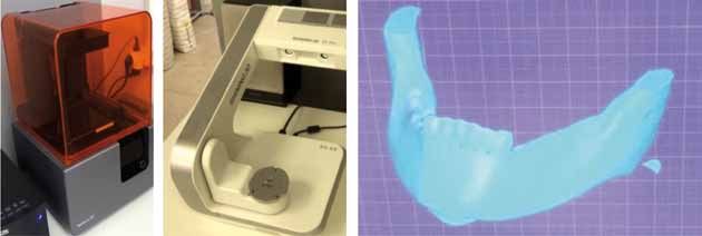
DE N TI S T R Y                                                                   | archiv euromedica | 202 1 | vol. 11 | num . 1 |
                                                                                                                                                         149

Fig. 2. Device for obtaining stereolithographic lower jaw: a — 3D printer; b — 3D scanner; c — 3D model of the lower jaw

Fig. 3. Orthopantomogram (а) and computed tomogram (b) with the possibility of 3D reconstruction of the lower jaw defect. Patient A., years old. D.S:
large follicular cyst of the mandibular body on the left

any sharp edges of the implant and allowed hermetic                                  The technology for producing engineered bone
insulation of the autobone chips through the first stage                       substitutes from macroporous titanium fiber material
of developing the bioengineered composition. The                               is based on the titanium chips cold pressing method.
technology for obtaining a medical implant is pro-                             Jaw implants are supposed to augment bone defects,
tected by a patent (Patent # 2733687).                                         which means they are to have a respectively designed
      To fabricate implants replacing the jaw defect,                          shape and size. To achieve these, a specially manufac-
chips with a thickness of 0.05-0.015 mm were used.                             tured mold was used. The individual mold was based
The spiral coil diameter was 0.8±0.2 mm, the distance                          on a dental flask used to produce plastic prostheses.
between the coils thus making 0.8±0.2 mm. The                                        In order to obtain an individual mold for a
chips were made on a numerically controlled machine                            mandibular defect with no disturbed continuity, the
(Fig. 4).                                                                      neoplasm was removed within healthy tissues using
      The engineered bone substitute is a combination                          a milling cutter and a drill, on a stereolithographic
of a non-woven titanium material with through poros-                           model. The resulting defect was filled with molten
ity fabricated from titanium chips and a spiral-shaped                         dental wax. Once cooled, the resulting wax model of
autobone. In order to obtain a spiral-shaped auto-                             the potential jaw implant was removed with the mold
bone, a spiral-shaped milling cutter was designed and                          to be made further (Fig. 7–9).
manufactured jointly with the KASKAD Machine-                                        Following a standard procedure, creamy die stone
Engineering Plant (Krasnodar, Russia, with a respec-                           was made, which was then poured into the flask up to
tive patent obtained (Patent # 2733687).                                       the middle of the side height. The wax composition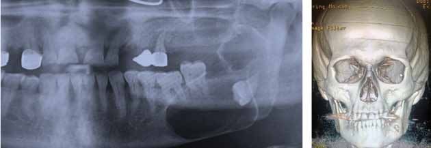
150   | archiv euromedica | 202 1 | vol. 11 | num . 1 |                                                                            DE N TI S T R Y

                                                                               Fig. 5. G eneral view of the manufactured cutter: a — shank;
                                                                               b — working part; с — cutter edge

         Fig. 4. Titanium shavings for the production of non-woven titanium
         through-porosity fabric

                                                                                                               Fig. 6. А — The stage of sampling of
                                                                                                               bone autochips from the iliac crest; В —
                                                                                                               View of bone chips following cut with
          A                                                     B                                              the original cutter

                                                                                                               Fig. 7. X -ray and lithographic model of
                                                                                                               the lower jaw of the affected tumor

                                                                                                               Fig. 8. R emoval of a neoplasm and
                                                                                                               formation of a bone cavity on a stere-
                                                                                                               olithographic model

         was placed in the die stone in the center to the middle               When the die stone was completely dissolved to make
         of its depth. Once the die stone was hard, a brush was                a creamy substance, it was poured into the flask up to
         used to cover its entire surface with a layer of paper                the upper edge. The flask was then covered with a lid
         glue, and then, once the glue dried, another portion                  and pressed down. When the die stone was completely
         of die stone was prepared, yet in salt water that time.               solid, the flask was opened in the reverse order. A jet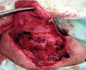
DE N TI S T R Y                                                     | archiv euromedica | 202 1 | vol. 11 | num . 1 |
                                                                                                                                         151

                                                                                            Fig. 9. Individual wax composition of the
                                                                                            jaw implant

of hot water helped melt the wax blank, and a mold                removed part of the lithographic model, the teeth were
for an individual implant was obtained. Knowing the               cut off along with the tumor-altered part of the lower
volume of the implant and the porosity to be obtained,            jaw.
we used a special formula to calculate the amount                      The obtained lithographic model was used to
of titanium chips required. In 85% of the implants,               bend a titanium reconstructive dynamic plate, in view
the porosity was 75±5% of the total implant volume.               of an additional section to be used for fixing the plate
After weighing the required amount of chips, we got               to the healthy stump of the lower jaw. On the upper
to laying them in the mold. Bone chips were obtained              part of the plate replacing the lower jaw branch, a tita-
from the retromolar area or from the ilium. The result            nium condylar process with a joint head was attached
was a pyramidal stack of titanium chips with layers of            using special screws.
autobone chips. The mold was assembled the right way                   Having completely covered the resulting litho-
based on the flask grooves. The cuvette flask placed              graphic model with wax, we set to producing a plaster
under a mechanical press to be further squeezed until             mold following the method described above. A
both halves were completely closed (Fig. 10).                     titanium reconstructive plate was placed in the center

                                                                                            Fig. 1 0. Mold for the stage of cold
                                                                                            pressing: a — finished mold; b — the
                                                                                            stage of laying the components of the
                                                                                            bioengineering composition; c — stage
                                                                                            of cold pressing

a                                    b                        c

     In cases where a bioengineered design was                    of the substitute depth. On all sides, the reconstruc-
required to replace the half of the lower jaw after its re-       tive plate was covered with a macroporous titanium
section, a slightly different technology was employed.            fiber material combined with autobone. Given its
     For this purpose, a large flask was made (115 mm             shape, it matched the resected part of the lower jaw
long, 35 mm wide and 70 mm high), while its compo-                (Fig. 11, 12).
nents and the disassembly specifics were similar to the                 The first (control) group included 76 patients
previous one.                                                     who were operated on following the standard conven-
     A lower jaw lithographic model was made based                tional methods. After sequestration and cystectomy,
on the CT data. Subject to the rules of oncology,                 the bone cavities were filled with blood clots or os-
retreating 2–2.5 cm from the tumor, resection of the              teoplastic material. 37 patients had lower jaw tumors,
affected area on the lower jaw was performed with its             of which 7 had true defects not only in the lower jaw
continuity disturbed. In the event that the condylar              yet also in the soft tissues. Patients with soft and bone
process was affected, resection was performed with                tissue defects were operated on in two stages. Through
the branch and the condylar process removed. On the               the first stage, soft tissues were repleted. Microsurgical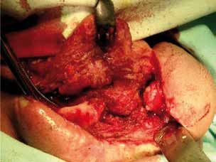
152   | archiv euromedica | 202 1 | vol. 11 | num . 1 |                                                                                   DE N TI S T R Y

                                                                                         Fig. 1 2. Bioengineering design

         Fig. 11. An intermediate stage in the fabrication of an engineered
         biomaterial                                                                           The remaining 30 patients who had bone defects
                                                                                         only were operated on using reconstructive titanium
                                                                                         plates combined with autobone.
         techniques were used in 4 patients, while Filatov-Gil-                                In the main group, in 31 patients with mandibu-
         lies tubed pedicle was used in the rest of the patients                         lar defects were augmented with the engineered bone
         (Fig. 13).                                                                      autograft fabricated by us.
               In the second stage, the bone defect, which devel-                              Of them, 9 persons were diagnosed with odon-
         oped following the surgery, was filled with a split rib                         togenic cysts, another 3 — with the lower jaw osteo-
         autograft or an ilium segment (Fig. 14).                                        myelitis. These patients developed the defects after sur-

         Fig. 13. Transplant-ready radial flap

         A                                                                             B

         Fig. 14. A — the stage of the collection of the parietal bone site; B — formed free bone autograft, ready for transplantation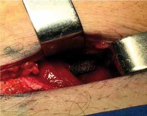
DE N TI S T R Y                                                           | archiv euromedica | 202 1 | vol. 11 | num . 1 |
                                                                                                                                     153

gical interventions that did not lead to any disturbance                       The incision line was applied strictly in the iliac
in the jaw continuity. Once the major focus removed,                     crest projection. The crest was detected by palpation.
the resulting bone cavity was profoundly washed                          The incision length of the was 9±3 cm. Once the iliac
with an antiseptic solution. A visual examination of                     crest was skeletonized, autobone chips or bone frag-
the resulting bone defect revealed altered bone tissue                   ments were collected (Fig. 16).
areas, which were removed with a milling cutter down                           For this purpose, a physiodispenser and the
to healthy tissues. The causative teeth were removed.                    specifically designed milling cutter were used to get a
Following respective indications, the root resection                     spiral-like autobone. Following our method, the tita-
was performed. Any sharp edges and resected roots                        nium and bone chips were stacked in a sterile plaster
were smoothed down with a cutter. The resulting bone                     mold in layers so as to make a pyramid. A dynamic
cavity was filled with a specially prepared bioengi-                     reconstructive perforated plate was placed in the chip
neered structure made of the NWTMTP based on the                         thickness, after which a manual press (pre-autoclaved
lithographic model produced subject to the specifi-                      and additionally wrapped with a sterile sheet) was used
cally designed method.                                                   to compress both halves of the mold until they were
      In 5 patients with true bone and soft tissue de-                   completely in contact. Further on, the bioengineered
fects, a full-layer muscle-skin grafting was used, which                 structure was extracted. To ensure its maturation,
contained inside a mature bioengineered structure of                     it was to be placed in the anterior abdominal wall.
the NWTMTP in combination with bone autochips.                           During that, the perforant vessels markings on the
      Of these patients with true bone and soft tissue                   abdomen skin were used as the reference points. The
defects, 3 had half-resected lower jaw with the condy-                   superficial abdominal muscle was dissected horizontal-
lar process exarticulation.                                              ly from the existing incision while the dissection went
      All other patients of the main group were treated                  from the anterior vaginal wall of the rectus abdominis
for mandibular defects using an autotransplantant                        muscle up to the volume so as to ensure a smooth
made by our method and harvested in the anterior                         passage through it and the desired positioning of the
abdominal wall. No skin was used there.                                  bioengineered structure. The latter was fixed to the
                                                                         surrounding soft tissues with 2–3 sutures of absorbable
      Harvesting and autotransplantation of engineered                   material (Fig. 17).
vascularized autotransplantant                                                 X-ray examination was performed on a monthly
      The stage of the harvesting the autotransplantant                  basis in order to monitor the biocomposition location
in all cases was performed in the anterior abdominal                     (Fig. 18).
wall. Prior to placing the autotransplantant in the an-                        We performed intraoperative ultrasound scanning
terior abdominal wall, surgical marking was performed                    in order to identify the location of axial blood vessels.
on the abdomen skin; the course of the perforant                               3.5 ± 0.5 months later, the next stage was
vessels was outlined with a surgical marker, the entire                  launched. Isolating the bioengineered structure from
process controlled by ultrasound (Fig. 15).                              the surrounding tissues without a vascular pedicle
                                                                         presented no serious issue. The end sections of the dy-
                                                                         namic plate with holes for the intraosseous screws were
                                                                         separated with gauze swabs. The sprouted part of the
                                                                         non-woven titanium material had to be isolated with a
                                                                         sharp tool (Fig. 19).
                                                                               Following the generally accepted method, the
                                                                         tumor-affected area of the jaw was skeletonized, which
                                                                         was resected retreating 2 cm from the tumor into the
                                                                         healthy jaw. 10 patients had the lower resection jaw
                                                                         with their condylar processes exarticulation. In 13
                                                                         patients, the joint was preserved (Fig. 20).
                                                                               Prior to installing the bioengineered structure,
                                                                         the remaining part of the jaw was placed in the central
                                                                         occlusion position while controlled by the splint
                                                                         (Fig. 21).
                                                                               A reconstructive dynamic titanium plate, along
                                                                         with the bioengineered structure, was attached to the
Fig. 15. Surgical marking of axial vessels on the skin of the abdomen   jaw stump with 3-4 standard bicortical intraosseous
                                                                         Stryker system screws (Fig. 22).
154   | archiv euromedica | 202 1 | vol. 11 | num . 1 |                                                                            DE N TI S T R Y

                                                                                                                 Fig. 16. A — Bone fragments from the
                                                                                                                 iliac crest; B — Bone chips taken from
                                                                                                                 the iliac crest
         A                                                     B

                                                                                        the surrounding skin and soft tissues (Fig. 23).
                                                                                              When working on a plastic surgery for a soft
                                                                                        tissue defect, a reasonable idea is to have two teams of
                                                                                        surgeons, for one of them to be responsible for prepar-
                                                                                        ing the recipient zone, the other team modeling the
                                                                                        grafting. For all cases within our study, a team of duly
                                                                                        certified microsurgeons were involved. In each clinical
                                                                                        case, a mandatory ultrasound Doppler examination of
                                                                                        the perforant and axial vessels was done (Fig. 24).
                                                                                              While controlling the bite, a dynamic reconstruc-
                                                                                        tive titanium plate was attached to the exposed lower
                                                                                        jaw stumps, which was ran, as reinforcement, into the
                                                                                        bioengineered structure depth (Fig. 25).
                                                                                              After that, microsurgeons used microscopes
                                                                                        to stitch the facial vessels with vessels feeding the
         Fig. 17. The engineered biomaterial is fixed in the thickness of the rectus   complex soft-tissue grafting with the bioengineered
         abdominis muscle (the wound is ready for suturing)                             structure inside, applying traditional vascular sutures.
                                                                                        The facial soft tissues and the transplanted grafting
                                                                                        were stitched in layers (Fig. 26).
                                                                                              15 patients of the main group had their bone de-
                                                                                        fects replaced with a bioengineered structure simulta-
                                                                                        neously with the lower jaw resection. All patients were
                                                                                        treated for ameloblastomas, often recurrent tumors of
                                                                                        the lower jaw. During that, no true soft tissue defects
                                                                                        were observed. Before the lower jaw resection, indi-
                                                                                        vidual molds were made based on our method, which
                                                                                        were sterilized and delivered to the operating room.
                                                                                              During the surgery, bone chips were taken from
                                                                                        the ilium using the specially designed cutter as men-
                                                                                        tioned earlier (Fig. 27).
                                                                                              As we started manufacturing a bioengineered
                                                                                        structure, layers of titanium and bone chips were
         Fig. 18.X-ray of the pelvic bones. The arrow indicates a bioengineering       placed in layers into the earlier prepared mold, to make
         structure                                                                      a pyramid and compressed. In the center of that the
                                                                                        reconstructive plate was to be found (Fig. 28).
                                                                                              The resulting bioengineered structure was placed
              In 5 patients with soft tissue and jaw defects, the                       in the lower jaw defect area that developed follow-
         bioengineered structure – taken as a single conglomer-                         ing the tumor resection, to be fixed afterwards, and
         ate – was transferred to the affected area together with                       then the structure was covered with surrounding soft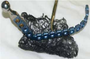
DE N TI S T R Y                                                                | archiv euromedica | 202 1 | vol. 11 | num . 1 |
                                                                                                                                                           155

 A                                                        B                                            C

 D                                                        E

                                                                                                                 Fig. 19. A — The stage of layered tis-
                                                                                                                 sue dissection using an electric knife;
                                                                                                                 B — The stage of isolating the inner
                                                                                                                 surface of a bioengineering structure;
                                                                                                                 C — Bioengineering construct extrac-
                                                                                                                 tion phase; D — completely removed
                                                                                                                 bioengineering structure; E — wound
                                                                                                                 tightly sutured with interrupted
                                                                                                                 sutures on the anterior abdominal wall

Fig. 20. Patient K., 34 years old. D.S. Ameloblastoma of the lower jaw on
the left. Resection of the affected area of the lower jaw performed
                                                                             Fig. 22. T he graft is fixed in the wound; the wound is ready for suturing

                                                                             tissues. The wound on the side of the mouth and skin
                                                                             was sutured tightly. Drainage was ensured with rubber
                                                                             drains (Fig. 29).

                                                                             RES U LTS AND DIS C U SSION
                                                                                  The data from evidence-based medicine research
                                                                             projects revealed a 47% reduction in absolute risk
Fig. 21. The stage of adjusting the bioengineering structure: a —           with a confidence interval of 3–59%. The number
matured bioengineering structure; b — titanium head of the condylar          of patients who need to be treated with our method
process; c — keys for bending                                                (number needed to treat / NNT) was 2 (CI — 2–3).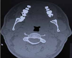
156   | archiv euromedica | 202 1 | vol. 11 | num . 1 |                                                                    DE N TI S T R Y

                                                                                                        Fig. 23. G eneral view of an autograft
                                                                                                        with a bioengineering structure inside

                                                                                                        Fig. 24. U ltrasound examination of
                                                                                                        intraoperatively perforated vessels

         A                                                                                                                                  B

                                                           Fig. 25. A — the stage of immersion of the bioengineering structure into the soft
                                                           tissues of the anterior abdominal wall; B — the stage of isolating the bioengineering
                                                           composition from the thickness of the rectus abdominis muscle; С — general view after
                                                     C     the end of the operation

         The relative risk reduction was at 94% with a CI of                  These results here reveal a fairly high rate of insuf-
         59–116%, which stands for a very high clinically               ficient aesthetic effectiveness in the control group if
         significant effect. The odds ratio was 0.03 with a CI          compared to the group treated using the specially de-
         of 0.01–0.26, whereas the risk of adverse outcomes             veloped method — 24 VS. 3%, respectively (χ2 — 4.99;
         proved very low (χ2 — 18.83; p = 0.0001).                      p = 0.026). The relative risk reduction was 84% with a
DE N TI S T R Y | archiv euromedica | 202 1 | vol. 11 | num . 1 |
157
confidence interval ranging from 2 to 133%. The abso-
lute risk reduction was 21% with a CI of 5 to 32%. The
number of patients who need to be treated in order to
prevent one adverse outcome (poor aesthetic effect)
is 5 with a CI of 3 to 19. The odds ratio of 0.11 with
a CI of 0.02–0.85 means that, when using the newly
developed method, the risk of an unfavorable outcome
was 5 times as low compared to the generally accepted
one (p = 0.026), this implying that the effectiveness of
the proposed technology in terms of ensuring aesthetic
effect is to be viewed significant both statistically and
clinically.
An unfavorable outcome (poor functional result)
Fig. 26. General view of sutured vessels (magnification ×6) was observed in many fewer cases — 3% and 42%,
Fig. 27. A — Skin incision with an
electric knife; B — Skeleton stage of the
A B
iliac crest
respectively. The absolute risk reduction was 39%
with a confidence interval of 22–50%. The number
of patients to be treated with the proposed interven-
tion was 3 (CI — 2–5). The relative risk reduction was
92% with a CI of 53–120%. Values exceeding 50%
correspond to a clinically significant effect. The odds
ratio was 0.05 with a CI of 0.01–0.36, i.e., the risk of an
adverse outcome in terms of failure to arrive at a posi-
tive functional effect while employing the proposed
method was very low.
The method was also associated with a high
statistically and clinically significant positive result in
Fig. 28. Bioengineered composition ready for transplantation into the terms of assessing the intervention effectiveness subject
area of the lower jaw after removal of the ameloblastoma: a — the body to factor like the patient’s psychoemotional satisfac-
of the lower jaw; b — the angle of the lower jaw; c — the branch of the tion following the surgical and orthopedic rehabilita-
lower jaw
Fig. 29. A — Surgical wound after
resection of a part of the lower jaw
body on the right and replaced with a
bioengineering composition; B — View
A B of a sutured and drained wound158 | archiv euromedica | 202 1 | vol. 11 | num . 1 | DE N TI S T R Y
Table 1. Key indicators of the intervention effect following the treatment of defects based on the specially developed method (Group I), compared to the
defect reconstruction based on conventional methods (Group II)
Indicators
Event Rate in Event Rate in Relative Risk Absolute Risk
NNT 95% CI Odds
Comparison groups Ratio
Treated Group Control Group Reduction % Reduction % х2 P
95% CI
% % 95% CI 95% CI
Poor aesthetic effect
84 21 5 0.11 4.9
Groups I and II 3 24 Р = 0.026
2-133 5-32 3-19 0.020.85 9
Poor functional effect
0.05
92 39 3 13.
Groups I and II 3 42 0.01 Р =0.00 01
53-120 22-50 2-5 84
0.36
Poor psychoemotional status following the final stage of the surgical and orthopedic rehabilitation
94 47 2 0.03 0.01 18.
Groups I and II 3 50 Р = 0.00 01
59 -116 3 - 59 2-3 0.26 83
tion stage. Unsatisfactory psychoemotional status after and the Krasnodar Regions of Russia revealed the
surgical and orthopedic rehabilitation based on the following: 32% of patients were not satisfied with the
proposed method, if matched against the conventional treatment outcomes, of which aesthetic dissatisfaction
treatment, was much lower — 3% and 50%, respec- accounted for 18%, whereas another 14% of patients
tively. were not satisfied with the orthopedic structures.
The absolute risk reduction was 47% with a 3. The study helped obtain experimental proof,
confidence interval of 3–59%, whereas the number protect by a patent, manufacture and implement into
of patients in need of treatment (NNT) was 2 (CI clinical practice a special cutter to be used for spiral-
2–3). The relative risk reduction was 94% at a CI of shaped bone sampling (Patent # 2733687).
59–116%, which stands proof to a very high clinically 4. A 3D prototyping-based method of sampling
significant effect. The odds ratio was 0.03 at a CI of and shaping a vascularized autograft combined with a
0.01-0.26, while the risk of adverse outcome proved non-woven titanium material with through-porosity
very low (χ2 — 18.83; = 0.0001). has been offered its theoretical explanation as well as
In view of the above, the key indicators used to implemented in clinical practice. If using the proposed
evaluate the intervention effectiveness in patients oper- method, the risk of adverse outcomes with no positive
ated on using the proposed method, if compared to functional result achieved remains very low, the odds
conventional methods, reveal a high clinical statistical ratio being at 0.05 with a CI of 0.01–0.36.
significance of the obtained outcomes as well as point 5. A new method of mandibular defects individ-
at the feasibility of employing the proposed treatment ual replacement has been improved relying on digital
methods in practical healthcare. technologies, as well as the method has been protected
The obtained outcomes allow recommending by a patent and introduced into clinical practice. In
these technologies to be scaled up thus embracing a case of using the proposed method, the rate of the pa-
wider clinical practice. tient’s unsatisfactory psychoemotional status following
the surgical and orthopedic rehabilitation stage was
C ON C L U SIONS much lower, if compared to the conventional treat-
1. Improved treatment effectiveness for patients ment — 3% and 50%, respectively.
with mandibular defects, which was due to employing 6. The specifically designed individual vascular-
a vascularized individual bioengineered composition ized autografts, while combined with a non-woven
based on non-woven titanium material with through titanium material with through porosity allowed
porosity. The risk of an unfavorable treatment out- increasing the aesthetic and the functional outcomes
come while using the proposed method was 5 times as as well as bring up the rate of the psychoemotional
low compared to the generally accepted ones. satisfaction following the treatment. Insufficient
2. Analysis of the treatment results in patients functional results were observed in 3% of the cases; the
with lower jaw defects, based on the data reported absolute risk reduction reached 47% with a confidence
by specialized maxillofacial hospitals of the Samara interval of 3–59%. The number of patients that neededDE N TI S T R Y | archiv euromedica | 202 1 | vol. 11 | num . 1 |
159
treatment (CNNT) was 2 (CI 2–3). The relative risk ridge augmentation. Russian Journal of Stomatology
reduction was 94% with a CI of 59–116%, which cor- = Stomatologiia. 2019;98(6):30–32. https://doi.
responds to a very high clinically significant effect. The org/10.17116/stomat20199806130
odds ratio was 0.03 with a CI of 0.01–0.26, while the 10. Kupryakhin S.V., Lepilin A.V., Kupryakhin V.A.
risk of arriving at an adverse outcome proved very low Optimization of dental implantation combined with
(χ2 — 18.83; p = 0.0001). closed sinus lift in patients with low maxillary sinus
floor // Archiv EuroMedica. 2019. Vol. 9; 2: 117–121.
https://doi.org/10.35630/2199-885X/2019/9/2/117
REFEREN C ES
1. Butsan S.B., Bulat S.G., Gileva K.S., Salikhov 11. Kupryakhin S.V., Lepilin A.V., Kupryakhin
K.S., Khokhlachev S.B., Arsenidze A.R., V.A., Postnikov M.A. Potential introduction of cell
Reshetun A.M. The use of autogenous free vascu- technologies to improve dental implant surface prepar-
larized fibula transplant for the correction of severe ing // Archiv EuroMedica. 2019. Vol. 9; 2: 122–129.
mandibular atrophy. Russian Journal of Stomatology https://doi.org/10.35630/2199-885X/2019/9/2/122
= Stomatologiya, 2019;98(5):32–45. https://doi. 12. Lee H.G., Kim Y.D. V olumetric stability of au-
org/10.17116/stomat20199805132 togenous bone graft with mandibular body bone:
2. Butsan S.B., Gileva K.S., Verbo E.V., cone-beam computed tomography and three-dimen-
Khokhlachev S.B., Abramian S.V., Smal A.A., sional reconstruction analysis. J Korean Assoc Oral
Bulat S.G. E volution of the mandibular defects Maxillofac Surg. 2015;41(5):232–239. https://doi.
reconstruction with free fibula flapx. Russian Journal org/10.5125/jkaoms.2015.41.5.232
of Stomatology = Stomatologiya, 2018;97(3):35–43. 13. Ozaki W., Buchman S.R. Volume maintenance
https://doi.org/10.17116/stomat201897335 of onlay bone grafts in the craniofacial skeleton:
3. Dasmah A, Thor A, Ekestubbe A, Sennerby micro-architecture versus embryologic origin. Plast
L, Rasmusson L. P articulate vs. block bone grafts: Reconstr Surg. 1998;102:291–299. https://doi.
three-dimensional changes in graft volume after recon- org/10.1097/00006534-199808000-00001
struction of the atrophic maxilla, a 2-year radiographic 14. Semkin V.A., Nadtochiy A.G., Kuzin A.V., Kol-
follow-up. J Craniomaxillofac Surg. 2012;40:654–659. otikov P.A. The effectiveness of tooth-preserving
https://doi.org/10.1016/j.jcms.2011.10.032 surgery in various forms of inflammatory and destruc-
4. Hern’andez, Alfaro F, Sancho-Puchades M, tive lesions in mandibular molars area. Russian Journal
Guijarro-Mart´ınez R. O ral reconstruction of of Stomatology = Stomatologiya, 2019;98(2):60–63.
the atrophic maxilla with intraoral bone grafts and https://doi.org/10.17116/stomat20199802160
biomaterials: a prospective clinical study with cone 15. Von Arx T., Buser D. Horizontal ridge aug-
beam computed tomography validation. Int J Oral mentation using autogenous block grafts and the
Maxillofac Implants. 2013;28(1):241–251. https:// guided bone regeneration technique with collagen
doi.org/10.11607/jomi.2405 membranes: a clinical study with 42 patients. Clin
5. Karpyuk V.B., Perova M.D., Gilevich I.V., Oral Implants Res. 2006;17:359–366. https://doi.
Sevostyanov I.A. Cell-potentiated regenerative org/10.1111/j.1600-0501.2005.01234.x
technologies for restoring jaw bone tissues in case of 16. Yu H, Chen L, Zhu Y, Qiu L. B ilamina cortical
odontogenic inflammatory & destructive process // tenting grafting technique for three-dimensional
Archiv EuroMedica. 2019. Vol. 9. № 2. P. 140–146. reconstruction of severely atrophic alveolar ridges in
https://doi.org/10.35630/2199-885X/2019/9/2/140 anterior maxillae: A 6-year prospective study. J Crani-
6. Karpyuk V.B., Perova M.D. Innovation-based ap- omaxillofac Surg. 2016;44(7):868–875.https://doi.
proach in reconstruction of reduced jaw alveolar ridge org/10.1016/j.jcms.2016.04.018
bone using cell regeneration technologies // Archiv
EuroMedica. 2019. Vol. 9. № 2. P. 147–155. https://
doi.org/10.35630/2199-885X/2019/9/2/147
7. Khoury F., Hanser T. M andibular bone block
harvesting from the retromolar region: a 10-year pro-
spective clinical study. Int J Oral Maxillofac Implants.
2015;30(3):688–697. https://doi.org/10.11607/
jomi.4117.
8. Kloss F.R., Offermanns V., Kloss-Brandstät-
ter A. Comparison of allogeneic and autogenous
bone grafts for augmentation of alveolar ridge defects
— A 12-month retrospective radiographic evaluation.
Clin Oral Implants Res. 2018;29(11):1163–75. ht-
tps://doi.org/10.1111/clr.13380.
9. Korzh D.G., Haritonov D.Yu., Stepanov I.V.,
Podoprigora A.V. Evaluation of autogenous man-
dibular bone block resorption in horizontal alveolarYou can also read