Pacemakers and MRI: A protocol in line with international guidelines and approved by the SFICV (French Society of Cardiovascular Imaging)
←
→
Page content transcription
If your browser does not render page correctly, please read the page content below
Diagnostic and Interventional Imaging (2017) 98, 203—215

RECOMMENDATIONS /Professional Information

Pacemakers and MRI: A protocol in line with
international guidelines and approved by
the SFICV (French Society of Cardiovascular
Imaging)
                                      Y. Cruypeninck a, B. Dubourg a,b, P. Michelin a,
                                      B. Godin c, C. Savoye-Collet a, E. Gérardin a,
                                      J.-N. Dacher a,b,∗

                                      a
                                        Department of Radiology, University Hospital of Rouen, 76031 Rouen, France
                                      b
                                        Inserm U1096, University of Rouen, 76000 Rouen, France
                                      c
                                        Department of Cardiology, University Hospital of Rouen, 76031 Rouen, France

                                      The rate of pacemaker implantation is increasing each year. It has been estimated that
                                      about 1 person in 1000 in the Western world has received a pacemaker worldwide [1].
                                      Despite major advances in pacemaker technology, notably with the advent of ‘‘magnetic
                                      resonance imaging (MR)-compatible’’ pacemakers, it should be borne in mind that these
                                      medical devices still contain ferromagnetic material and remain a relative contraindication
                                      for MRI. Nonetheless, when the risk—benefit ratio is favorable, imaging may be performed
                                      under specific safety conditions. In all cases, MRI in a patient with a pacemaker must be
                                      closely supervised by cardiology (cardiac rhythm disorder specialists) and radiology teams
                                      [2]. To ensure such conditions are fulfilled, this kind of imaging should only be performed in
                                      hospitals that have implemented efficient interdepartmental protocols and collaboration
                                      procedures.

 ∗   Corresponding author at: Department of Radiology, University Hospital of Rouen, 1, rue de Germont, 76031 Rouen cedex, France.
     E-mail address: jean-nicolas.dacher@chu-rouen.fr (J.-N. Dacher).

http://dx.doi.org/10.1016/j.diii.2016.06.024
2211-5684/© 2016 Editions françaises de radiologie. Published by Elsevier Masson SAS. All rights reserved.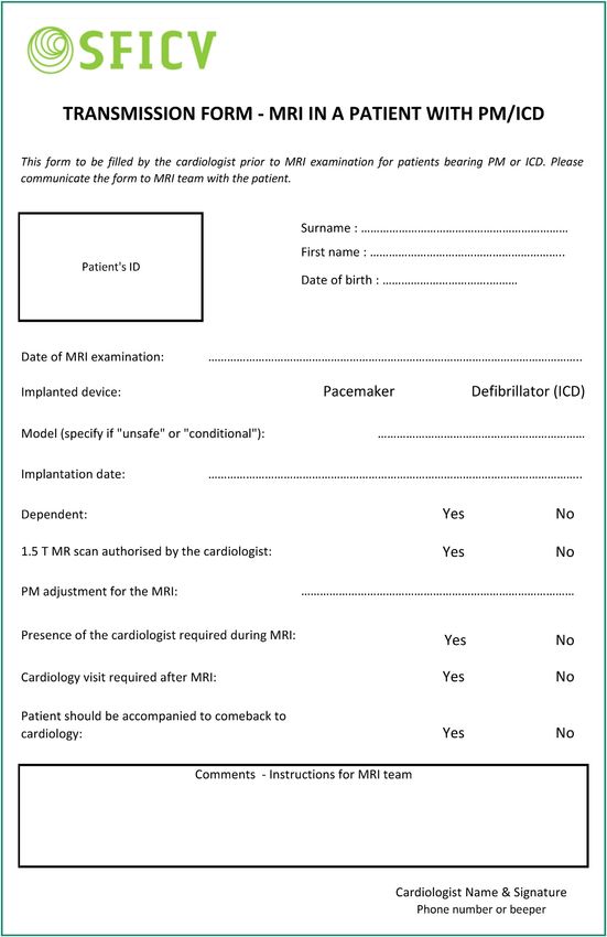
204                                                                                                       Y. Cruypeninck et al.

    At the same time, the use and scope of MRI has expanded          MRI-compatible pacemakers are not non-magnetic;
greatly with, for example, 5.3 million MRI scans performed       therefore, they are categorized as ‘‘MR conditional’’ by the
in France in 2012 [3]. To continue considering pacemakers        Foods and Drugs Administration, not ‘‘MR Safe’’. This implies
as an absolute contraindication for MRI when this modal-         maintaining a high safety standard. All pacemaker manufac-
ity’s indications are steadily expanding, could result in        turers provide their own recommendations for MRI on their
missed opportunities for these patients. A Japanese study        websites (Table 1).
demonstrated that 17% of pacemaker patients were likely to           In practice, MRI teams should implement the same
require MRI in the year following implantation [4]. Similarly,   safety conditions, whether the patient has a MRI-compatible
Roguin et al. estimated that the likelihood of pacemaker         pacemaker or not. The main differences are in fact the indi-
patients requiring MRI during the lifespan of the device         cations for MRI and the applicable legal framework. The
ranged from 50% to 75% [5]. Finally, a two-fold increase in      manufacturer’s warranty guaranteeing the absence of inci-
the likelihood of needing MRI is observed in patients aged       dents means that the indications for MRI can be extended.
over 65.                                                         For example, a patient with a MRI-compatible pacemaker
    The indications for MRI keep expanding. For the pace-        with dementia, brain metastases, extrapyramidal symptoms
maker patient age group, the main indications are for            or meniscus injury may benefit from MRI, even if the proce-
neurological disorders (stroke, dementia, Parkinson’s dis-       dure is not vital for patient care. However, MRI would most
ease, etc.) and neurosurgery (tumors, hematoma, abscess,         likely be contraindicated in the same clinical setting if the
etc.), although MRI examinations for cancer and cardiovas-       pacemaker is not MRI-compatible.
cular disorders are also steadily increasing [6,7].                  Contraindications for implantable defibrillators are more
    The aim of the present article is to suggest rules of        stringent than for pacemakers, except for the new ‘‘MRI-
good practice when performing MRIs in pace maker patients        conditional’’ models. Still, imaging may be performed in the
regardless the device’s compatibility. This recommendation       same rigorous safety conditions as for pacemakers, if the
was endorsed by the SFICV (French Society of Cardiovascular      expected benefit exceeds by far the potential risk.
Imaging).

                                                                 Potential risks
Pacemakers, implantable automatic
defibrillators: basic principles                                  The risk of harm to the patient or device during a MRI scan
                                                                 is not negligible. Multiple interactions occur when a patient
Schematically, pacemaker insertion is indicated for patients     with a pacemaker is placed within a MRI machine. A large
with atrioventricular conduction abnormalities. An inter-        number of patient, pacemaker and MRI parameters need to
national three-letter code describes the pacing mode and         be taken into account to minimize the risks associated with
settings. The first letter indicates which chamber(s) are         MRI scans. Each additional sequence, each additional minute
stimulated [A atrium, V ventricle, D (dual) atrium and ven-      that the scan lasts represents a new risk, hence the need to
tricle, O none], the second which chamber(s) are detected        keep the procedure to the bare minimum.
(A, V, D, O), and the third the mode of response of the pace-        Potentially interfering factors are: B0 static field and gra-
maker (I inhibited, T triggered, D inhibited and triggered, O    dient intensities, the spatial magnetic field gradient, the
none). For example, a pacemaker programmed to the VOO            radiofrequency used (pulse type), the specific absorption
mode performs simple asynchronous ventricular pacing.            rate (SAR), the anatomical region investigated, the position
    An implantable automatic defibrillator is a pacemaker         inside the magnet, the implanted material (leads and pac-
that can deliver shocks. Such pacemakers are indicated           ing box), the patient’s dependence on the pacemaker, lead
for patients at risk for ventricular rhythm disorders, cer-      length, loops and direction, and the duration of the scan.
tain patients with cardiomyopathy or cardiac failure with a
reduced ejection fraction of less than 30—35%.
                                                                 Risks for the pacemaker
The new ‘‘MRI-compatible’’ pacemakers                            Placing a pacemaker in a MRI system may result in dam-
                                                                 age to the pacing box or leads. Potential risks include
These recently marketed devices consist in a pacing box
                                                                 premature deterioration of the battery, heating of stimu-
and leads that are immediately recognizable on X-ray exam-
                                                                 lation leads with possible endocardial burns, device switch
inations due to the radiopaque markers they contain. In
                                                                 to asynchronous mode, stimulator deprogramming or inhi-
2010, a study on the first commercially released device con-
                                                                 bition, permanent device failure, and stimulator or lead
firmed that MRI could be performed without harm to the
                                                                 migration.
patients or damage to the device [8]. The study included 464
patients with this pacemaker from 41 different sites. Two
hundred and forty-four of these patients underwent non-
cardiac MRI 9 to 12 weeks after pacemaker implantation.          Risks for the patient
The control group included 206 patients. No immediate or
delayed complications attributable to MRI were observed.         The potential risks for the patient are variable, depending on
No differences were found between the threshold param-           how much the patient relies on his/her pacemaker. Although
eters measured for the pacemakers having undergone MRI           the risks are different, both dependent and non-dependent
and control pacemakers.                                          patients are subject to potentially dangerous incidents, and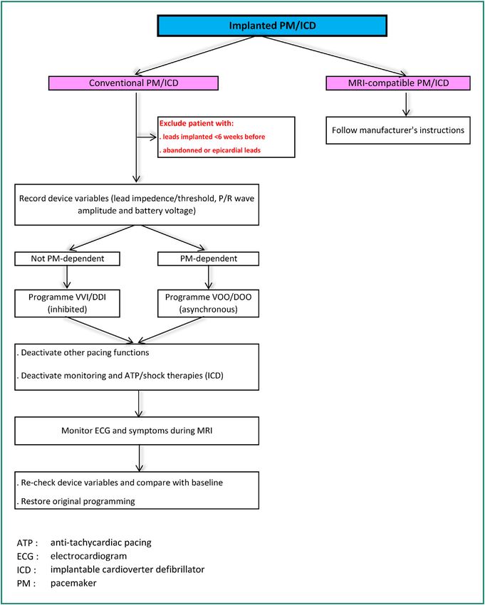
Pacemakers and MRI
Table 1   MRI-compatible pacemakers.
                Models         Field        Patient    Exclusion zone   SAR                Slew rate      Spatial    Total          Leads
                                            position                                                      gradient   exposure
                                                                                                                     duration
                                                                                                                     allowed
BIOTRONIK       Evia SR-DR     1.5 T or 3 T Strict     Isocenter of     Full body          Slew rate ≤    Not        30 min total   Safio S 53,
                               (for some    supine     the reel         ≤ 2 W/kg           216 T/m/s      specified   active         S60
                               combina-     position   excluded                            per axis                  imaging by     Siello S 53,
                Entovis        tion                    between                             Up to the                 session        S 60, S45
                SR-DR          pace-                   the level of                        serial                                   Solia S 45,
                               maker/leads)            eyes and                            number                                   S & T 53, S
                                                       top of hips                         66237094                                 & T 60
                                                                                           included,                                Setrox S 53,
                                                                                           maximum                                  S60
                                                                                           slew rate of
                                                                                           125 T/m/s
                                                                                           per axis
                Estella                                Over                                Slew rate ≤
                SR-DR                                  exclusion                           200 T/m/s
                Ecuro                                  zone from                           per axis
                SR-DR                                  the serial
                                                       number
                                                       66237095
                Eluna 8                                Full body        Head < 3.2 W/kg                              10 hours for
                Epyra 6 et 8                                                                                         the life of
                Etrinsa 6 et                                                                                         the system
                8
BOSTON          Accolade       1.5 T        Prone or   Full body        Full               Slew          Not         Not            INGEVITY
  SCIENTIFIC    MRI                         supine                      body ≤ 4 W/kg if   rate ≤ 200 T/m/s
                                                                                                         specified    specified       MRI,
                Essensio                    position                    INGEVITY leads     per axis                                 FINELINE II
                MRI                                                     otherwise                                                   Sterox,
                Proponent                                               2 W/kg                                                      FINELINE II
                MRI                                                     Head < 3.2 W/kg                                             Sterox EZ
                Advantio                                                Full
                MRI                                                     body ≤ 2 W/kg
                Formio MRI                                              Head < 3.2 W/kg
                Ingenio MRI
                Vitalio MRI

                                                                                                                                                   205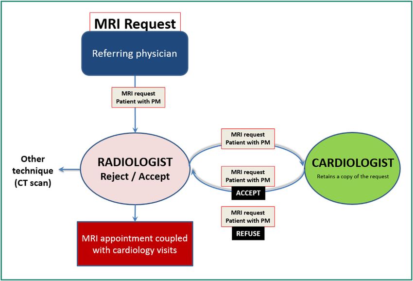
206
Table 1   (Continued)
                Models        Field         Patient    Exclusion zone                 SAR           Slew rate   Spatial        Total          Leads
                                            position                                                            gradient       exposure
                                                                                                                               duration
                                                                                                                               allowed
St JUDE         Assurity      1.5 T         Strict     Consult SJM    Full          Slew             Not        30 min of      Tendril MRI,
  MEDICAL       MRI                         supine     web site       body ≤ 2 W/kg rate ≤ 200 T/m/s specified   active         Tendril STS,
                Accent MRI                  position   Full body      or ≤ 4 W/kg per axis                      imaging by     IsoFlex
                Endurity                               or             based on                                  session.       Optim
                MRI                                    Isocenter of   PM/leads                                  30 min of
                                                       the reel       association                               wait
                                                       excluded       Head < 3.2 W/kg                           between
                                                       between                                                  the sessions
                                                       the eyes (or
                                                       10 cms
                                                       above) and
                                                       the
                                                       vertebra L4
                                                       depends on
                Endurity                               the
                Endurit                                PM/leads
                Core                                   association
                Nanostim      1.5 T         Prone or   Full body                                                Full body ≤    ≤ 19 T/m or    None
                leadless                    supine                                                              4 W/kg         1900
                                            position                                                            Head ≤         gauss/cm
                                                                                                                3.2 W/kg
SORIN           Kora 100      1.5 T         Prone or   Isocenter of
                                            supine     the reel
                                            position   excluded
                                                       between
                                                       the level of
                                                       eyes and
                                                       top of hips

                Full body ≤   Slew rate ≤   Not        40 min of      Beflex
                2 W/kg        200 T/m/s     specified   active         52 cm
                Head <        per axis                 imaging        RF45D — 58 cm

                                                                                                                                                      Y. Cruypeninck et al.
                3.2 W/kg                               except         RF46D
                                                       thorax by
                                                       session.
                                                       20 min
                                                       thoracic
                                                       region
Pacemakers and MRI
Table 1    (Continued)
                  Models          Field           Patient        Exclusion zone                  SAR                 Slew rate       Spatial         Total          Leads
                                                  position                                                                           gradient        exposure
                                                                                                                                                     duration
                                                                                                                                                     allowed
                  Kora 250                                       Full body                       Full
                                                                                                 body ≤ 2 W/kg
                                                                                                 (1 W/kg in
                                                                                                 thoracic region
                                                                                                 if body mass
                                                                                                 index < 23)
                                                                                                 Head < 3.2 W/kg
MEDTRONIC         Revo            1.5 T           Prone or       Isocenter of                    Full body ≤         Slew rate ≤     ≤ 20 T/m or
                                                  supine         the reel                        2 W/kg              200 T/m/s       2000
                                                  position       excluded                        Head < 3.2 W/kg     per axis        gauss/cm
                                                                 between C1
                                                                 et T12
                  Not             CapSureFix
                  specified        Novus
                                  MRITM
                                  SureScan®
                                  5076
                                  (models
                                  5076-35,
                                  5076-45,
                                  5076-
                                  52,5076-58,
                                  5076-65,
                                  5076-85)
                  Enrythm                                        No
                  Advisa                                         restrictions
                  Ensura
                  Micra           1.5 or 3 T                                                     Full                                ≤ 25 T/m or     None
                                                                                                 body ≤ 4 W/kg                       2500
                                                                                                 Head < 3.2 W/kg                     gauss/cm
MRI: magnetic resonance imaging. These recommendations are for illustration only and do not exclude to check accurately the recommendations given by the manufacturers.

                                                                                                                                                                            207
208                                                                                                       Y. Cruypeninck et al.

non-dependent patients are not apparently at less risk than       All three kinds of devices are considered to be relative con-
dependent patients [9].                                           traindications for MRI with a field of not more than 1.5 Tesla.
    For dependent patients, the risk is over-detection result-        The decision to go ahead with MRI must be discussed
ing in inhibition of stimulation (bradycardia, cardiac arrest).   among the physician asking for the scan, the cardiac rhythm
Indeed, the pacemaker can wrongly detect radiofrequency           specialist and the radiologist. These three clinicians should
waves as a signal mimicking cardiac activity and stop stim-       review the indication and consider alternatives to MRI.
ulating the patient’s ventricles. The clinical outcome is             If MRI is to be performed, the following conditions must
bradycardia, or even cardiac arrest.                              be fulfilled without fail:
    For non-dependent patients, the risk is inappropriate         • presence of a clinician at the patient’s bedside during the
asynchronous stimulation that can result in tachycardia or           scan;
ventricular fibrillation. This basically means that the stimu-     • quickly available crash cart and external defibrillator;
lator no longer receives external input and stimulates the        • permanent ECG monitoring during the scan with a MRI-
heart regardless of its real physiological activity.                 compatible ECG system;
    However, many studies have reported successful MRI            • correct pacemaker programming prior to entering the MRI
scans for patients with standard pacemakers without any              environment and pacemaker operation checked before
harm to either the patient or the device. Martin et al.              and after the scan by the cardiologist;
reported a series of 62 MRI scans for 49 patients without         • pacemaker implanted at least 6 weeks before the MRI
any clinical incidents [10]. In the same way, Nazarian et al.        scan;
reported that no complications had occurred during the            • if necessary, chest X-ray to verify the absence of dam-
555 scans they performed on 438 patients with pacemak-               aged, abandoned or epicardial leads;
ers. It should be noted nevertheless that devices implanted       • immediate discontinuation of the scan upon occurrence
before 1998 were excluded. In three cases, the pacemakers            of an incident.
reverted to transient back-up programming mode without
consequences either for the patients or the devices [11]. In          In the AFSSAPS guidelines, 3 Teslas MRI scans were
the study by Roguin et al., no thermal injury was observed        strictly contraindicated. However, certain new generation
and the levels of magnetic attraction and torsion remained        MRI-compatible pacemakers are eligible for 3-Tesla static
low. However, the authors reported that devices manufac-          fields. The American Heart Association (AHA) advises against
tured before 2000 were more subject to damage than more           performing MRI for patients with pacemakers, with a higher-
recent models [12].                                               level contraindication for pacemaker-dependent patients,
    Devices implanted after 2000 are smaller in size and          unless the benefit of MRI be significantly higher than the
contain less ferromagnetic components explaining their            risk [13].
improved MRI-compatibility. So, the date the device was               Pacemaker-dependent and non-dependent patients are
implanted should also be taken into account when making           managed in the same way as regards to MRI (Fig. 1) [14],
decisions. The year 2000, a date that is easy to remember,        the only difference is how the pacemaker is programmed.
is a key element of the decision tree.
    In the same way, the anatomical region that requires
investigation and pacemaker manufacturer recommenda-              Pacemakers and MRI: a strictly monitored
tions should also be taken into account, since the risk is        process
higher if the pacemaker is located in the imaged region.
    Any clinical incident (malaise, palpitations or even          The present protocol for managing pacemaker patients is in
patient asking to stop the scan), electrical anomaly (espe-       line with international guidelines and has been approved by
cially bradycardia or tachycardia) should result in immediate     the executive committee of the Société française d’imagerie
discontinuation of the MRI scan and removal of the patient        cardiovasculaire (SFICV, French society for cardiovascular
from within the magnet as quickly as possible. ECG readout        imaging).
quality should be monitored, in particular during the emis-           MRI scans for patients with pacemakers should only be
sion of radiofrequency waves. ECG artifacts can occur and         considered in public or private hospitals for which the imag-
must not be mistaken for heart rhythm problems. We suggest        ing department and cardiology department, as well as a
using both external monitoring and the imager’s onboard           cardiac emergency unit, are located on the same campus.
monitoring system (ECG, heart rate monitoring).                   MRI-compatible vital signs monitoring equipment, an exter-
                                                                  nal defibrillator and an adequately stocked crash cart must
                                                                  be available in the MRI environment to deal with poten-
                                                                  tial emergency situations. MRI staff must be appropriately
French and international guidelines                               trained in basic resuscitation techniques.
                                                                      Given the significant rise in the number of requests for
In 2005, the French Agency for the Safety of Health Prod-         MRI scans for patients with pacemakers and the relative
ucts (AFSSAPS now known as ANSM) issued guidelines on the         confusion regarding ‘‘MRI-compatible’’ and ‘‘non-MRI-
interactions between MRI and active implantable devices           compatible’’ devices, healthcare facilities should define
(pacemakers, implantable defibrillators, neurostimulators).        standardized procedures for performing MRI scans in
Some of the contraindications for these devices are the           patients with pacemakers, regardless MR compatibility.
same, such as the minimum time after implantation and                 Such procedures should describe responsibilities and
the maximum magnetic field intensity. As far as possible,          resource persons, how the patient is prepared before,
computed tomography (CT) should be preferred over MRI.            during and after the scan, and should provide technical
Pacemakers and MRI 209
Figure 1. Recommendations regarding the use of magnetic resonance imaging (MRI) for patients with implantable cardiac devices. PM:
pacemaker; ICD: implantable cardioverter defibrillator. Adapted from reference [14].
recommendations as regards to how the scan is performed, In order to differentiate requests for patients with
how the patient is monitored and how MRI is prescribed, as pacemakers from the usual MRI request procedure, a spe-
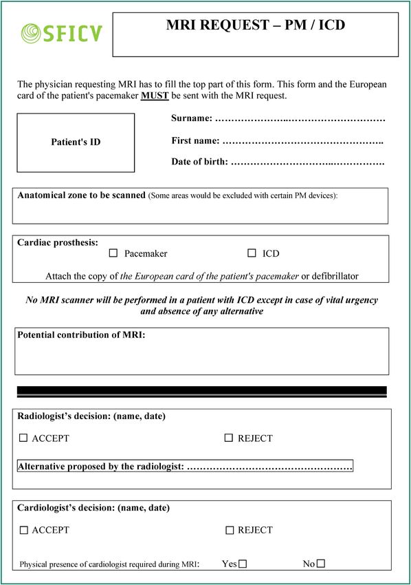
well as information on the patient circuit. cific pacemaker patient form (Fig. 4) should be used and
The document can be made available to all staff using should be processed separately (Fig. 5). Without a specific
a hospital document management system. Drawing up such process, pacemaker patients may make it to their appoint-
procedures requires multidisciplinary cooperation. All the ment only to see the scan postponed and rescheduled
information provided in the procedure must be in line with subsequently.
ANSM and AHA guidelines. The specific request form, to be completed by the physi-
In addition to this procedure, we propose a communica- cian requesting the MRI, will provide specific details as to
tion sheet to be filled in by the cardiac rhythm specialist why MRI is requested and the potential clinical benefit for
(Fig. 2). This sheet is completed when the patient consults the patient. It also involves the requesting physician person-
prior to MRI and is then sent to the imaging department ally in the decision to proceed with MRI. For both cardiac
(Fig. 3). It provides proof that the patient has actually rhythm specialists and radiologists, the form ensures trace-
attended the pre-MRI visit. The cardiac rhythm specialist ability from the very start of the process. It also provides
can also provide, via this sheet, specific recommendations them with the possibility of reviewing the patient record
as to patient care after the MRI scan and before the post-MRI prior to imaging, to discuss the request’s relevance and per-
visit. haps suggest an alternative method. The request must be210 Y. Cruypeninck et al.
Figure 2. Magnetic resonance imaging (MRI)-cardiology communication sheet.
validated sequentially first by the radiologist, then by the • a communication sheet to liaise between the cardiology
cardiologist, before MRI can be performed. department and the medical imaging department.
To summarize, healthcare facilities with a MRI service
should use: The previously described procedure is only feasible as
• a specific MR imaging request form;
part of a scheduled process (non-emergency). In emergency
• a standardized procedure stating all the necessary rec-
cases, tripartite dialogue (requesting physician, cardiolo-
ommendations on prescribing MRI, the patient circuit, gist, and radiologist) is required to assess the benefit—risk
patient care and technical recommendations for scanning; ratio. If MRI is vital, the cardiologist, radiologist and on-callPacemakers and MRI 211
Figure 3. Pacemaker patient circuit on the day of the magnetic resonance imaging (MRI) scan.
MRI operator can proceed with imaging in accordance with are adequate medical grounds for MRI and that it cannot be
our recommendations (see Clinical case). replaced by another imaging modality. He/she monitors the
patient during the scan, limits the acquisition scans to the
strict minimum, and ensures the patient’s safety and compli-
Role of each staff member ance of the procedure with the pacemaker manufacturer’s
recommendations.
The successful management of patients with pacemakers is The MRI technician reviews the cardiac rhythm spe-
conditional upon multidisciplinary collaboration. The multi- cialist’s instructions on the MRI-Cardiology communication
disciplinary team including prescribing physician, radiologist sheet and ensures that appropriate medical attention is
and cardiologist, assesses the benefit—risk ratio and, if available throughout the scan. He/she prepares the MRI-
possible, considers using an alternative modality. The car- compatible monitoring equipment, resuscitation material
diologist evaluates the feasibility of imaging based on the (crash cart) and external defibrillator. During acquisition,
patient’s cardiac status. He/she examines the patients dur- he/she ensures that the SAR is as low as possible (nor-
ing the pre- and post-MRI visits and fills in the MRI-Cardiology mal operating mode or level 0). After the scan, he/she
communication sheet. If necessary, the cardiologist monitors ensures that the patient returns safely to the cardiology
the patient during MRI. The radiologist ensures that there department.212 Y. Cruypeninck et al. Figure 4. Request form for magnetic resonance imaging (MRI) for patients with pacemakers or defibrillators.
Pacemakers and MRI                                                                                                                  213

Figure 5.   Discussion/approval process for requests for magnetic resonance imaging (MRI) for patients with pacemakers or defibrillators.

Conclusion
Although MRI is still nowadays contraindicated for patients
with pacemakers, it may be performed under specific
conditions, following multidisciplinary assessment and orga-
nization of the scan by a three-member team comprising
the radiologist, a cardiac rhythm specialist and the physi-
cian requesting the scan. MRI is facilitated in patients with
the new ‘‘MRI-compatible’’ pacemakers, provided that the
whole device (pacing box and leads) be MRI-compatible.
Nevertheless, MRI scans of patients with such new gener-
ation pacemakers must still be performed with the same
highest level of precaution as for patients with standard
pacemakers. Whatever the case, MRI should only be per-
formed in specialized facilities where all the potentially
required resources are immediately available. In no instance          Figure 6. Antero-posterior chest X-ray prior to magnetic
should MRI of patients with pacemakers be considered as               resonance imaging (MRI) showing the pacemaker and leads (non-
trivial.                                                              compatible).

Clinical case                                                         epidural injury with central iso-signal intensity and high
                                                                      peripheral signal intensity, and T2-weighted images show
This 55-years old man with a non-MRI-compatible pacemaker             widespread high signal intensity from C5 to T2 (Figs. 8 and 9)
(Fig. 6) complains of brutal onset neurological deficit fol-           as well as anterior displacement of the cord with high cen-
lowing recent implementation of anticoagulation therapy.              tromedullar signal intensity. Taken together, these findings
Clinical examination reveals incomplete paresis of all four           suggest subacute compressive epidural hematoma compli-
limbs and a sensory level C6. After discussion between the            cated by spinal cord injury.
prescribing physician, radiologist and cardiologist, MRI of              The patient did not complain about pacemaker heating
the cervical spine is approved. Close cardiac monitoring is           during the procedure. No rhythm or conduction anomalies
ensured during the scan and the patient’s vital signs are             were observed. A neurosurgical approach, guided by MRI,
monitored using a MRI-compatible system.                              was attempted and confirmed diagnosis. Unfortunately, the
   The artifacts caused by the pacing box are clearly visible         patient did not recover neurological function. No cardiac
on the scout image in the axial plane (Fig. 7). T2-weighted           complications were observed, either immediately or some-
images and T1-weighted images of the cervical spine are               time after MRI, for this patient with a non-MRI-compatible
acquired in the sagittal plane. T1-weighted images reveal             pacemaker.
214 Y. Cruypeninck et al.
Figure 7. Metal artifacts caused by the pacing box on the scout
image (arrows).
Figure 9. Magnetic resonance imaging (MRI) of spinal cord:
T1-weighted image in the axial plane. Right posterior epidural
hematoma (arrows) with iso-signal intensity on T1-weighted images
except for the peripheral rim that shows high signal intensity.
Disclosure of interest
The authors declare that they have no competing interest.
References
[1] Ellenbogen KA, Kay GN, Chu-Pak L, Wilkoff BL. Clinical car-
diac pacing, defibrillation, resynchronization therapy. 4th ed.
Philadelphia: Saunders Elsevier; 2011.
[2] Dacher JN, Caudron J. Stimulateur cardiaque et IRM. L’avis du
radiologue. In: Boyer L, Guéret P, editors. Imagerie en coupe du
cœur et des vaisseaux. Paris: Springer-Verlag; 2013. p. 23—8.
[3] The Organisation For Economic Co-Operation and Devel-
opment (OECD). Magnetic Resonance Imaging (MRI) exams
in Hospitals. Health/ Key Tables from the OECD; 2014
http://www.oecd-ilibrary.org/social-issues-migration-health/
examens-avec-imagerie-par-resonance-magnetique-irm-total-
2014-1 mri-exam-total-table-2014-1-fr.
[4] Sakakibara Y, Mitsui T. Concerns about sources of electromag-
netic interference in patients with pacemakers. Jpn Heart J
1999;40:737—43.
[5] Roguin A, Schwitter J, Vahlhaus C, et al. Magnetic resonance
imaging in individuals with cardiovascular implantable elec-
Figure 8. Magnetic resonance imaging (MRI) of spinal cord: mid- tronic devices. Europace 2008;10:336—46.
line T2-weighted image in the sagittal plane. Posterior epidural [6] Barral M, Cornud F, Neuzillet Y, et al. Characteristics of unde-
hematoma (arrows). tected prostate cancer on diffusion-weighted MR imaging at
3-Tesla with a b-value of 2000 s/mm2 : imaging-pathologic cor-
relation. Diagn Interv Imaging 2015;96:923—9.
[7] Dallaudière B, Lecouvet F, Vande Berg B, et al. Diffusion-
weighted MR imaging in musculoskeletal diseases: current
concepts. Diagn Interv Imaging 2015;96:327—40.
[8] Wilkoff BL, Bello D, Taborsky M, et al. Magnetic resonance
imaging in patients with a pacemaker system designed for the
MR environment. Heart Rhythm 2011;8:65—73.Pacemakers and MRI 215
[9] Irnich W, Irnich B, Bartsch C, Stertmann WA, Gufler H, Weiler imaging safe: in vitro and in vivo assessment of safety and
G. Do we need pacemakers resistant to magnetic resonance function at 1.5 T. Circulation 2004;110:475—82.
imaging? Europace 2005;7:353—65. [13] Hundley WG, Bluemke DA, Finn JP, et al. ACCF/ACR/
[10] Martin ET, Coman JA, Shellock FG, Pulling CC, Fair R, Jenkins AHA/NASCI/SCMR 2010 expert consensus document on cardio-
K. Magnetic resonance imaging and cardiac pacemaker safety vascular magnetic resonance: a report of the American College
at 1.5-Tesla. J Am Coll Cardiol 2004;43:1315—24. of Cardiology Foundation Task Force on Expert Consensus Doc-
[11] Nazarian S, Hansford R, Roguin A, et al. A prospective eval- uments. J Am Coll Cardiol 2010;55:2614—62.
uation of a protocol for magnetic resonance imaging of [14] Brignole M, Auricchio A, Baron-Esquivias G, et al. 2013 ESC
patients with implanted cardiac devices. Ann Intern Med guidelines on cardiac pacing and cardiac resynchronization
2011;155:415—24. therapy: the task force on cardiac pacing and resynchronization
[12] Roguin A, Zviman MM, Meininger GR, Rodrigues ER, Dickfeld therapy of the European Society of Cardiology (ESC). Developed
TM, Bluemke DA, et al. Modern pacemaker and implantable in collaboration with the European Heart Rhythm Association
cardioverter/defibrillator systems can be magnetic resonance (EHRA). Europace 2013;15:1070—118.You can also read