Choroidal and peripapillary changes in high myopic eyes with Stickler syndrome
←
→
Page content transcription
If your browser does not render page correctly, please read the page content below
Xerri et al. BMC Ophthalmology      (2021) 21:2
https://doi.org/10.1186/s12886-020-01777-3

 RESEARCH ARTICLE                                                                                                                                  Open Access

Choroidal and peripapillary changes in high
myopic eyes with Stickler syndrome
Olivia Xerri1†, Federico Bernabei2†, Elise Philippakis3, Cyril Burin-Des-Roziers4,2, Pierre-Olivier Barale5, Olivier Laplace5,
Claire Monin5, Dominique Bremond-Gignac1,4, Gilles Guerrier6, Sophie Valleix4,7, Antoine Brezin4,2 and
Pierre-Raphaël Rothschild4,2*

  Abstract
  Background: To compare different clinical and Spectral-Domain Optical Coherence Tomography (SD-OCT)
  features of high myopic eyes with Stickler syndrome (STL) with matched controls.
  Methods: Patients with genetically confirmed STL with axial length ≥ 26 mm and controls matched for axial length
  were included. The following data were obtained from SD-OCT scans and fundus photography: choroidal and
  retinal thickness (respectively, CT and RT), peripapillary atrophy area (PAA), presence of posterior staphyloma (PS).
  Results: Twenty-six eyes of 17 patients with STL and 25 eyes of 19 controls were evaluated. Compared with
  controls, patients with STL showed a greater CT subfoveally, at 1000 μm from the fovea at both nasal and temporal
  location, and at 2000 and 3000 μm from the fovea in nasal location (respectively, 188.7±72.8 vs 126.0±88.7 μm,
  172.5±77.7 vs 119.3±80.6 μm, 190.1±71.9 vs 134.9±79.7 μm, 141.3±56.0 vs 98.1±68.5 μm, and 110.9±51.0 vs 67.6±
  50.7 μm, always P< 0.05). Furthermore, patients with STL showed a lower prevalence of PS (11.5% vs 68%, P< 0.001)
  and a lower PAA (2.2±2.1 vs 5.4±5.8 mm2, P=0.03), compared with controls.
  Conclusions: This study shows that high myopic patients with STL show a greater CT, a lower PAA and a lower
  prevalence of PS, compared with controls matched for axial length. These findings could be relevant for the
  development and progression of myopic maculopathy in patients with STL.
  Keywords: Choroidal thickness, Congenital myopia, Hereditary vitreopathy, High myopia, Stickler syndrome

Background                                                                            collagen, namely II, IX, and XI [3–6]. The most com-
Stickler syndrome (STL) is an inherited connective tis-                               mon pathogenic variants associated with STL are found
sue disorder, that leads to a broad spectrum of manifes-                              in the COL2A1 and COL11A1 gene, that account re-
tations including facial, skeletal, ear, and ocular                                   spectively for the 80–90% and 10–20% of cases [7].
abnormalities [1, 2]. The disease has an estimated inci-                              Common ocular findings include congenital myopia,
dence of 1: 7.500 to 9.000 newborns and is caused by                                  vitreous abnormalities, and early onset cataract [2].
mutations in the genes encoding for different types of                                Furthermore, the disease represents a serious sight-
                                                                                      threating condition, due to a high risk of developing
                                                                                      retinal detachment that seems to be related to an ab-
* Correspondence: pierreraphaelrothschild@hotmail.com
†
 Olivia Xerri and Federico Bernabei are the Authors contributed equally to            normal vitreoretinal interface as well as a complication
the work and should be considered co-first Authors.
4
                                                                                      of myopia [8, 9].
 Université de Paris, Centre de Recherche des Cordeliers, INSERM, UMR_1138,
                                                                                        Few studies have reported the prevalence of high
F-75006 Paris, France
2
 Service d’Ophtalmologie, Ophtalmopôle de Paris, Hôpital Cochin, AP-HP, 27            myopia (HM) in STL, and it is estimated to occur in 76
rue du Faubourg Saint Jacques, 75014 Paris, France                                    to 80% of patients [8, 10]. Moreover HM is typically
Full list of author information is available at the end of the article

                                       © The Author(s). 2021 Open Access This article is licensed under a Creative Commons Attribution 4.0 International License,
                                       which permits use, sharing, adaptation, distribution and reproduction in any medium or format, as long as you give
                                       appropriate credit to the original author(s) and the source, provide a link to the Creative Commons licence, and indicate if
                                       changes were made. The images or other third party material in this article are included in the article's Creative Commons
                                       licence, unless indicated otherwise in a credit line to the material. If material is not included in the article's Creative Commons
                                       licence and your intended use is not permitted by statutory regulation or exceeds the permitted use, you will need to obtain
                                       permission directly from the copyright holder. To view a copy of this licence, visit http://creativecommons.org/licenses/by/4.0/.
                                       The Creative Commons Public Domain Dedication waiver (http://creativecommons.org/publicdomain/zero/1.0/) applies to the
                                       data made available in this article, unless otherwise stated in a credit line to the data.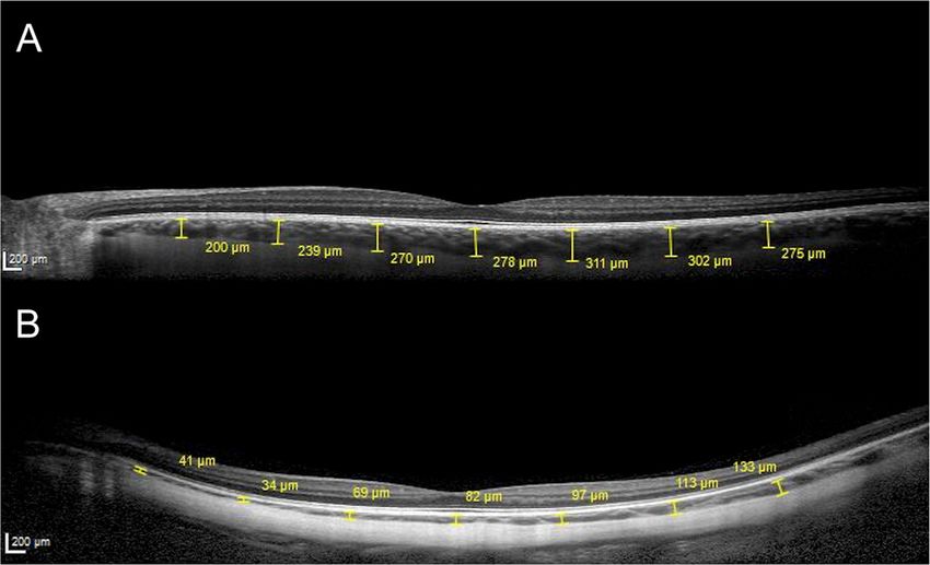
Xerri et al. BMC Ophthalmology   (2021) 21:2                                                                     Page 2 of 7

present at birth and has a non-progressive course [11].        from medical records. Furthermore, in order to evaluate
This is a highly distinctive feature of the disease, because   the status of the retina and the choroid in the absence of
it is often the only manifestation during infancy. In idio-    macular complications related to myopia, eyes with
pathic HM the elongation of the axial length and the           lacquer cracks, myopic CNV, myopic traction maculopa-
progressive increase in the curvature of the posterior         thy, dome-shaped macula and macular hole were
pole occurs later in life and are associated with the de-      excluded from both groups.
velopment of sight-threatening chorioretinal complica-
tions including choroidal neovascularization (CNV),            Data collection
myopic traction maculopathy and macular hole [12, 13].         The following data were extrapolated from medical
To date, except for retinal detachment, no other severe        records: age, sex, axial length (IOL Master700®, Carl
complications related to HM have been described in             Zeiss Meditec, Jena, Germany), lens status, fundus pho-
patients with STL. The improvements in spectral do-            tography of the posterior pole encompassing optic nerve
main - optical coherence tomography (SD-OCT) tech-             and macula (Canon CR2 plus AF®, Canon, Tokyo, Japan
nology, such as enhanced-depth imaging (EDI) mode,             and/or Optos® California, Optos, Marlborough, MA,
allow a better visualization of retinal and choroidal          USA), SD-OCT scans and infra-red (IR), fundus images
structures along with a more precise characterization of       (Spectralis®, Heidelberg Engineering Inc., Heidelberg,
quantitative parameters such as choroidal and retinal          Germany).
thickness (respectively, CT and RT), [14]. Thus, the aim         SD-OCT horizontal 30° line scan, passing through the
of this study was to investigate clinical and SD-OCT           fovea, were acquired with EDI and high-resolution
characteristics of highly myopic eyes in patients with         mode, obtaining an average of 60 scans with a quality
genetically confirmed STL and to compare them with             rate superior to 25. On SD-OCT scan, CT was defined
those of highly myopic patients without STL.                   as the vertical distance from the hyperreflective line of
                                                               the Bruch’s membrane to the hyperreflective line of the
Methods                                                        inner surface of the sclera. CT at the sub-foveal location
Design and patients                                            and at 1000, 2000 and 3000 μm from the fovea, in the
This retrospective observational study was conducted in        nasal and temporal locations were measured.
the setting of a collaborative project named French              On SD-OCT scan, total RT was defined as the vertical
vitreoretinopathy study group (FVSG). Patients were            distance from the hyperreflective line of the vitreoretinal
identified from databases of the retina service of differ-     interface to the hyperreflective line of the retinal pig-
ent tertiary eye care centers and were subsequently            ment epithelium. RT at the fovea, at the nasal and tem-
examined at least once by one of the authors (PRR) at          poral clivus and at 3000 μm from the fovea in the nasal
the OphtalmoPole de Paris, Hôpital Cochin (Paris,              and temporal locations were measured. The peripapillary
France). Referring hospitals included: Hôpital des             atrophy area (PPA) was obtained using a previously
Quinze-Vingts (Paris, France), Hôpital Lariboisière,           described modified technique [15]. In brief, the optic
(Paris, France) and Hôpital Necker-Enfants Malades,            nerve area was subtracted to the area bounded by the
(Paris, France). Institutional review board approvals for      edge of atrophy based on IR images [15]. All the mea-
retrospective chart reviews were obtained commensurate         surements were performed independently by two oph-
with the respective institutional requirements prior to        thalmologists (OX and FB), both blinded to patient’s
the beginning of the study. Described research was             characteristics, by using the built-in calipers of the soft-
approved by the ethics committee of the French society         ware. The average of the 2 measurements was used for
of ophthalmology and adhered to the tenets of the dec-         analysis.
laration of Helsinki. Fully written informed consent was         The presence and the location of posterior staphy-
obtained for all patients. Patients with genetically con-      loma (PS) were evaluated by both fundus photography
firmed STL were identified at the retina service of the        and SD-OCT scan and classified as follows: 0) absence
participating centers and those with HM, defined by the        of PS, i) PS with macular involvement, ii) PS without
presence of an axial length of 26 mm or longer, were in-       macular involvement, iii) other [16].
cluded in the study group. Subjects with HM, not sus-
pected to have SLT based on family history and on the          Statistical analysis
absence of ocular and extraocular features, were               Data analysis was conducted with XLSTAT Version
matched for axial length and included as controls. Exclu-      2017.02.43358 (Addinsoft, Paris, France). Quantitative
sion criteria for both groups were as follows: history of      data are expressed as mean ± standard deviation (SD)
retinal detachment and other retinal diseases, glaucoma,       and qualitative data are expressed as percentages, with a
any previous retinal laser photocoagulation or surgical        confidence interval of 95% [CI 95%]. The Shapiro-Wilk’s
procedure except for cataract surgery and missing data         test was used to assess normality of data. An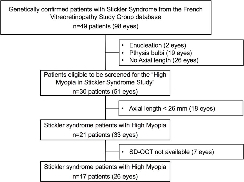
Xerri et al. BMC Ophthalmology (2021) 21:2 Page 3 of 7
independent sample t-test was used to compare nor- presented HM. Seven eyes were subsequently excluded
mally distributed variables between two groups, while from this cohort for the lack of SD-OCT scans. Finally,
Mann-Whitney U test was used for not normally distrib- 26 eyes of 17 patients with HM and genetically con-
uted variables. Qualitative variables were compared be- firmed SLT were included (STL group). The flow chart
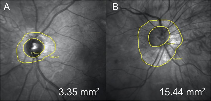
tween the two groups using chi-square test or Fisher of the inclusion process is represented in Fig. 1.
exact test when necessary. The correlations of sub-foveal The demographic and clinical characteristics of
CT with age, axial length, foveal RT, and PPA, and of patients with STL and control subjects are reported in
PPA with age and axial length were examined using Table 1.
Pearson’s correlation analysis. A P value < 0.05 was con- No significant differences were found in axial length
sidered statistically significant. between the two groups (P > 0.05). A significantly lower
prevalence of PS was found in STL group compared
Results with control group (P < 0.01). In particular, PS was
Forty-nine patients (98 eyes) with genetically confirmed present in 3 eyes (11.5% [0–23.8]) of the STL group and
STL were identified from the FVSG database. All the in 17 eyes (68.0% [49.7–86.3]) of the control group. In
cases a had a positive family history for STL and all had the 3 eyes of STL group PS involved the macula, while
a pathogenic mutation in the COL2A1 gene. Forty-seven in the control group, 13 eyes presented PS that involved
eyes were excluded for the following reasons: enucle- the macula, and in 4 eyes PS involved the peripapillary
ation (n = 2), phthisis bulbi (n = 19), absence of axial region.
length measurement (n = 26). Of the remaining 30 The choroidal and retinal parameters of patients with
patients (51 eyes), 21 (70.0%, [56.6–86.4]) (33 eyes) STL and control subjects are reported in Table 2.
Fig. 1 Inclusion flow chart of patients with Stickler syndrome and high myopiaXerri et al. BMC Ophthalmology (2021) 21:2 Page 4 of 7
Table 1 Demographic and clinical characteristics of patients P > 0.05). A significantly lower value of mean PPA
with stickler syndrome and control subjects was found in patients with STL compared to controls
Characteristics Stickler group Control group P (2.2 ± 2.1 vs. 5.4 ± 5.8 mm2, P = 0.03) (Fig. 3).
Patients (n) 17 19 In the two groups sub-foveal CT showed a significant
Eyes (n) 26 25 correlation with age (R= − 0.3, P = 0.02), axial length
(R= − 0.7, P < 0.01) and PPA (R = − 0.5, P < 0.01). In
Phakic (n) 11 18
addition, PPA showed a significant correlation with axial
Sex (m/f) 7/10 11/8 0.3
length (R=0.5, P < 0.01).
Age (years ± SD) 34.5 ±13 39.5 ±11.1 0.16
Axial Length (mm ± SD) 28.0 ± 2.0 29.2 ± 2.5 0.07 Discussion
SD Standard deviation In the present study we evaluated different SD-OCT
parameters and clinical characteristics in high myopic
patients with STL and we compare them with controls,
In brief, mean CT at the sub-foveal location was matched for axial length. Surprisingly, in the STL group,
higher in patients with STL compared to control sub- eyes presented a lower prevalence of PS compared with
jects (188.7 ± 72.8 vs. 126.0 ± 88.7 μm, P = 0.01) (Fig. 2). the control group, that showed values that are in agree-
Moreover, significantly higher values of mean CT ment with the literature [17]. In addition, patients with
measured at 1000 μm from the fovea at both nasal STL presented a significantly greater CT subfoveally and
and temporal location and at 2000 and 3000 μm from in all the analyzed locations, except for the two more
the fovea in nasal location, were found in the STL distant from the fovea at the temporal side.
group compared to control group (respectively, 172.5 The development of myopia is typically associated with
± 77.7 vs. 119.3 ± 80.6 μm, P = 0.03; 190.1 ± 71.9 vs. a progressive increase of axial length along with the
134.9 ± 79.7 μm, P = 0,03; 141.3 ± 56 vs. 98.1 ± myopic refractive error [18]. The association of HM with
68.5 μm, P = 0.02 and 110.9 ± 51 vs. 67.6 ± 50.7 μm, a thin CT has been well established and it seems to be
P = 0.01). Conversely, no significant differences were in part related to the increased axial length [19]. How-
found between the two groups in mean CT at 2000 ever, a recent meta-analysis failed to prove axial length
and 3000 μm from the fovea in the temporal location as an independent risk factor for the thinning of the
and in RT at all measured locations including the choroid, suggesting the role of other variables in the CT
fovea, the nasal and temporal clivus and the 3000 μm decreasing process [20]. Interestingly, a recent report
from the fovea in the nasal and temporal location (all from the Beijing Eye Study cohort, showed that the
Table 2 Choroidal and retinal parameters of patients with stickler syndrome and control subjects
Parameter Stickler group Control group P
Choroidal Thickness (μm)
Sub-foveal 188.7 ± 72.8 126.0 ± 88.7 0.01
Nasal 1000a 172.5 ± 77.7 119.3 ± 80.6 0.03
Nasal 2000 141.3 ± 56.0 98.1 ± 68.5 0.02
Nasal 3000 110.9 ± 51.0 67.6 ± 50.7 0.01
Temporal 1000 190.1 ± 71.9 134.9 ± 79.7 0.03
Temporal 2000 182.8 ± 61.3 140.8 ± 76.0 0.06
Temporal 3000 186.0 ± 62.6 142.9 ± 78.5 0.08
Total Retinal Thickness (μm)
Foveal 224.5 ± 43.9 230.9 ± 36.8 0.26
Nasal clivus 313.4 ± 68.8 325.6 ± 42.8 0.82
Nasal 3000 265.9 ± 41.4 264.8 ± 50.1 0.93
Temporal clivus 299.8 ± 62.0 318.5 ± 36.2 0.42
Temporal 3000 237.0 ± 38.6 244.8 ± 32.0 0.76
Peripapillary atrophy (mm2) 2.2 ± 2.1 5.4 ± 5.8 0.03
Posterior Staphyloma (n (%)) 3 (11.5% [0–23.8]) 17 (68.0% [49.7–86.3]) < 0.001
SD Standard deviation. a Denotes the position 1000 μm nasal to the fovea. The same naming convention is used for the subsequent entries. Significant P values
(< 0.05) are in boldXerri et al. BMC Ophthalmology (2021) 21:2 Page 5 of 7 Fig. 2 Representative macular SD-OCT scans of a 36 years old male with Stickler syndrome, presenting an axial length of 27.3 mm (a) and of 35 years old male with high myopia, presenting an axial length of 28.1 and posterior staphyloma (b). In both patients, choroidal thickness measurements were performed at sub-foveal location and at 1000, 2000 and 3000 μm from the fovea, in the nasal and temporal locations presence of PS was the most important factor affecting preserved choroid and a lower prevalence of PS. In CT in highly myopic patients [21]. Our results are in addition, Ellabban and collaborators investigated the CT agreement with those of Zhou and coauthors, supporting in eyes with PS involving the macula and found a thin- the strong association between these two parameters ning of the choroid in the whole macular area except for [21]. In particular, we found that control subjects pre- a relatively well-preserved choroid in the temporal area, sented a high prevalence of PS along with a thinner CT, that seemed to be less prone to be affected by PS [22]. while patients with STL showed a relatively well- This finding could help explain the absence of difference Fig. 3 Representative infra-red optic nerve images of 34 years old female with Stickler syndrome (a) and of 33 years old female enrolled as control (b). Peripapillary atrophy area were respectively 3.35 mm2 and 15.44 mm2
Xerri et al. BMC Ophthalmology   (2021) 21:2                                                                                      Page 6 of 7

in CT in the area temporal to the fovea between STL           confirmation for all patients with STL, the sample size is
and control group, given that, in almost the totality of      considerable. Future studies with larger sample size may
patients, PS was localized at the posterior pole.             warrant further consideration. Finally, the absence of
   It is recognized that both PS and a thin choroid are       genetic testing in the control group is another limit.
risk factors for the development of complications related     However, the exclusion of subjects with family history or
to HM [23, 24]. Several studies have indeed reported          any manifestations of STL makes it unlikely that these
that the presence of PS and choroidal thinning are asso-      patients have been included as controls.
ciated with a higher rate of diffuse chorioretinal atrophy,
CNV and lacquer cracks in eyes with HM and are con-           Conclusions
sidered as predictive markers for retinal complications       In conclusion, highly myopic patients with STL showed
[20, 23–25]. Furthermore, it is now admitted that the         a significantly thicker choroid along with a lower PAA
presence of PS is associated with the development of          and a lower prevalence of PS compared with controls,
myopic macular lesions and that it is a cause of pro-         after accounting for axial length. Further prospective
gression of myopic maculopathy rather than a conse-           studies are required to better characterize the features of
quence [16].                                                  myopia in patients with STL. These findings help to
   To the best of our knowledge, no complications             shed light on the pathogenetic mechanisms underlying
related to myopic maculopathy have been described in          pathologic myopia and could be relevant for the devel-
the setting of STL. The low rate of PS and the relatively     opment and progression of myopic maculopathy in
well-preserved choroid might help explain the apparent        patients with STL.
absence of macular complications related to HM in this
                                                              Abbreviations
peculiar subset of patients.                                  CT: Choroidal thickness; CNV: Choroidal neovascularization; EDI: Enhanced-
   Although there is no proven explanation for the lower      depth imaging; FVSG: French vitreoretinopathy study group; HM: High
rate of PS and the subsequent preservation of the chor-       myopia; IR: Infra-red; PPA: Peripapillary atrophy area; PS: Posterior staphyloma;
                                                              RT: Retinal thickness; SD-OCT: Spectral domain - optical coherence
oid, one of the main differences between STL and non-         tomography; STL: Stickler syndrome
STL patients resides in the age of onset of the elongation
of the eye. Indeed, patients with STL typically born with     Acknowledgements
HM, the so-called “congenital myopia”. Conversely, in         We thank the Association pour l’enseignement et la recherche en
                                                              ophtalmologie (AERO) for the support to this study as well as the members
non-STL subjects the elongation of the eye occurs sev-        of the French Vitreoretinopathy Study Group (FVRSG) and family members
eral years after birth, commonly at the school age [18].      for their participation. We also thank Valérie Beunardeau from the
It can be speculated that the elongation of an immature       Bibliothèque Javal - Centre de Documentation Ophtalmologique de la
                                                              Société Française d’Ophtalmologie for reference management assistance.
tissue in utero may result in a better tissue-adaptation.
Conversely, in a post-mature state, a worse adaptation        Authors’ contributions
could occur, leading to scleral weakness (staphyloma)         OX: Conceptualization, Methodology, Writing - Original Draft, Validation. FB:
                                                              Conceptualization, Methodology, Writing - Original Draft, Data curation,
and to a subsequent choroidal thinning and eventually         Validation. EP: Investigation, Writing. CBDR: Investigation. POB: Investigation,
to complications such as lacquer cracks and CNV.              Formal analysis. OL: Investigation. CM: Investigation. DBG: Investigation. SV:
   In line with this hypothesis, we also found that patient   Investigation, Review & Editing. GG: Writing - Review & Editing, Validation,
                                                              Supervision, Data curation. AB: Review & Editing, Supervision, Project
with STL presented a lower PPA. Several studies have          administration. PRR: Writing - Review & Editing, Validation, Supervision, Data
suggested that PPA could represent a reliable marker for      curation. All authors read and approved the final manuscript.
monitoring the progression of HM and it has been
                                                              Funding
shown that PPA is positively correlated with age and          None.
axial length [15, 26]. Our results in agreement with
those of Liu et al. also showed a correlation between         Availability of data and materials
                                                              The datasets used and/or analysed during the current study are available
PPA and axial length [15]. No difference was found in         from the corresponding author on reasonable request.
RT in all the measured locations between STL and con-
trol group. These results are in keeping with a previous      Ethics approval and consent to participate
study suggesting that RT is not influenced by CT [27].        Described research was approved by the ethics committee of the French
                                                              society of ophthalmology and adhered to the tenets of the declaration of
   We acknowledge some limitations in the design of our       Helsinki.
study. First, a notable number of eyes were excluded for
enucleation or phthisis bulbi, presumably due to retinal      Competing interests
                                                              The authors declare that they have no competing interests.
detachment. It can not be excluded that they may also
differ from other STL eyes in posterior pole findings.        Author details
                                                              1
Second, the small sample size represents other limita-         Service d’Ophtalmologie, Hôpital Necker-Enfants Malades, AP-HP, F-75014
                                                              Paris, France. 2Service d’Ophtalmologie, Ophtalmopôle de Paris, Hôpital
tion. However, given the rarity of the disease and the        Cochin, AP-HP, 27 rue du Faubourg Saint Jacques, 75014 Paris, France.
stringent inclusion criteria along with the genetic           3
                                                               Service d’Ophtalmologie, Hôpital Lariboisière, AP-HP, F-75014 Paris, France.
Xerri et al. BMC Ophthalmology (2021) 21:2 Page 7 of 7
4
Université de Paris, Centre de Recherche des Cordeliers, INSERM, UMR_1138, myopia. Am J Ophthalmol. 2008;146:102–10. https://doi.org/10.1016/j.ajo.
F-75006 Paris, France. 5Service d’Ophtalmologie, Hôpital des Quinze-Vingts, 2008.03.010.
Paris, France. 6Anaesthetic and Intensive Care Department, Hôpital Cochin, 18. Morgan IG, Ohno-Matsui K, Saw SM. Myopia. Lancet. 2012;379:1739–48.
Paris Descartes university, 75014 Paris, France. 7Laboratoire de Génétique https://doi.org/10.1016/S0140-6736(12)60272-4.
Moléculaire, Faculté de Médecine Paris, Hôpital Necker-Enfants Malades, 19. Hirata M, Tsujikawa A, Matsumoto A, Hangai M, Ooto S, Yamashiro K, et al.
Université de Paris, AP-HP; Inserm, U_1163, Institut IMAGINE, F-75014 Paris, Macular Choroidal thickness and volume in Normal subjects measured by
France. swept-source optical coherence tomography. Invest Opthalmol Vis Sci.
2011;52:4971. https://doi.org/10.1167/iovs.11-7729.
Received: 1 October 2020 Accepted: 15 December 2020 20. Wang S, Wang Y, Gao X, Qian N, Zhuo Y. Choroidal thickness and high
myopia: a cross-sectional study and meta-analysis. BMC Ophthalmol. 2015;
15:70. https://doi.org/10.1186/s12886-015-0059-2.
21. Zhou LX, Shao L, Xu L, Wei WB, Wang YX, You QS. The relationship
References between scleral staphyloma and choroidal thinning in highly myopic eyes.
1. Stickler GB, Belau PG, Farrell FJ, Jones JD, Pugh DG, Steinberg AG, et al. The Beijing Eye Study. Sci Rep. 2017;7:9825. https://doi.org/10.1038/s41598-
Hereditary progressive arthro-ophthalmopathy. Mayo Clin Proc. 1965;40: 017-10660-z.
433–55 http://www.ncbi.nlm.nih.gov/pubmed/14299791. Accessed 25 22. Ellabban AA, Tsujikawa A, Matsumoto A, Yamashiro K, Oishi A, Ooto S, et al.
Aug 2019. Macular Choroidal thickness measured by swept source optical coherence
2. Donoso LA, Edwards AO, Frost AT, Ritter R, Ahmad N, Vrabec T, et al. Clinical tomography in eyes with inferior posterior Staphyloma. Invest Opthalmol
variability of Stickler syndrome: role of exon 2 of the collagen COL2A1 Vis Sci. 2012;53:7735. https://doi.org/10.1167/iovs.12-9952.
gene. Surv Ophthalmol. 2003;48:191–203 http://www.ncbi.nlm.nih.gov/ 23. Wang N-K, Lai C-C, Chou CL, Chen Y-P, Chuang L-H, Chao A-N, et al.
pubmed/12686304. Accessed 25 Aug 2019. Choroidal thickness and biometric markers for the screening of lacquer
3. Printzlau A, Andersen M. Pierre Robin sequence in Denmark: a retrospective cracks in patients with high myopia. PLoS One. 2013;8:e53660. https://doi.
population-based epidemiological study. Cleft Palate Craniofacial J. 2004;41: org/10.1371/journal.pone.0053660.
47–52. https://doi.org/10.1597/02-055. 24. Steidl SM, Pruett RC. Macular complications associated with posterior
4. Van Camp G, Snoeckx RL, Hilgert N, van den Ende J, Fukuoka H, Wagatsuma staphyloma. Am J Ophthalmol. 1997;123:181–7. https://doi.org/10.1016/
M, et al. A new autosomal recessive form of Stickler syndrome is caused by s0002-9394(14)71034-7.
a mutation in the COL9A1 gene. Am J Hum Genet. 2006;79:449–57. https:// 25. Ohno-Matsui K, Lai TYY, Lai C-C, Cheung CMG. Updates of pathologic
doi.org/10.1086/506478. myopia. Prog Retin Eye Res. 2016;52:156–87. https://doi.org/10.1016/j.
5. Richards A, Yates JR, Williams R, Payne SJ, Pope FM, Scott JD, et al. A family preteyeres.2015.12.001.
with Stickler syndrome type 2 has a mutation in the COL11A1 gene 26. Hayashi K, Ohno-Matsui K, Shimada N, Moriyama M, Kojima A, Hayashi W,
resulting in the substitution of glycine 97 by valine in alpha 1 (XI) collagen. et al. Long-term pattern of progression of myopic maculopathy.
Hum Mol Genet. 1996;5:1339–43. https://doi.org/10.1093/hmg/5.9.1339. Ophthalmology. 2010;117:1595–1611.e4. https://doi.org/10.1016/j.ophtha.
6. Baker S, Booth C, Fillman C, Shapiro M, Blair MP, Hyland JC, et al. A loss of 2009.11.003.
function mutation in the COL9A2 gene causes autosomal recessive Stickler 27. Ikuno Y, Tano Y. Retinal and Choroidal biometry in highly myopic eyes with
syndrome. Am J Med Genet Part A. 2011;155:1668–72. https://doi.org/10. spectral-domain optical coherence tomography. Invest Opthalmol Vis Sci.
1002/ajmg.a.34071. 2009;50:3876. https://doi.org/10.1167/iovs.08-3325.
7. Richards AJ, McNinch A, Martin H, Oakhill K, Rai H, Waller S, et al. Stickler
syndrome and the vitreous phenotype: mutations in COL2A1 and COL11A1. Publisher’s Note
Hum Mutat. 2010;31:E1461–71. https://doi.org/10.1002/humu.21257. Springer Nature remains neutral with regard to jurisdictional claims in
8. Parma ES, Körkkö J, Hagler WS, Ala-Kokko L. Radial perivascular retinal published maps and institutional affiliations.
degeneration: a key to the clinical diagnosis of an ocular variant of Stickler
syndrome with minimal or no systemic manifestations. Am J Ophthalmol.
2002;134:728–34. https://doi.org/10.1016/S0002-9394(02)01646-X.
9. Fincham GS, Pasea L, Carroll C, McNinch AM, Poulson AV, Richards AJ, et al.
Prevention of retinal detachment in Stickler syndrome. Ophthalmology.
2014;121:1588–97. https://doi.org/10.1016/j.ophtha.2014.02.022.
10. Matsushita I, Nagata T, Hayashi T, Kimoto K, Kubota T, Ohji M, et al. Foveal
hypoplasia in patients with Stickler syndrome. Ophthalmology. 2017;124:
896–902. https://doi.org/10.1016/j.ophtha.2017.01.046.
11. Popkin JS, Polomeno RC. Stickler’s syndrome (hereditary progressive arthro-
ophthalmopathy). Can Med Assoc J. 1974;111:1071–6 http://www.ncbi.nlm.
nih.gov/pubmed/4429933. Accessed 1 Sep 2019.
12. Tseng G-L, Chen C-Y. Evaluation of high myopia complications prevention
program in university freshmen. Medicine. 2016;95:e5093. https://doi.org/10.
1097/MD.0000000000005093.
13. Lichtwitz O, Boissonnot M, Mercié M, Ingrand P, Leveziel N. Prevalence of
macular complications associated with high myopia by multimodal
imaging. J Fr Ophtalmol. 2016;39:355–63. https://doi.org/10.1016/j.jfo.
2015.11.005.
14. Margolis R, Spaide RF. A pilot study of enhanced depth imaging optical
coherence tomography of the choroid in Normal eyes. Am J Ophthalmol.
2009;147:811–5. https://doi.org/10.1016/j.ajo.2008.12.008.
15. Liu W, Gong L, Li Y, Zhu X, Stewart JM, Wang C. Peripapillary atrophy in
high myopia. Curr Eye Res. 2017;42:1308–12. https://doi.org/10.1080/
02713683.2017.1307992.
16. Ohno-Matsui K, Kawasaki R, Jonas JB, Cheung CMG, Saw S-M, Verhoeven
VJM, et al. International photographic classification and grading system for
myopic maculopathy. Am J Ophthalmol. 2015;159:877–883.e7. https://doi.
org/10.1016/j.ajo.2015.01.022.
17. Hsiang HW, Ohno-Matsui K, Shimada N, Hayashi K, Moriyama M, Yoshida T,
et al. Clinical characteristics of posterior staphyloma in eyes with pathologicYou can also read