MiRNA-1183-targeted regulation of Bcl-2 contributes to the pathogenesis of rheumatic heart disease
←
→
Page content transcription
If your browser does not render page correctly, please read the page content below
Bioscience Reports (2020) 40 BSR20201573
https://doi.org/10.1042/BSR20201573

Research Article

miRNA-1183-targeted regulation of Bcl-2 contributes
to the pathogenesis of rheumatic heart disease
Ni Li1,2,* , Linwen Zhu1,* , Hua Zhou1 , Dawei Zheng1 , Guodong Xu1 , Lebo Sun1 , Jianqing Gao2 and
   Guofeng Shao1
1 Departmentof Cardiothoracic Surgery, Ningbo Medical Centre Lihuili Hospital, Ningbo University, Ningbo, Zhejiang 315041, China; 2 Institute of Pharmaceutics, College of

                                                                                                                                                                                           Downloaded from http://portlandpress.com/bioscirep/article-pdf/40/11/BSR20201573/896723/bsr-2020-1573.pdf by guest on 20 November 2020
Pharmaceutical Sciences, Zhejiang University, Hangzhou, China
Correspondence: Jianqing Gao (gaojianqing@zju.edu.cn) or Guofeng Shao (sgf1958@sina.com)

                                           To determine whether up-regulation of miR-1183 targeting the gene for anti-apoptotic factor,
                                           B-cell lymphoma 2 (BCL-2) contributes to apoptosis in patients with rheumatic heart disease
                                           (RHD). Peripheral blood samples were isolated for miR-1183 characterization. The function
                                           of miRNA-1183 in RHD using miRNA mimic on PBMCs and THP-1 cell models. The binding
                                           of miR-1183 and Bcl-2 gene was confirmed by luciferase activity test. We also measured
                                           expression levels of BCL-2 in heart valve tissue from patients with RHD using ELISA and
                                           immunohistochemistry. In silico analysis and reporter gene assays indicated that miR-1183
                                           directly targets the mRNA encoding BCL-2. It is found that miR-1183 binds directly to the
                                           3 UTR of the BCL-2 mRNA and down-regulates the mRNA and protein levels of BCL-2.
                                           Overexpression of miR-1183 in RHD patients and cell lines down-regulated BCL-2 expres-
                                           sion and induced apoptosis. With the progression of the disease, the expression of BCL-2 in
                                           the heart valve tissue of patients with RHD decreased. MiRNA-1183 is up-regulated in RHD
                                           and induces cardiac myocyte apoptosis through direct targeting and suppression of BCL-2,
                                           both of which might play important roles in RHD pathogenesis. During the compensatory pe-
                                           riod of RHD, up-regulated miR-1183 destroyed the balance of apoptosis proteins (Bax and
                                           BAK) in Bcl-2 family, enhance the apoptosis cascade reaction and reduce the anti apop-
                                           tosis effect. The significantly higher expression levels of miR-1183 appear to play distinct
                                           roles in RHD pathogenesis by regulation BCL-2, possibly affecting myocardial apoptosis
                                           and remodeling in the context of RHD.

                                         Introduction
                                         Rheumatic heart disease (RHD) is an autoimmune disease induced by group A hemolytic streptococcus
                                         infections, primarily affecting people under 40 years old, and often damaging endocardium, pericardium,
                                         and myocardium [1,2]. Many studies have confirmed that the pathogenesis of RHD is related to immune
                                         and autoimmune responses after viral or bacterial infections; nevertheless, the pathogenesis of its patho-
                                         genesis remains unclear [3]. Streptococcus cross-reactions act on heart tissue, driving CD4+ T cells to
                                         expand and produce autoimmune damage, which is considered to be the most likely pathogenesis of RHD
* These authors contributed              [4,5]. Rheumatic carditis can involve the endocardium, myocardium, and pericardium. It can occur re-
equally as first authors.                 peatedly and acutely during the course of the disease, causing valve congestion, swelling, fibrosis, and
Received: 20 May 2020
                                         adhesion of tendon papillary muscle. All these effects produce permanent pathological changes in the
Revised: 13 October 2020                 myocardium after becoming chronic, which is a characteristic feature of heart failure in RHD patients [6].
Accepted: 14 October 2020                The anti-apoptotic factor, B-cell lymphoma 2 (BCL-2), was found to be decreased in RHD patients. The
                                         accumulating data indicate likely undergo apoptotic processes.
Accepted Manuscript online:
19 October 2020                             At present, the most effective and direct treatment of RHD is valve replacement surgery; however, most
Version of Record published:             patients need to take warfarin anticoagulation for a long time after surgery, and their quality of life is
02 November 2020                         significantly reduced. RHD involves complex pathological changes. MicroRNA is a hot topic in the field

© 2020 The Author(s). This is an open access article published by Portland Press Limited on behalf of the Biochemical Society and distributed under the Creative Commons Attribution   1
License 4.0 (CC BY).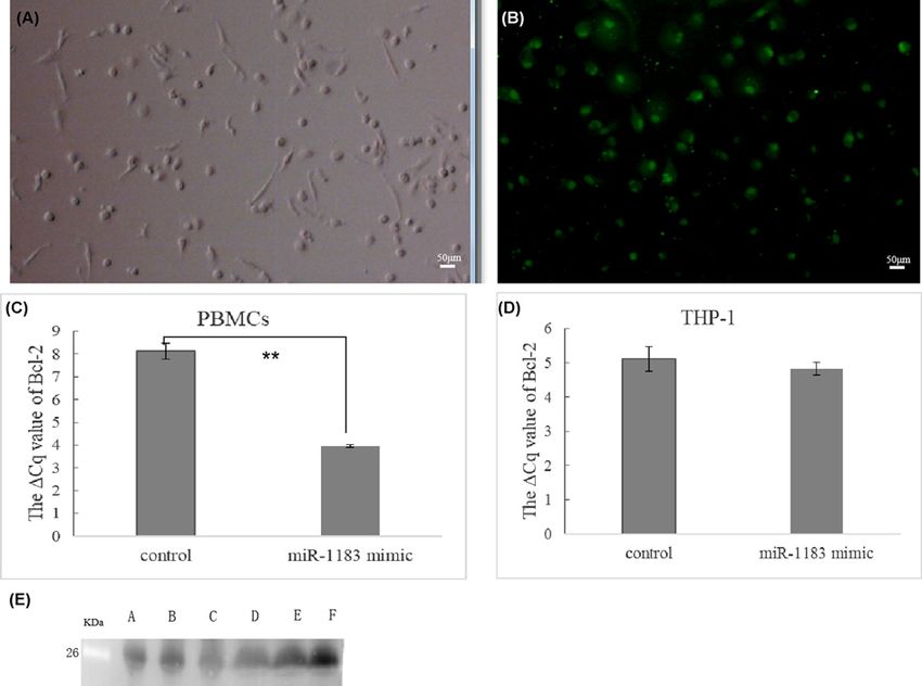
Bioscience Reports (2020) 40 BSR20201573
                                                                                                                                                  https://doi.org/10.1042/BSR20201573

Table 1 Clinical characteristics of patients with RHD and normal controls
                                                                                     RHD group                                                    Healthy controls

                     Blood samples                                                         20                                                               20
                   Sex (Male/Female)                                                     7:13                                                            10:10
                           Age                                                        56.7 +
                                                                                           − 6.0                                                       49.9 +
                                                                                                                                                            − 6.2
                        Smoking                                                        5(25%)                                                           4(20%)
                      Hypertension                                                       5(25%)                                                              0
                        Diabetes                                                            0                                                                0
                  Hereditary diseases                                                       0                                                                0
                   Immune diseases                                                          0                                                                0
                         Tumor                                                              0                                                                0

                                                                                                                                                                                                    Downloaded from http://portlandpress.com/bioscirep/article-pdf/40/11/BSR20201573/896723/bsr-2020-1573.pdf by guest on 20 November 2020
                Other vascular diseases                                                     0                                                                0
                         Trauma                                                             0                                                                0
                        Gestation                                                           0                                                                0
         Cardiac function grade (NYHA grade)                                             II∼IV                                                               /
                          EF(%)                                                      59.22 +− 7.14                                                           /
                      LVEDD (mm)                                                     48.65 +− 4.48                                                           /
                     PASP (mmHg)                                                     51.1 +− 4.96                                                            /

 Notes: LVEDD, left ventricular end-diastolic size; EF, ejection fraction. Cardiac function grades: NYHA divided into Grade II, Grade III, Grade IV. PASP,
 pulmonary artery pressure.

                    of cardiovascular research in recent years [7–9]. Nevertheless, there are few reports of microRNA in the research of
                    RHD [10,11].
                       Many studies have reported that microRNA is involved in myocardial fibrosis. It was shown that miRNA directly
                    inhibited the expression of various extracellular matrix proteins, and it regulated a variety of signal pathways related to
                    fibrosis, especially fibrosis occurring after necrosis caused by myocardial infarction [12–14]. At present, there is little
                    research on the role of miRNA in RHD, and the molecular biology of RHD may prove fruitful. Molecular techniques
                    can detect RHD early, allowing for early treatment. It is hoped that, in the near future, miRNA will become a tool for
                    early diagnosis of RHD. Furthermore, it may provide new ways to treat valvular fibrosis. Nevertheless, the molecular
                    mechanisms through which miRNAs influence myocardial apoptosis and remodeling of RHD have not yet been fully
                    elucidated.
                       Therefore, in the present study, we focused on the functions of miR-1183 in RHD models, specifically those induc-
                    ing apoptosis. BCL-2 might act as a target gene of miR-16-5p. Here, we investigated whether levels of miR-1183 were
                    altered in patients with RHD, as well as in primary human peripheral blood mononuclear cells (PBMCs) and human
                    myeloid leukemia monocyte (THP-1) cells. The aim was to determine whether alteration of BCL-2 expression levels
                    might contribute to the pathophysiology of RHD.

                    Methods
                    Experimental specimen and clinical characteristics
                    From March 2017 to December 2019, we enrolled 20 patients with RHD and 20 healthy controls without medical
                    problems. All experiments were carried out in accordance with relevant guidelines and regulations. The inclusion
                    criteria of the RHD group were as follows: (i) RHD diagnosed by echocardiography, including diagnosis of mitral
                    valve prolapse because of mitral chordae tendineae fracture and mitral insufficiency, scheduled for mitral valve re-
                    placement, aortic insufficiency scheduled for aortic valve replacement, or tricuspid regurgitation or insufficiency of
                    closure scheduled for valve replacement or valvuloplasty; and (ii) left ventricular ejection fraction (EF) >50%. Pul-
                    monary arterial pressure was measured by echocardiography and divided into low, mild and severe conditions: low
                    PAH (PASP < 30 mmHg), mild PAH (30 mm Hg ≤ PASP < 55 mmHg), and severe PAH (PASP ≥ 55 mmHg).
                    Exclusion criteria were as follows: valve damage caused by congenital, syphilitic heart valve disease; infective endo-
                    carditis; papillary muscle dysfunction; tendon rupture; myxoid degeneration; and other autoimmune diseases. The
                    characteristics of the cases and controls are shown in Table 1.
                       In the healthy control group, blood lipid levels, blood pressure, and blood glucose were all within normal ranges,
                    and heart function and pulmonary artery pressures were normal; no heart disease was detected. We collected 5 ml of
                    venous blood from the normal and case groups into tubes containing EDTA. After centrifugation at 4◦ C for 15 min,

2            © 2020 The Author(s). This is an open access article published by Portland Press Limited on behalf of the Biochemical Society and distributed under the Creative Commons Attribution
             License 4.0 (CC BY).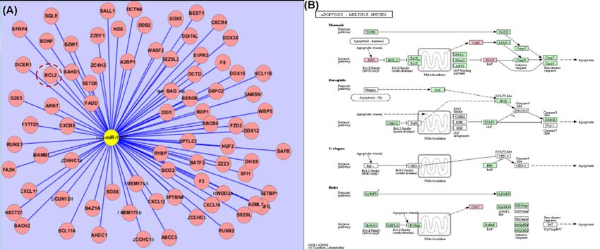
Bioscience Reports (2020) 40 BSR20201573
https://doi.org/10.1042/BSR20201573

the supernatants were removed and stored at −80◦ C. We obtained the approval of the medical ethics committee of
Li Huili Hospital, and written informed consent was obtained from all patients.

RNA extraction
The reverse transcription kit and real-time PCR kit were purchased from TaqMan and assays were performed accord-
ing to the manufacturer’s instructions. Using the ultramicro spectrophotometer k5600, 1 μl RNA sample was used
for determination. We recorded the concentration and purity of each sample. The ratio of absorbance at 260 nm to
that at 280 nm was between 1.8 and 2.0, which can be used in gene chip detection and PCR.

Construction of the miR-1183 expression vector and the target gene
3 UTR double luciferase expression vector, transfection and luciferase

                                                                                                                                                                                           Downloaded from http://portlandpress.com/bioscirep/article-pdf/40/11/BSR20201573/896723/bsr-2020-1573.pdf by guest on 20 November 2020
assays
Plasmid transfections for luciferase assays in 293T cells were performed with 3 UTR luciferase 0.1 μg, miRNA ex-
pression plasmid 0.4 μg, Renilla plasmid 0.02 μg in a 24-well plate as described by the manufacturer. The binding
between miRNA and target gene 3 UTR was verified using double fluorescent enzyme reporter gene technology. Lu-
ciferase activity was measured 48 h post transfection using the Dual Luciferase Reporter Assay System as described
by the manufacturer (Promega).
   The names of plasmids in the detection system were as follows: Mir1183-NC: microRNA empty plasmid, as
the negative control of the target microRNA plasmid; Mir-1183: miRNA vector plasmid, expressing the target mi-
croRNA (hsa-mir-1183 (21031-1)); 3 UTR-NC: 3 UTR empty plasmid as negative control of target gene 3 UTR
plasmid; 3 UTR: target gene 3 UTR plasmid (BCL2 (21010-1)); 3 UTR-MU: target gene 3 UTR mutant plasmid
(BCL2 (21011-1)); posotive control 3 UTR: TRAF6 gene 3 UTR plasmid; Vector plasmid of posotive control miRNA:
hsa-mir-146b. Hsa-miR-1183 mature miRNA sequence is CACUGUAGGUGAUGGUGAGAGUGGGCA. The con-
trol RNU6b sequence is CGCAAGGATGACACGCAAATTCGTGAAGCGTTCCATATTTTT.

Isolation, culture, and identification of primary cells
Peripheral blood mononuclear cells (PBMCs) were separated using Ficoll density gradient centrifugation. The pe-
ripheral blood samples and d-Hanks solutions were collected and diluted at 1:1. The volume ratio of diluted blood to
lymphocytes was 1:1. Samples were centrifuged at 2000 rpm/min at room temperature for 20 min. After centrifuga-
tion, the fluid was divided into four layers. The monocytes in the cloud layer were removed using a capillary dropper,
and then centrifuged repeatedly by adding d-Hanks solution to prepare monocyte suspensions. The cell precipitates
were cultured in RPMI 1640 complete medium, and cultured for 24 h. We added macrophage colony stimulating
factor to continue the culture. The medium was changed every 72 h and RPMI 1640 containing 10 ng/ml M-CSF +
10% fetal bovine serum was added. The subsequent experiment was carried out 7 days later.

Human peripheral blood mononuclear (THP-1) cells
THP-1 cells were cultured in RPMI 1640 medium containing 10% FBS and double antibodies. Macrophages were
induced by propylene glycol methyl ether acetate (PMA). After 3 days of continuous culture, the cells were replaced
by 1640 cells without PMA.

Cell transfection, RT-PCR
Lipo2000 was used as the transfection vector (50 μl + 2500 μl opti) in the process of cell transfection. Mimic-1183,
control group (40 μl + 1000 μl opti), 100 μl per well, mixing for 5 min, standing for 20 min. After 48 h of transfection,
the supernatants were removed, some of the extracted RNA was used for RT-PCR follow-up experiments. The cDNA
was prepared using the TaqMan miRNA qRT PCR kit as previous study.

Measurement of BCL-2 expression in heart valve tissue and plasma using
ELISA
We added 300 μl washing solution and soaked it for 30 s. We then added 100 μl 2× diluted standard into the standard
well. We added 100 μl standard diluent or culture medium into the blank wells. We then added 80 μl × detection
buffer and 20 μl serum sample into the sample wells. Cell culture supernatant: we added 100 μl cell culture supernatant
into sample wells, and added 50 μl 1:100 diluted BCL-2 antibody to each well. The plates were sealed and incubated
at room temperature for 2 h, after which they were washed six times. We added 100 μl of chromogenic substrate into

© 2020 The Author(s). This is an open access article published by Portland Press Limited on behalf of the Biochemical Society and distributed under the Creative Commons Attribution   3
License 4.0 (CC BY).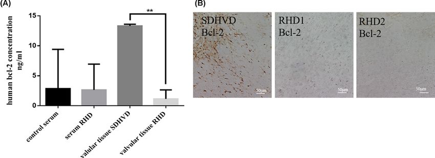
Bioscience Reports (2020) 40 BSR20201573
                                                                                                                                                  https://doi.org/10.1042/BSR20201573

Table 2 quantitative analysis and quality assurance of 6 samples for miRNA gene chip

                                                                                                                                                                      Up-regulation of
                                                                                               concentration                                                            gene chip
      Group                   No.                OD260/280               OD260/230                (ng/μl)               Capacity (μl)            Quality (ng)            detection

    Control group               1                     1.74                    1.87                   28.93                     10                    289.30                    0.13
                                2                     1.79                    1.88                   21.69                     10                    216.90                    0.11
                               N3                     1.80                    1.91                   29.88                     10                    298.80                    0.08
     RHD group                  4                     1.71                    1.96                   26.19                     10                    261.90                     2.5
                                5                     1.73                    1.86                   24.33                     10                    243.30                    1.36
                                6                     1.79                    1.80                   23.23                     10                    232.30                    1.36

                                                                                                                                                                                                    Downloaded from http://portlandpress.com/bioscirep/article-pdf/40/11/BSR20201573/896723/bsr-2020-1573.pdf by guest on 20 November 2020
                    each well, in the dark, and incubated at room temperature for 5–30 min. Finally, we added 100 μl of termination
                    solution to each well. Within 30 min, the optical density was measured at 450 nm.

                    Immunohistochemistry (paraffin section IHC)
                    Sections were baked for 40 min, and placed into the reagent tank for dewaxing, hydration, and incubation with
                    3% hydrogen peroxide (diluted with pure water) for 10 min to eliminate the activity of endogenous peroxidase.
                    Paraffin-sections were then deparaffinized and rehydrated. Antigen retrieval was performed using heat-incubation in
                    epitope retrieval solution (IHC World, U.S.A.) for 30 min. The primary antibodies used for immunohistochemistry
                    were anti-bcl-2 (1:200).

                    He staining of tissue sections
                    After baking, the above tissue sections were successively infiltrated into xylene 1 and dewaxed with xylene II for 10
                    min. Immerse in anhydrous ethanol I, anhydrous ethanol II, 95% alcohol, 80% alcohol, and 70% alcohol for 2 min
                    respectively. Hematoxylin was dyed for 3 min, then immersed in 50%, 70% and 80% alcohol for 2 min respectively,
                    then immersed in eosin for 5 s. Mirror inspection after neutral gum sealing.

                    Statistical analysis
                    For pyrosequencing, DNA quality was assessed using GraphPad Prism 6 analyzer software. The Student’s t-test and
                    ANOVA were used to test for differences in methylation levels for categorical variables. For qRT-PCR results, we used
                    the paired t-test or the χ2 test as appropriate. Data are shown as means +
                                                                                             − SD. P
Bioscience Reports (2020) 40 BSR20201573
https://doi.org/10.1042/BSR20201573
Downloaded from http://portlandpress.com/bioscirep/article-pdf/40/11/BSR20201573/896723/bsr-2020-1573.pdf by guest on 20 November 2020
Figure 1. has-mir-1183 target gene prediction
(A) Miranda, miRBase and targetscan jointly predict target genes (target gene bcl-2, cxcr4, etc). (B) KEGG analysis on apoptosis
pathway.
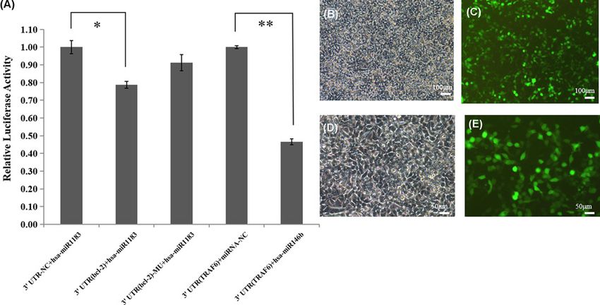
Figure 2. The binding of mir-1183 to Bcl-2 by the method of double luciferase reporter gene
(A) Detection of 3 UTR reporter gene (luciferase). (B–E) Fluorescent photos of GFP plasmid transfected. (B) GFP 100× B; (C) GFP
100× G; (D) GFP 200× B; (E) GFP 200× G (*PBioscience Reports (2020) 40 BSR20201573
https://doi.org/10.1042/BSR20201573
Downloaded from http://portlandpress.com/bioscirep/article-pdf/40/11/BSR20201573/896723/bsr-2020-1573.pdf by guest on 20 November 2020
Figure 3. Functional acquisition study of miR-1183 on primary peripheral blood mononuclear cells (PBMCs) and THP-1 cells
(A and B) Effect of mir-1183 transfection by primary peripheral blood mononuclear cells (PBMCs); (A) under ordinary microscope
and (B) under fluorescence microscope. (C and D) Expression of Bcl-2 mRNA in PBMCs and THP-1 cells transfected with mir-1183
mimic. (C) PBMCs Bcl-2 mRNA expression by PCR; (D) THP-1 Bcl-2 mRNA expression by PCR (**PBioscience Reports (2020) 40 BSR20201573
https://doi.org/10.1042/BSR20201573
Downloaded from http://portlandpress.com/bioscirep/article-pdf/40/11/BSR20201573/896723/bsr-2020-1573.pdf by guest on 20 November 2020
Figure 4. Detection of Bcl-2 on heart valve and serum in patients with rheumatic heart disease
(A) ELISA determination. (B) Comparison of the expression of Bcl-2 between one case of mild degenerative heart disease and two
cases of rheumatic heart disease by immunohistochemistry (scale bar: 50 μm) (**PBioscience Reports (2020) 40 BSR20201573
https://doi.org/10.1042/BSR20201573
to predict the target genes of miR-1183. Taken together, the present and previous findings suggest that increased
miR-1183 expression promotes apoptosis, thereby becoming involved in RHD.
We then sought to identify apoptosis-related target genes of miR-1183. Hundreds of target genes including BCL-2,
EGFR, and CXCR4 were found. The target genes bcl2A1, EGFR, and Bax were found by miR-1183 on target gene
prediction software. We found that apoptosis was involved in the pathogenesis of RHD. The BCL-2 gene was closely
related to the pathological changes of RHD. The relationship between miR-1183 and related target genes was studied
by functional acquisition and functional deletion. The expression levels of bcl2A1 gene transfected with miR-1183
in human peripheral blood mononuclear cells decreased significantly. Combined with RT-PCR experiments, these
results suggest that apoptosis is involved in the pathogenesis of RHD, and the bcl-2 gene was involved in the reg-
ulation of apoptosis [17–19]. During the compensatory period of RHD, high expression levels of miR-1183 trigger
decrease in bcl-2 gene expression levels, thereby regulating apoptosis. For further study, we selected BCL-2, which
Downloaded from http://portlandpress.com/bioscirep/article-pdf/40/11/BSR20201573/896723/bsr-2020-1573.pdf by guest on 20 November 2020
is an anti-apoptotic protein that prolongs cell survival. Aberrant down-regulation of BCL-2 disrupts mitochondrial
membrane integrity, induces the mitochondrial release of pro-apoptotic proteins, and triggers caspase activation, and
cytoskeletal degradation, resulting in apoptosis. In the present study, we found that miR-1183 binds directly to the
3 UTR of the BCL-2 mRNA and down-regulates the mRNA and protein levels of BCL-2 in 293T cells. These findings
suggest that BCL-2 plays an important role in regulation of apoptosis in RHD.
We speculate that the mechanism might be, up-regulated miR-1183 promote/inhibit the balance of apoptosis pro-
teins (Bax, BAK) in Bcl-2 family, promote the release of cytochrome c, enhance the apoptosis cascade reaction, and
reduce the anti apoptosis effect. The up-regulation of mir-1183 may affect the expression of Bcl-2 and the level of
cardiomyocyte apoptosis, which may be one of the reasons for the further development of rheumatic heart disease.
The purpose of the present study was to explore the application of differentially expressed miRNAs as biomarkers
in the diagnosis of RHD, and to help elucidate the mechanisms. As a preliminary study, we used real-time quantitative
PCR to measure CT value differences of miR-1183 and verified the results using a gene chip assay. To date, there are few
reports of miRNA in RHD. MiR-1183 screened by microarray has not been found to be related to the cardiovascular
system.
There are still limitations in the present study. The sample size needs to be further expanded: only 40 samples
were selected for PCR verification, which is far from sufficient. There is no gold standard clinical diagnosis of RHD.
Rheumatic antibody cannot diagnose patients with RHD in the inactive period; the possibility of RHD can only
be tested using color Doppler echocardiography and medical history. However, combined with relevant research
involving miRNA, we can find that the specific expression of miRNA in patients with RHD is due to the hyperplasia
and fibrosis of heart valve, which leads to the increase of miRNA expression in patients with rheumatic heart disease.
This prediction requires experimental verification. No primary cardiomyocytes were cultured from patients with
rheumatic heart disease,this should be a better cell model, but due to the limited conditions, the cells could not be
effectively analyzed. In the present study, we selected PBMCs [20,21] and THP-1 cells [22] and human miRNA mimics
for functional acquisition and miRNA inhibitors for functional deletion. We focused on mRNAs, simulating the role of
miRNA in vivo, to determine whether the progression of RHD was related to immunity; nevertheless, there are some
defects in design. It is necessary to carry out further biological function analysis to determine the pathophysiological
mechanisms of these miRNAs in RHD.
Conclusions
In conclusion, we demonstrated that miR-1183 is differentially expressed in RHD. The significantly higher expression
levels of miR-1183 appears to play a role in pathogenesis RHD by regulation of the anti-apoptotic protein, BCL-2,
which might affect myocardial apoptosis and remodeling in RHD.
Data Availability
All data included in the present study are available upon request by contact with the corresponding author.
Competing Interests
The authors declare that there are no competing interests associated with the manuscript.
Funding
The research was supported by the grants of National Natural Science Foundation of China [grant number 82000365]; Basic
public welfare projects in Zhejiang province [grant number LGD20H020001, LGF19H020004]; Zhejiang Province Medical and
8 © 2020 The Author(s). This is an open access article published by Portland Press Limited on behalf of the Biochemical Society and distributed under the Creative Commons
Attribution License 4.0 (CC BY).Bioscience Reports (2020) 40 BSR20201573
https://doi.org/10.1042/BSR20201573
Health Project [grant number 2017RC026, 2020KY273]; Natural Science Foundation of Ningbo [grant number 2019C50069,
202003N4269]; Ningbo Health Branding Subject Fund [grant number PPXK2018-01].
Author Contribution
Ni Li and Guofeng Shao conceived the study, participated in its design and coordination, and helped draft the manuscript. Ni Li
and Dawei Zheng performed the experients. Ni Li, Linwen Zhu, Hua Zhou, Dawei Zheng, Jianqing Gao analyzed the data. Con-
tributed reagents/materials/analysis tools: Lebo Sun, Guodong Xu. Ni Li wrote the manuscript, and all authors read and approved
the final draft.
Abbreviations
bcl-2, B-cell lymphoma 2; miR-1183, hsa-microRNA-1183; PBMC, human peripheral blood mononuclear cell; RHD, rheumatic
Downloaded from http://portlandpress.com/bioscirep/article-pdf/40/11/BSR20201573/896723/bsr-2020-1573.pdf by guest on 20 November 2020
heart disease; SDHVD, mild degenerative heart disease; THP-1, human myeloid leukemia monocyte cell.
References
1 Abdallah, E.A. (2012) Rheumatic heart disease associated with secondary renal amyloidosis. Arab. J. Nephrol. Transplant. 5, 149–152
2 Azimian, H., Dayyani, M., Toossi, M.T.B. and Mahmoudi, M. (2018) Bax/bcl-2 expression ratio in prediction of response to breast cancer radiotherapy.
Iran. J. Basic Med. Sci. 21, 325–332
3 Chen, W. and Li, S. (2017) Circulating microrna as a novel biomarker for pulmonary arterial hypertension due to congenital heart disease. Pediatr.
Cardiol. 38, 86–94, https://doi.org/10.1007/s00246-016-1487-3
4 Christodoulou, M.I., Kontos, C.K., Halabalaki, M., Skaltsounis, A.L. and Scorilas, A. (2014) Nature promises new anticancer agents: Interplay with the
apoptosis-related bcl2 gene family. Anticancer Agents Med. Chem. 14, 375–399, https://doi.org/10.2174/18715206113139990089
5 Dong, H., Sun, Y., Shan, F., Sun, Q. and Yang, B. (2015) Down-regulation of mir-101 contributes to rheumatic heart disease through up-regulating tlr2.
Med. Sci. Monit. 21, 1500–1506
6 Dougherty, S., Beaton, A., Nascimento, B.R., Zuhlke, L.J., Khorsandi, M. and Wilson, N. (2018) Prevention and control of rheumatic heart disease:
Overcoming core challenges in resource-poor environments. Ann. Pediatr. Cardiol. 11, 68–78, https://doi.org/10.4103/apc.APC˙135˙17
7 Guilherme, L., Dulphy, N., Douay, C., Coelho, V., Cunha-Neto, E., Oshiro, S.E. et al. (2000) Molecular evidence for antigen-driven immune responses in
cardiac lesions of rheumatic heart disease patients. Int. Immunol. 12, 1063–1074, https://doi.org/10.1093/intimm/12.7.1063
8 Guilherme, L. and Kalil, J. (2013) Rheumatic heart disease: Molecules involved in valve tissue inflammation leading to the autoimmune process and
anti-s. Pyogenes vaccine. Front. Immunol. 4, 352, https://doi.org/10.3389/fimmu.2013.00352
9 Guilherme, L., Oshiro, S.E., Fae, K.C., Cunha-Neto, E., Renesto, G., Goldberg, A.C. et al. (2001) T-cell reactivity against streptococcal antigens in the
periphery mirrors reactivity of heart-infiltrating t lymphocytes in rheumatic heart disease patients. Infect. Immun. 69, 5345–5351,
https://doi.org/10.1128/IAI.69.9.5345-5351.2001
10 Hu, M., Wei, X., Li, M., Tao, L., Wei, L., Zhang, M. et al. (2019) Circular rna expression profiles of persistent atrial fibrillation in patients with rheumatic
heart disease. Anatol. J. Cardiol. 21, 2–10
11 Kameny, R.J., He, Y., Zhu, T., Gong, W., Raff, G.W., Chapin, C.J. et al. (2018) Analysis of the microrna signature driving adaptive right ventricular
hypertrophy in an ovine model of congenital heart disease. Am. J. Physiol. Heart Circ. Physiol. 315, H847–H854,
https://doi.org/10.1152/ajpheart.00057.2018
12 Li, N., Lian, J., Zhao, S., Zheng, D., Yang, X., Huang, X. et al. (2015) Detection of differentially expressed micrornas in rheumatic heart disease:
Mir-1183 and mir-1299 as potential diagnostic biomarkers. Biomed. Res. Int. 2015, 524519
13 Li, Y., Zhang, S., Xu, W., Guo, X., Xu, Q. and Chen, Y. (2015) A normal polymorphism site of tlr2 3 untranslated region is related to rheumatic heart
disease by up-regulating tlr2 expression. Ann. Clin. Biochem. 52, 470–475, https://doi.org/10.1177/0004563214564581
14 Lin, F., Chen, H.W., Zhao, G.A., Li, Y., He, X.H., Liang, W.Q. et al. (2020) Advances in research on the circrna-mirna-mrna network in coronary heart
disease treated with traditional chinese medicine. Evid. Based Complement Alternat. Med. 2020, 8048691, https://doi.org/10.1155/2020/8048691
15 Lin, J., Jiang, J., Zhou, R., Li, X. and Ye, J. (2018) Microrna-451b participates in coronary heart disease by targeting vegfa. Open Med. (Wars) 15, 1–7
16 Lu, Q., Sun, Y., Duan, Y., Li, B., Xia, J., Yu, S. et al. (2018) Comprehensive microrna profiling reveals potential augmentation of the il1 pathway in
rheumatic heart valve disease. BMC Cardiovasc. Disord. 18, 53, https://doi.org/10.1186/s12872-018-0788-2
17 Luo, M., Wang, G., Xu, C., Zeng, M., Lin, F., Wu, J. et al. (2019) Circulating mir-30c as a predictive biomarker of type 2 diabetes mellitus with coronary
heart disease by regulating pai-1/vn interactions. Life Sci. 239, 117092, https://doi.org/10.1016/j.lfs.2019.117092
18 Nieters, A., Conde, L., Slager, S.L., Brooks-Wilson, A., Morton, L., Skibola, D.R. et al. (2012) Prrc2a and bcl2l11 gene variants influence risk of
non-hodgkin lymphoma: Results from the interlymph consortium. Blood 120, 4645–4648, https://doi.org/10.1182/blood-2012-05-427989
19 Nulu, S., Bukhman, G. and Kwan, G.F. (2017) Rheumatic heart disease: The unfinished global agenda. Cardiol. Clin. 35, 165–180,
https://doi.org/10.1016/j.ccl.2016.08.006
20 Toor, D. and Sharma, N. (2018) T cell subsets: An integral component in pathogenesis of rheumatic heart disease. Immunol. Res. 66, 18–30,
https://doi.org/10.1007/s12026-017-8978-z
21 Zheng, D., Xu, L., Sun, L., Feng, Q., Wang, Z., Shao, G. et al. (2014) Comparison of the ventricle muscle proteome between patients with rheumatic
heart disease and controls with mitral valve prolapse: Hsp 60 may be a specific protein in rhd. Biomed. Res. Int. 2014, 151726,
https://doi.org/10.1155/2014/151726
© 2020 The Author(s). This is an open access article published by Portland Press Limited on behalf of the Biochemical Society and distributed under the Creative Commons 9
Attribution License 4.0 (CC BY).Bioscience Reports (2020) 40 BSR20201573
https://doi.org/10.1042/BSR20201573
22 Zloto, K., Tirosh-Wagner, T., Bolkier, Y., Bar-Yosef, O., Vardi, A., Mishali, D. et al. (2020) Preoperative mirna-208a as a predictor of postoperative
complications in children with congenital heart disease undergoing heart surgery. J. Cardiovasc. Transl. Res. 13, 245–252,
https://doi.org/10.1007/s12265-019-09921-1
Downloaded from http://portlandpress.com/bioscirep/article-pdf/40/11/BSR20201573/896723/bsr-2020-1573.pdf by guest on 20 November 2020
10 © 2020 The Author(s). This is an open access article published by Portland Press Limited on behalf of the Biochemical Society and distributed under the Creative Commons Attribution
License 4.0 (CC BY).You can also read