A 14-year-old Male Who Has Fever and Rash - Pediatrics in ...
←
→
Page content transcription
If your browser does not render page correctly, please read the page content below
visual diagnosis
A 14-year-old Male Who
Has Fever and Rash
William M. Stauffer, MD, MSPH, DTM&H,* Angela D. Siwek,
MD,† Deepak Kamat, MD, PhD,‡ Erika Kempler-Meyer, MD§
Presentation
A 14-year-old boy presents with rash, fever, chills, and
difficulty walking due to right hip pain. The patient had
been healthy until 2 weeks ago when he developed
muscle pain, headaches, sore throat, cough, and de-
creased appetite. Ten days ago, he was started on a course
of azithromycin for “bronchitis.” Rapid streptococcal
antigen and mononucleosis antibody tests were negative
at that time. Three days later, the antibiotic regimen was
changed to a second-generation cephalosporin for a pre-
sumed urinary tract infection after red blood cells ap-
peared in the urine. Urine nitrite and leukocyte esterase
were negative.
Three days later (and 4 days prior to presentation), the
patient developed right shoulder pain, fever, “sores” in
his mouth, blood-streaked sputum, frequent episodes of
nosebleeding, “pain with deep breaths,” a rash on his
lower legs, and sore and swollen feet. At that time, his
white blood cell (WBC) count was 5.2⫻103/mcL
(5.2⫻109/L), with a normal differential count; findings
on chest radiograph were normal. The patient currently
has difficulty walking due to right hip pain. He denies any
history of exposure to infectious diseases.
The boy’s past medical history is remarkable for a
ventricular septal defect (VSD) discovered during early
infancy. He has received prophylaxis against bacterial
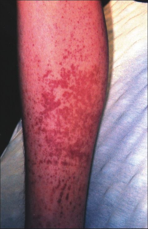
Figure 1. Raised red-purple rash on the lower extremities. endocarditis before every dental procedure. His growth
and development are normal, and he has no allergies to
food or medicine. His immunizations are up to date, and
current medications include cefprozil, acetaminophen
with codeine, and ibuprofen. He is in eighth grade, lives
with his parents, and has no siblings. He denies sexual
activity and use of tobacco, alcohol, or drugs.
On physical examination, the patient appears pale and
weak. His temperature is 101.3°F (38.5°C), blood pres-
sure is 135/78 mm Hg, respiratory rate is 22 breaths/
min, and heart rate is 104 beats/min. Raised, red-purple,
nonblanching skin lesions cover both distal lower ex-
*Center for International Health & International Travel Clinic, Regions Hospital,
Pediatric Emergency Medicine, University of Minnesota, Minneapolis, MN. tremities (Fig. 1). The oropharynx is mildly erythema-
†
Department of Pediatrics, University of Minnesota, Minneapolis, MN. tous. The neck is supple without lymphadenopathy. Car-
‡
Director, Institute of Medical Education, Children’s Hospital of Michigan, diac examination reveals normal first and second sounds
Detroit, MI.
§
Doernbecher Children’s Hospital, Oregon Health & Science University, Portland, with a grade II/VI harsh, holosystolic murmur along the
OR. left lower sternal border. Chest auscultation reveals fine
424 Pediatrics in Review Vol.24 No.12 December 2003
Downloaded from http://pedsinreview.aappublications.org/ by guest on January 11, 2021
visual diagnosis

crackles in both lungs. The abdomen is benign, and there
is no hepatosplenomegaly. He has full active and passive
motion of the joints, marked tenderness and erythema
over the right greater trochanter, and edema of the nail
fold of the right great toe. Results of the neurologic
examination are normal.
    The WBC count is 24.5⫻103/mcL (24.5⫻109/L),
with 85% polymorphonuclear neutrophils. The erythro-
cyte sedimentation rate (ESR) is 107 mm/h and
C-reactive protein (CRP) is 14.5 mg/dL (145 g/L).
Several blood cultures are drawn. A chest radiograph
reveals multiple scattered patchy, almost nodular infil-
trates involving the right upper and lower lobes, the right
perihilar region, and the left lower lobe (Fig. 2).

                                                                      Figure 2. Chest radiograph of multiple scattered patchy
                                                                      infiltrates.

                                                                                              Pediatrics in Review Vol.24 No.12 December 2003 425
                        Downloaded from http://pedsinreview.aappublications.org/ by guest on January 11, 2021
visual diagnosis
Diagnosis: Infective
Endocarditis
Infective endocarditis (IE) is
strongly suspected; a transthoracic
echocardiogram reveals a 2-cm veg-
etation within an aneurysm of the
membranous septum associated
with a 5-mm VSD (Fig. 3). The
next day, four sets of blood cultures
grow Streptococcus viridans and
Staphylococcus aureus.
Discussion
Differential Diagnosis Based
on Rash
Characterizing a rash may narrow
the differential diagnosis signifi-
cantly for a patient who presents
with fever and rash. In this case, the
skin finding is palpable purpura.
Purpura is characterized by red-
purple nonblanchable skin discol-
orations that are greater than
0.5 cm in diameter. Palpable pur-
pura results from vasculitic lesions
or embolic phenomena. Examples
of vasculitic disorders associated
with palpable purpura are Henoch-
Schönlein purpura, Kawasaki dis-
Figure 3. Echocardiogram showing a 2-cm vegetation and aneurysm in the membranous
ease, juvenile rheumatoid arthritis, septum with a 5-mm ventricular septal defect.
systemic lupus erythematosus, and
polyarteritis nodosa. Infectious em-
boli are due most commonly to gram-negative cocci undergone surgical repair of congenital heart disease,
(meningococci, gonococci), gram-negative rods (Enter- patients receiving immunosuppressant therapy, and pa-
obacteriacae), and gram-positive cocci (staphylococci, tients who have chronic indwelling intravascular cathe-
streptococci). Other causes of rash and fever include ters.
Rickettsia sp (Rocky Mountain spotted fever), drug re-
actions (sulfonamides), cytomegalovirus, sarcoidosis, Pathophysiology
tumors (leukemia, lymphoma), hemolytic uremic syn- It is hypothesized that any valvular lesion causing either
drome, thrombocytopenic purpura, and cryoglobuline- high-velocity or turbulent flow may lead to thickening or
mia (frequently caused by hepatitis B or C infections). disruption of the endocardium. Eventually, a sterile fi-
In this patient, the presence of a VSD and an infec- brin and platelet thrombus may form at the site of
tious source (paronychia of the right great toe) strongly endocardial breakdown. During bacteremia, circulating
suggested an underlying IE. pathogens may infect these thrombi, particularly bacteria
capable of adhering to the surfaces of thrombi, such as
Infective Endocarditis Streptococcus sp. Other factors that contribute to the
The incidence of IE is approximately 1 in 1,280 among development of IE include the size of the microbial
hospitalized children. In recent years, the incidence in inoculum and the genetic predisposition of the individ-
patients who have underlying rheumatic heart disease has ual patient.
decreased remarkably, and a new high-risk group has The congenital heart defects associated most com-
emerged. This new group includes patients who have monly with IE are left-sided obstructive lesions, stenotic
426 Pediatrics in Review Vol.24 No.12 December 2003
Downloaded from http://pedsinreview.aappublications.org/ by guest on January 11, 2021visual diagnosis
or regurgitant valvular lesions, systemic-pulmonary arte- ious gram-negative organisms: Haemophilus sp, Acti-
rial communicating lesions, and any condition requiring nobacillus actinomycetemcomitans, Cardiobacterium
artificial valves or prosthetic conduits. Additional risk hominis, Eikenella sp, and Kingella kingae. Fungal infec-
factors are poor dental hygiene, intravenous drug abuse, tions are rare and usually occur after cardiac surgery or
central venous catheters, and open heart surgery. therapy with multiple antibiotics. In 5% to 10% of IE
Complications of IE among children include conges- cases, blood cultures are negative, and in fewer than 3%
tive heart failure, coronary artery emboli with secondary of cases, the vegetation is polymicrobial.
myocardial infarction, cardiac dysrhythmias, valvular ring Some pathogens are present more commonly in cer-
abscesses, ventricular or atrial septal perforation, mural tain clinical conditions. Staphylococcal endocarditis is
rupture and hemorrhage, and immune complex- seen among patients who have indwelling vascular cath-
mediated diffuse glomerulonephritis. Once established, eters or prosthetic valves; S viridans infection is most
sections of the infected vegetation may break off and common among patients who have native valves or have
enter the circulation, eventually lodging elsewhere, caus- undergone recent dental procedures. Enterococcal (group
ing infarction, localized infection, or both. Embolization D) endocarditis is associated with recent gastrointestinal or
may occur either to the lungs from right-sided endocar- genitourinary manipulation. Pseudomonas aeruginosa and
ditis or to other organs and parts of the body through the Serratia marcescens endocarditis most often affects patients
systemic circulation from left-sided endocarditis. The who have a history of intravenous drug abuse.
patient described here has emboli shedding into both the
pulmonary and systemic circulations, as demonstrated by Clinical and Laboratory Findings
lung infarctions (pulmonary circulation) (Fig. 2) and the The initial presentation of endocarditis varies from an
palpable purpura (systemic circulation) (Fig. 1). Chronic insidious onset with prolonged low-grade fevers to an
exposure to bacterial or other foreign protein within the acute onset with severe symptoms. The insidious course
vegetation leads to development of antibodies and result- usually is caused by penicillin-sensitive strains of S viri-
ant circulating immune complex disease. Patients who dans. Penicillin-resistant organisms, such as staphylo-
have IE and develop arthralgias and arthritis, splenomeg- cocci, usually cause acute-onset disease. The most com-
aly, Roth spots, glomerulonephritis, and thrombocyto- mon signs and symptoms of IE are chest pain, abdominal
penia frequently have higher circulating immune com- pain, arthralgia, myalgia, dyspnea, malaise, night sweats,
plex levels than patients who have IE without these weight loss, nausea, and vomiting. A small number of
findings. patients develop hematuria.
Many organisms can cause endocarditis. The patho- Physical examination may reveal new or changing
gens seen most frequently include S viridans, S aureus, heart murmurs. About 50% to 60% of patients who have
Enterococcus sp, S bovis, and the HACEK group of fastid- IE demonstrate splenomegaly, and 30% have petechiae.
Other skin manifestations of IE include Osler nodes,
splinter hemorrhages, and Janeway lesions. Osler nodes
are red, painful, nodular lesions of the finger; splinter
Definitions hemorrhages are linear hemorrhages under the nails; and
Infective endocarditis—Infection and inflammation of Janeway lesions are small, red lesions of the palms or
the endocardium soles. Roth spots are retinal hemorrhages that show
Embolus—A blood clot or other particulate material central clearing. Osler nodes, splinter hemorrhages,
carried by the blood stream from one site to another Janeway lesions, and Roth spots develop late in the
Paronychia—Inflammation involving the tissue around course of IE, particularly among patients who are not
the nailbed treated appropriately.
Purpura—Purplish or brownish-red discoloration of For patients who have IE, the WBC count may be
the skin that is greater than 0.5 cm in diameter normal, but neutrophilia is common, and most patients
Vegetation—A pathologic growth of the tissue or a have elevated concentrations of acute-phase reactants
blood clot composed largely of fused blood platelets, (eg, ESR, CRP). Anemia and hematuria are frequent
fibrin, and sometimes bacteria that is adherent to findings. The electrocardiogram usually is normal but
diseased endocardium. may show changes caused by an underlying anatomic
Fungating vegetation—A spongy vegetation that has cardiac disorder. Multiple blood cultures of adequate
the appearance of a fungus volume, drawn at different times, are necessary to estab-
lish the diagnosis. Timing the blood collection with the
Pediatrics in Review Vol.24 No.12 December 2003 427
Downloaded from http://pedsinreview.aappublications.org/ by guest on January 11, 2021visual diagnosis
Vegetations that are small or obscured by unusual anat-
Abbreviated Duke
Table 1 omy in cases of complex congenital heart disease fre-
quently are missed.
Clinical Criteria for Currently, the Duke Criteria are the most sensitive
Infective Endocarditis and specific clinical criteria for diagnosing IE in adults
and children. The diagnosis is determined to be “definite,”
Major Criteria “possible,” or “rejected” based on pathologic (microbio-
● Positive blood culture for infective endocarditis (IE) logic or histologic identification of the pathogen within
● Evidence of endocardial involvement vegetations) and clinical criteria. The clinical criteria are
Positive echocardiogram for IE
OR subdivided into major and minor categories (Table). The
New valvular regurgitation (worsening or changing diagnosis of IE is definite when the patient has the patho-
of pre-existing murmur not sufficient) logic criteria plus two major clinical criteria, one major and
Minor Criteria three minor clinical criteria, or five minor criteria. Some
authors suggest that splenomegaly, a particularly common
● Predisposition: Predisposing heart condition or
intravenous drug use
finding in children who have IE, should be added as a
● Fever: Temperature >38.0°C (100.4°F) clinical criterion in the pediatric population.
● Vascular phenomena: Major arterial emboli, septic
pulmonary infarcts, mycotic aneurysm, intracranial Treatment
hemorrhage, conjunctival hemorrhages, and Janeway Unless the patient requires immediate treatment, antimi-
lesions
● Immunologic phenomena: Glomerulonephritis, Osler
crobial therapy should be withheld until the diagnosis of
nodes, Roth spots, and rheumatoid factor IE is confirmed by laboratory examination. Once the
● Microbiologic evidence: Positive blood culture but diagnosis is established, treatment with bactericidal
does not meet a major criterion as noted above or rather than bacteriostatic antibiotics should be started
serologic evidence of active infection with organism without delay. Initial antibiotic therapy consists of two
consistent with IE
● Echocardiographic findings: Consistent with IE but
synergistic antibiotics, thereby decreasing the emergence
do not meet a major criterion as noted above of resistant organisms. Once the offending organisms are
identified and sensitivities are available, the antibiotic
Adapted with permission from Durack DT, Lukes AS, Bright DK. New
criteria for diagnosis of infective endocarditis: utilization of specific therapy is adjusted accordingly. Antibiotic levels are
echocardiographic findings: Duke Endocarditis Service. Am J Med. maintained at a much higher level (5- to 20-fold higher)
1994;96:200 –209.
than the in vitro minimum inhibitory concentration for
4 to 8 weeks because organisms causing IE grow in high
concentration at a low metabolic rate in a relatively
onset of fever is not important because affected patients avascular site. Staphylococcal endocarditis may require
usually have constant bacteremia. The causative organism prolonged antimicrobial therapy. Indications for surgical
may be recovered from the first two blood cultures in 90% intervention include severe valvular involvement with
of patients. Negative blood cultures may be observed in intractable heart failure, heart block from a periaortic
patients who have been receiving antibiotic therapy or who abscess, myocardial abscess, recurrent embolic phenom-
have unusual pathogens that are difficult to culture. ena, and medical treatment failure.
Echocardiography is an invaluable tool for diagnosing
IE, studying cardiac structure and function, and predict- Complications
ing complications. For example, fungating vegetations The most common complications of IE are heart failure,
and vegetations larger than 1 cm in diameter are associ- conduction disorders, and central nervous system and
ated strongly with embolization. Transesophageal echo- pulmonary emboli. Approximately 50% to 60% of pa-
cardiography (TEE) is the most sensitive technique for tients have serious morbidity, with a mortality rate ap-
identifying vegetations in adults, although its superiority proaching 25% despite the availability of effective an-
in children is debated. Children generally have thinner tibiotic therapy. Patients who have IE caused by S
chest walls and frequently have right-sided cardiac le- aureus have the poorest prognosis compared with
sions, making transthoracic echocardiography (TTE) a patients whose disease is due to other bacteria. Al-
more sensitive study for children. Neither TEE nor TTE though rare, fungal IE has an extremely high mortality
has 100% sensitivity, so negative echocardiographic find- rate despite use of antifungal medications and surgical
ings do not necessarily exclude the possibility of IE. treatment.
428 Pediatrics in Review Vol.24 No.12 December 2003
Downloaded from http://pedsinreview.aappublications.org/ by guest on January 11, 2021visual diagnosis
Prevention Suggested Reading
The American Heart Association recommends antibi- Dajani AS, Taulert KA, Wilson W, et al. Prevention of bacterial
otic prophylaxis for any person who has a heart defect endocarditis: recommendation by the American Heart Associa-
(except secundum atrial septal defect and mitral valve tion. JAMA. 1997;277:1794 –1801
prolapse without mitral regurgitation) and is undergo- Danilowicz D. Infective endocarditis. Pediatr Rev. 1995;16:
148 –154
ing a procedure likely to cause bacteremia. Examples
Del Pont JM, De Cicco LT, Vartalitis C, et al. Infective endocarditis
include dental procedures and surgery involving the in children: clinical analysis and evaluation of two diagnostic
upper respiratory, gastrointestinal, or genitourinary criteria. Pediatr Infect Dis J. 1995;14:1079 –1086
tracts. Patients at high risk for IE should receive Ferrieri P, Gewitz MH, Gerber MA, et al. Unique features of
proper dental care and, with their caregivers, be able to infective endocarditis in childhood. Pediatrics. 2002;109:
recognize the early signs and symptoms of IE to 931–943
Martin JM, Neches WH, Wald ER. Infective endocarditis: 35 years
initiate treatment of any local or systemic infections
of experience at a children’s hospital. Clin Infect Dis. 1997;24:
promptly. 669 – 675
Thank You!
We are very grateful to the following people (those
other than our PIR Board members) who reviewed
articles for us during 2003:
Robert L. Brent, MD, PhD, DSc
S. Jean Emans, MD
Richard E. Kreipe, MD
Kenneth J. Lindahl, MD
John T. McBride, MD
David M. Siegel, MD, MPH
Michael Weitzman, MD
Erratum
Alert readers noticed that there is no PIR Quiz question #5 in the
October issue, although there is an answer for question #5 in the answer
key. During the production process, quiz questions were misnum-
bered. All quiz answers in the answer key are correct for the questions
with which they are identified; there simply is no question #5 in this
issue. We apologize for the confusion and inconvenience this error has
created.
In the Fluoride article in the same issue, the caption that accompa-
nies Figure 4 (page 333) is incorrect. It should read: “A ‘pea-size’
amount of toothpaste. The quantity of 1,100 ppm toothpaste pictured
here weighs 0.4 g and provides 0.44 mg of fluoride.”
Pediatrics in Review Vol.24 No.12 December 2003 429
Downloaded from http://pedsinreview.aappublications.org/ by guest on January 11, 2021Visual Diagnosis: A 14-year-old Male Who Has Fever and Rash
William M. Stauffer, Angela D. Siwek, Deepak Kamat and Erika Kempler-Meyer
Pediatrics in Review 2003;24;424
DOI: 10.1542/pir.24-12-424
Updated Information & including high resolution figures, can be found at:
Services http://pedsinreview.aappublications.org/content/24/12/424
References This article cites 5 articles, 2 of which you can access for free at:
http://pedsinreview.aappublications.org/content/24/12/424.full#ref-li
st-1
Subspecialty Collections This article, along with others on similar topics, appears in the
following collection(s):
Cardiology
http://classic.pedsinreview.aappublications.org/cgi/collection/cardiol
ogy_sub
Cardiovascular Disorders
http://classic.pedsinreview.aappublications.org/cgi/collection/cardiov
ascular_disorders_sub
Permissions & Licensing Information about reproducing this article in parts (figures, tables) or
in its entirety can be found online at:
https://shop.aap.org/licensing-permissions/
Reprints Information about ordering reprints can be found online:
http://classic.pedsinreview.aappublications.org/content/reprints
Downloaded from http://pedsinreview.aappublications.org/ by guest on January 11, 2021Visual Diagnosis: A 14-year-old Male Who Has Fever and Rash
William M. Stauffer, Angela D. Siwek, Deepak Kamat and Erika Kempler-Meyer
Pediatrics in Review 2003;24;424
DOI: 10.1542/pir.24-12-424
The online version of this article, along with updated information and services, is
located on the World Wide Web at:
http://pedsinreview.aappublications.org/content/24/12/424
Pediatrics in Review is the official journal of the American Academy of Pediatrics. A monthly
publication, it has been published continuously since 1979. Pediatrics in Review is owned,
published, and trademarked by the American Academy of Pediatrics, 345 Park Avenue, Itasca,
Illinois, 60143. Copyright © 2003 by the American Academy of Pediatrics. All rights reserved.
Print ISSN: 0191-9601.
Downloaded from http://pedsinreview.aappublications.org/ by guest on January 11, 2021You can also read