DIAGNOSIS OF HAIR DISORDERS - KATHRIN HILLMANN, MD, AND ULRIKE BLUME-PEYTAVI, MD, PHD
←
→
Page content transcription
If your browser does not render page correctly, please read the page content below
Diagnosis of Hair Disorders
Kathrin Hillmann, MD, and Ulrike Blume-Peytavi, MD, PhD

                      Hair disorders include hair loss, increased hair growth, and hair structure defects with
                      increased breakage, as well as unacceptable cosmetic appearance, such as reduced shine,
                      strength, curliness, and elasticity. It is the task of the dermatologist to choose the right
                      diagnostic tool depending on the suspected clinical diagnosis. Moreover, certain tools are
                      best suited for diagnosis in private practice, whereas others can only be used to monitor
                      hair growth under treatment in clinical studies. The techniques can be classified as either
                      invasive (eg, biopsies in scarring alopecia), semi-invasive (trichogram, unit area tri-
                      chogram), or noninvasive (eg, global hair counts, phototrichogram, electron microscopy,
                      laser scanning microscopy) methods. Further, one must differentiate between subjective
                      and objective techniques. For the practicing dermatologist, body and scalp hair distribution
                      by use of different grading systems, the hair pull test, and dermoscopy belong in the
                      category of basic diagnostic tools. Basic techniques may be extended by computer-
                      assisted phototrichogram and, in selected cases, by use of the trichogram and/or scalp
                      biopsies. For research purposes optical coherent tomography, electron microscopy, bio-
                      chemical methods, atomic force microscopy, and confocal laser scanning microscopy are
                      optional tools. For clinical studies global photographs (global expert panel), hair weighing,
                      phototrichogram, and different clinical scoring systems have proven to be objective tools
                      for documentation and evaluation of hair growth and hair quality.
                      Semin Cutan Med Surg 28:33-38 © 2009 Elsevier Inc. All rights reserved.

T    he precise and reliable diagnosis of hair growth disorders is
     mandatory to develop a successful therapeutic and cosmetic
management strategy. Although the diagnosis of hair disorders is
                                                                              History
                                                                              An evaluation of the patient’s personal history, as well as family and
significantly based on the clinical experience and trichological              drug history, gynecological problems, and general internal medical
knowledge of the dermatologist, there is a broad spectrum of diag-            disorders, as well as a discussion and patient’s expectations and
nostic techniques that are very helpful in diagnostic work-up and             wishes should be performed. The disorder’s impact on quality of life
for monitoring hair growth cycle disturbances                                 as well as any psychological disturbances, also are important and
                                                                              contributing factors for developing a management approach.
   For the practicing dermatologist, two types of diagnostic tools
should be distinguished: obligate and facultative procedures. Obli-
gate or basic diagnostic procedures are composed of a thorough                Clinical Examination
evaluation of the patient’s and his/her family’s history, supported by
a complete and thorough clinical examination. Basic examination               The clinical examination should begin with complete inspection of
techniques include grading of hair loss or excessive hair growth, a           scalp and body hair distribution and the whole skin examination.
                                                                              When undertaking an in-depth inspection of the scalp, the physi-
pull test, and dermoscopy of the scalp, especially for the examina-
                                                                              cian should look for signs of inflammation, scaling, erythema, and
tion of hair follicle openings and hair shaft irregularities. Depending
                                                                              scarring. The presence of follicular openings, exclamation mark
on the suspected diagnosis, facultative techniques, such as micro-
                                                                              hairs, or tufted hairs is important. For further classification, the hair
scopic examination of hair roots or hair shafts, phototrichogram, or          loss pattern and density should be analyzed. In addition, hair shaft
other techniques may be added.                                                quality should be evaluated based on caliber, fragility, length, and
                                                                              elasticity.
                                                                                  Grading systems have been established, particularly for androge-
                                                                              netic alopecia (AGA) and for alopecia areata. For grading male pat-
Clinical Research Center for Hair and Skin Science (CRC), Department of
                                                                              tern baldness, the Hamilton-Norwood scale is the most-used clas-
    Dermatology and Allergy, Charité–Universitätsmedizin Berlin, Berlin,
    Germany.                                                                  sification. In women the best-known scale for female-pattern AGA is
Address correspondence to Kathrin Hillmann, MD, Clinical Research Center      the Ludwig scale which is graded on a 3-point scale. The Gan-
    for Hair and Skin Science (CRC), Department of Dermatology and Al-        Sinclair scale has 5 grades and the Savin-scale has 8 differentiation
    lergy, Charité–Universitätsmedizin Berlin, Charitéplatz 1, Berlin, Ger-   classes and are more accurate for specification and have become
    many. E-mail: kathrin.hillmann@charite.de                                 accepted.1-3

1085-5629/09/$-see front matter © 2009 Elsevier Inc. All rights reserved.                                                                           33
doi:10.1016/j.sder.2008.12.005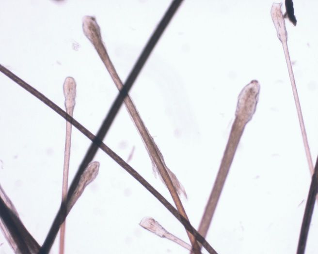
34                                                                                                                 K. Hillmann and U. Blume-Peytavi

Table 1 Different Types of Effluvium in Trichogram
Suspected Diagnosis                   Plucking Sides               Anagen Catagen Telogen Dysplastic Dystrophic Broken
Normal                    Frontal ⴙ occipital                       70-90%
Diagnosis of hair disorders 35
technology enables the investigator to objectively quantify hair growth
rate, density of hair follicles, and hair shaft thickness, as well as the
number of vellus and terminal hairs. For differences in evaluation of
hair growth parameters see Table 3.
Contrast-Enhanced PTG (CE-PTG)
The CE-PTG is a potent method for analysis of hair growth and loss.
The photographs of the scalp areas are taken twice, at intervals of 2
to 5 days. Depending on the clinical demand and trial protocol,
usually 2 to 3 areas are shaved, dyed, and photographed (see
Table 2).12
At the first visit (day 0), all hairs in the defined areas (1 cm2) are
trimmed 1 mm from the skin surface. At both visits, the hair sites are
covered with a transient (brown or black) hair dye for contrast
enhancement. Afterward, photographs are taken with a macro cam-
era by the use of a scalp immersion proxigraphy method. In other
PTG methods the use of gel-like intermediate-substances is de-
scribed.
Figure 1 Trichogram: telogen hair roots, epilated, and embedded In hair loss studies as well as in the measurement of body hair
hair roots under magnification. density, it is fundamental that exactly the same area must be
visualized on both photos. This area can be guaranteed by the use
of a semipermanent tattoo on the investigational area.
To obtain optimal and reliable results, experienced technicians
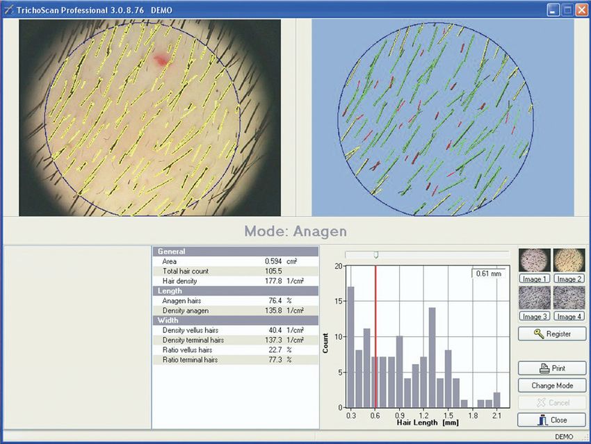
and examiners are needed. For the patient, it is an unpleasant pro- Automated Phototrichogram: TrichoScan
cedure, mainly because of the 5 days without hair washing, as well The TrichoScan® is a GCP-validated, investigator-independent,
as the discomfort of the hair plucking. The trichogram also has automated software program for the analysis of hair growth. It
significance when the evaluation of the hair root is necessary, for combines standard epiluminescence microscopy with automatic
example, in the diagnosis of loose anagen hair and in anagen-dys- digital image analysis for the measurement of all important hair
plastic effluvium. parameters.13,14 The defined scalp areas are trimmed with a stan-
dardized hair clipper (Hairliner, Wella, Germany) and exactly 3
Unit Area Trichogram days later are dyed with a commercially available solution (e.g.,
The unit area trichogram is a semi-invasive quantitative method that Goldwell top chic, black 2N, Darmstadt, Germany). After the
uses hair plucking to estimate the following hair growth parameters: extra color is removed with an alcoholic solution and, the area is
hair follicle density, proportion of anagen fibers, and hair shaft still wet, images are obtained with a digital camera with a rigid
diameter. It is based on plucking hairs in a defined area (usually contact lens, which ensures that the images are always taken at
⬎30 mm2), which are then counted and measured. Microscopic the same distance from the scalp. As an automated image analysis
analysis enables one to differentiate hair growth phases and mea- tool, accurate TrichoScan results strongly depend on the image
surement of hair length.11 The unit area trichogram can be used for quality; therefore, stray hairs, excessive hair dye, or air bubbles
follow-up of scalp hair changes in a study cohort for observing hair and other confounders have to be eliminated.
growth cycling and for monitoring topical or systemic drug effect; The TrichoScan is used to support clinical diagnosis in hair consul-
nevertheless, it is rather time-consuming and is unsuitable for large- tation as well as in clinical studies (Figure 2). During follow-up, it is
scale clinical trials. fundamental that exactly the same area is seen on both photos; other-
wise, accurate results will not be achieved.
Phototrichogram
The phototrichogram (PTG) is a noninvasive, reproducible method of
taking close-up photographs of certain defined, shaved scalp areas to Dermoscopy and Videodermoscopy
follow hair growth during a given time period. Several manual, semi- Today, hair diagnostic consultation is impossible to imagine with-
automatic, and automatic PTG methods exist. All have in common the out dermoscopy of the scalp. With handheld systems, videodermos-
dyeing of hair a black or brown color to enhance hair/skin contrast copy has the advantage of storing the hair and skin findings for use
before taking photos. One automated phototrichogram technique is as further controls. The magnification enhances the images and
known as the TrichoScan, a well-established method in many derma- detects the hair shaft in the follicle (if present) and its length, diam-
tology offices and trichological centers in Europe.1 The use of PTG eter, and possible anomalies. It is useful for the differential diagnosis
Table 2 Investigational Sites for Trichogram; Phototrichogram; TrichoScan®
Suspected Diagnosis Investigational Site A Investigational Site B
Male pattern hair loss Frontal regression hair line and/or 2 cm besides occiput region (Protuberantia
vertex region occipitalis externa)
Female pattern hair loss, diffuse effluvium, Approx. 2 cm behind frontal hair 2 cm besides occiput region (Protuberantia
alopecia areata diffusa, loose anagen line occipitalis externa)
hair
Alopecia areata circumscripta, Border of the alopecic patch Contralateral
trichotillomania36 K. Hillmann and U. Blume-Peytavi
Table 3 Evaluation Parameters for Phototrichogram Methods
TrichoScan CE-PTG
Number of all hairs and hair Note: TrichoScan cannot pick up hairs that are ⴙ
density (n/cm2) too short or too fine (approx.Diagnosis of hair disorders 37
Figure 2 Typical screenshot of the TrichoScan result presentation. With precise clipping, anagen hairs can be analysed.
For this purpose the hair in the target area must first be clipped completely to the skin surface with a standard single
use hair clipper. After three days the hair in the area is dyed and analysed. As only anagen hairs grow considerably
within these three days, by mathematical approximation the TrichoScan counts only anagen hairs.
Confocal Laser Scanning Microscopy investigate the roughness and the weathering of the cuticle and to
Confocal laser scanning microscopy noninvasively generates a measure the lifting of the scales. AFM is, however, limited to mea-
three-dimensional image of the surface structure of a hair as well as surement of the topographic morphology perpendicular to the sam-
different internal structures of hair (cortex and medulla fibers) and ple plane, meaning that re-entrant surfaces (i.e., spaces obscured by
the emission spectrum. It is a useful, noninvasive method for exam- the main surface) and subsurface information cannot be detected, in
ining objects with curved surfaces and does not require any sample contrast to the results available with scanning electron micrsocopy
preparation; the hair can be observed in its natural environment or confocal microscopy using fluorescence.18
with less damage than produced by other microscopic methods AFM be used as an imaging technique as well as a tool for quan-
such as scanning electron microscopy.17 It also provides fluorescent titative assessment of the effects of human hair treatment.19 As a
images either by exploiting the natural fluorescence of keratin or by noninvasive method, it requires no special or extensive sample
adding different fluorescent dyes as markers of various structures. preparation, like single-electron microscopy, and provides accurate
Confocal laser scanning microscopy is useful in obtaining “dynamic topographic information.
studies,” such as the routes of penetration of fluorochromes into the
cortex and “optical sections” of the specimen.17 Optical Coherence Tomography
OCT is able to provide highly reproducible in vivo and ex vivo
Atomic Force Microscopy measurements of hair shaft thickness, including the inner-hair vari-
Atomic force microscopy (AFM) supplies three-dimensional images ation of diameter and shape.20 It produces a two-dimensional image
(profilometry) with high resolution at the nanometer scale, produc- of optical scattering from internal tissue microstructures in a way
ing qualitative and quantitative measurements of the sample. The that is analogous to the pulse-echo image seen with ultrasound.21
method requires no preparation of the sample, avoiding contact OCT can be used for research purposes in trichological examina-
between the tip probe and the sample surface. It can be used to tion, especially for measuring the hair diameter, cross section sur-38 K. Hillmann and U. Blume-Peytavi
face, and hair shape. In the future, it may be used to help investigate 3. Olsen EA: Current and novel methods for assessing efficacy of hair growth pro-
moters in pattern hair loss. J Am Acad Dermatol 48:253-262, 2003
the influence of hair growth promoting agents to follow in vivo hair 4. DeUgarte CM, Woods KS, Bartolucci AA, et al: Degree of facial and body terminal
shaft changes over time. hair growth in unselected black and white women: Toward a populational defi-
nition of hirsutism. J Clin Endocrinol Metab 91:1345-1350, 2006
5. Shapiro J, Wiseman M, Lui H: Practical management of hair loss. Can Fam Phys
46:1469-1477, 2000
Scalp Biopsy 6. Rebora A, Guarrera M, Baldari M, et al: Distinguishing androgenetic alopecia from
chronic telogen effluvium when associated in the same patient. Arch Dermatol
The scalp biopsy, mostly performed with a 4-mm cylindrical punch, 143:1243-1245, 2005
is an important tool in the diagnosis of cicatricial, but also of certain 7. Guarrera M, Semino MT, Rebora A: Quantitating hair loss in women: A critical
cases of noncicatricial, alopecia. The selection of the correct biopsy approach. Dermatology 194:12-16, 1997
8. Price VH, Menefee E: Quantitative estimation of hair. Growth I. Androgenetic
site, depending on the disease, is crucial for successful histologic alopecia in women: Effect of minoxidil. J Invest Dermatol 95:677-682, 1990
findings. In nonscarring alopecia (e.g., trichotillomania, AGA) a 9. Price VH, Menefee E, Sanchez M, et al: Changes in hair weight in men with
punch in the center of the lesion is appropriate; in scarring alopecia, androgenetic alopecia after treatment with finasteride, 1 mg daily: Three- and
4-year results. J Am Acad Dermatol 55:71-74, 2006
the sample has to be taken from the active peripheral margin. 10. Blume-Peytavi U, Orfanos CE: Microscopy of the hair—The trichogram, In Serup
Generally, two biopsies should be carried out, one for transverse J, Jemec GBE, Grove GL, eds: Handbook of Non-Invasive Methods and the Skin
and the other for vertical sectioning. For transverse sectioning, the (ed 2). Boca Raton, FL, CRC Press Taylor & Francis Group, 2006, pp 875-881
11. Rushton DH, de Brouwer B, de Coster W, et al: Comparative evaluation of
punch is embedded and cut horizontally, allowing a quick overview scalp hair by phototrichogram and unit area trichogram analysis within the
of the hair follicle quantity, diameter, grouping, and morphometric same subjects. Acta Derm Venereol 73:150-153, 1993
data.22 The vertical specimen should be cut in half longitudinally to 12. Van Neste DJ: Assessment of hair loss: Clinical relevance of hair growth evaluation
methods. Clin Exp Dermatol 27:358-365, 2002
hair growth direction, one part is for hematoxylin-eosin stain the 13. Hoffmann R, Trichoscan: What is new? Dermatology 211:54-62, 2005
other for immunofluorescence. The vertical slices show hair follicle 14. Hoffmann R, Trichoscan: Combining epiluminescence microscopy of hair growth
histology for the examination of whole hair shafts and their struc- in vivo. Eur J Dermatol 11:362-368, 2001
15. Ross EK, Vincenzi C, Tosti A: Videodermoscopy in the evaluation of hair and scalp
tures as well as potential infiltrates. disorders. J Am Acad Dermatol 55:799-806, 2006
Since it is an invasive technique, the indication for a scalp biopsy 16. Canfield D: Photographic documentation of hair growth in androgenetic alopecia.
should be carefully considered but, when necessary, the interven- Dermatol Clin 14:713-721, 1996
17. Hadjur C, Daty G, Madry G, et al: Cosmetic assessment of the human hair by
tion should not be delayed. This technique is used in atrophic as
confocal microscopy. Scanning 24:59-64, 2002
well as cicatricial alopecia, nonspecific inflammatory scalp diseases, 18. Gurden SP, Monteiro VF, Longo E: Quantitative analysis and classification of
scalp tumors, nonspecific differential diagnosis of noncicatricial al- AFM images of human hair. J Microsc 215:13-23, 2004
opecia, and in clinical studies. 19. You H, Yu L: Atomic force microscopy as a tool for study of human hair. Scanning
19:431-437, 1997
20. Blume-Peytavi U, Vieten J, Knüttel A, et al: Optical coherent tomography (OCT):
A new method for online-measurement of hair shaft thickness. J Dtsch Dermatol
References Ges 2:546, 2004
1. Blume-Peytavi U, Hillmann K, Guarrera M: Hair growth assessment techniques, In 21. Huang D, Swanson EA, Lin CP, et al: Optical coherence tomography. Science
Blume-Peytavi U, Tosti A, Whiting DA, et al (eds): Hair Growth and Disorders. 22:1178-1181, 1991
Berlin-Heidelberg, Germany, Springer-Verlag, 2008, pp 125-157 22. Elston DM, McCollough ML, Angeloni VL: Vertical and transverse sections of
2. Bouhanna P: Multifactorial classification of male and female androgenetic alope- alopecia biopsy specimens: Combining the two to maximize diagnostic yield. J Am
cia. Dermatol Surg 26:555-561, 2000 Acad Dermatol 32:454-457, 1995You can also read