YOUNG MAN WITH TYPICAL CHEST PAIN HOMEM JOVEM COM PRECORDIALGIA TÍPICA HOMBRE JOVEN CON DOLOR PRECORDIAL TÍPICO - Raimundo Barbosa Barros MD "The ...
←
→
Page content transcription
If your browser does not render page correctly, please read the page content below
YOUNG MAN WITH TYPICAL CHEST PAIN
  HOMEM JOVEM COM PRECORDIALGIA TÍPICA
HOMBRE JOVEN CON DOLOR PRECORDIAL TÍPICO
         Raimundo Barbosa Barros MD
       (“The Fox” “A raposa” “El zorro”)
           Fortaleza-Ceará- Brazil&
   Andrés Ricardo Pérez-Riera MD “El Potro”
                    PART I
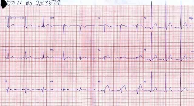
Prezado Professor Andrés y queridos colegas/amigos:

Homem, 47 anos, com história de precordial tipica. Pometo a coronariografia
em 24 horas

Raimundo Babarbosa Barros. “ A raposa”

Qual é a artéria culpada?

-----------------------------------------------------------------------------------------------------------------
Dear Professor Andrés and dear colleagues/friends: Man, 47yo, with a history of
typical chest pain. I promise the coronary angiography within 24 hours
Whats is the culprit artery?

Raimundo Barbosa Barros MD “Te Fox”
--------------------------------------------------------------------------------------------------------------------------
Estimado Profesor Andrés y queridos colegas/amigos: Hombre, de 47 años. Con
historia de dolor precordial típico. Prometo la cinecoronariografia dentro de 24
horas.

Cual es la arteria culpada?

Raimundo Barbosa Barros “ El zorro”
COLLEAGUES OPINIONS
 OPINIÃO DOS COLEGAS
OPINIÓN DE LOS COLEGAS

Me parece que este paciente tiene un infarto agudo inferior. Creo que en este caso esta
comprometido el septo interventricular tanto izquierdo cuanto derecho por oclusión
subita de la arteria coronaria derecha a nivel de la marginal primera,
Saludos Pancho Femenia
----------------------------------------------------------------------------------------------------------------------------
I think this patient has an acute inferior myocardial infarction. I think in this case is
committed both left interventricular septum as a right by sudden occlusion of right
coronary artery(RCA) at the level of the marginal first
Regards
Francisco FemeniaMD Mendoza – Argentina
------------------------------------------------------------------------------------------------------------------------------
Looks like LAD occlusion, of note is short ST segment ,is hypercalcemia present? Acute
pancreatitis cam mimic MI. Is this a trick?
-------------------------------------------------------------------------------------------------------------
Parece oclusión de la arteria descendente anterior. El segmento ST es corto. Está
presente hipercalcemia? Pancreatitis aguda puede imitar IM. ¿Es esto un truco?

Melvin M Scheinman, MD PhD
Department of Cardiac Electrophysiology, University of California San
Francisco, San Francisco, California, USA. scheinman@medicine.ucsf.edu
Professor of Medicine
Address: UCSFElectrophysiology Service 500 Parnassus Avenue San
Francisco, CA 94143-1354. Telephone/FAX/E-mail: Phone: (415) 476-5706
Fax: (415) 476-6260
email: scheinman@medicine.ucsf.edu
Dear Andres, The ECG during this young man's acute coronary syndrome suggests a mid
or distal LAD lesion, perhaps distal to the 1st diagonal. I believe this is because there is
no ST segment depression in the inferior leads.
Regards,
Frank
--------------------------------------------------------------------------------------------------------------
Querido Andrés, el ECG en este síndrome coronario agudo de este hombre joven sugiere
una lesión media o distal de la LAD, tal vez distal a la primera diagonal porque no hay
depresión del segmento ST en las derivaciones inferiores.
Un cordial saludo,

Frank
-----------------------------------------------------------------------------------------------------------------------------
Caro Andrés, O ECG durante a síndrome aguda do jovem homem sugere uma lesão
média ou distal da LAD, talvez distal a 1ra diagonal. Eu acredito isto porque não há
depressão do segmento ST nas derivações inferiores.
Atenciosamente,
Frank
-------------------------------------------------------------------------------------------------------------------------------
LAD proximal third
AB
------------------------------------------------------------------------------------------------------------
1/ proximal da Descendente Anterior
  3
AB
-----------------------------------------------------------------------------------------------------------------
Hello. Everything else than LAD would be surprising as the maximal ST elevation is
in V2-V3.
Regards
Kjell Nikus
Tampere, Finland
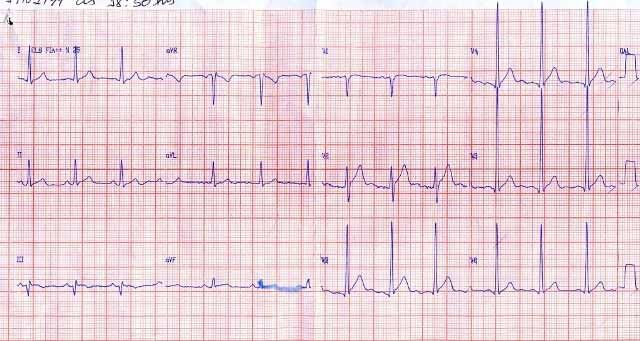
The changes in the extremity leads are rather mild. In the first ECG, there seems to be slight
elevation of the ST segment in the inferior leads, which would point to a distal occlusion in a
"wrap-around-the-apex" LAD. However, in the second recording, where there is more severe
ischemia, aVL is elevated, while LIII is slightly depressed ("reciprocal" ST depression).
Anatomical information should be analyzed from the ECG with maximal ischemia. The
findings indicate that there is a medium-sized or large diagonal branch distal to the occlusion.
I am not sure about the QRS duration, but there seems to be at least partial RBBB, which also
points to a proximal occlusion (proximal to the second septal branch, which usually is a large
septal, subtending the right bundle branch). We have no previous ECG, so we don't know if
the mild intraventricular conduction defect is old or new. But, summa summarum, I would
expect a proximal LAD occlusion in a wrapping LAD. Anatomic prediction is influenced by
inter-individual variation of coronary anatomy
Kind regards Kjell Nikus Tampere Finland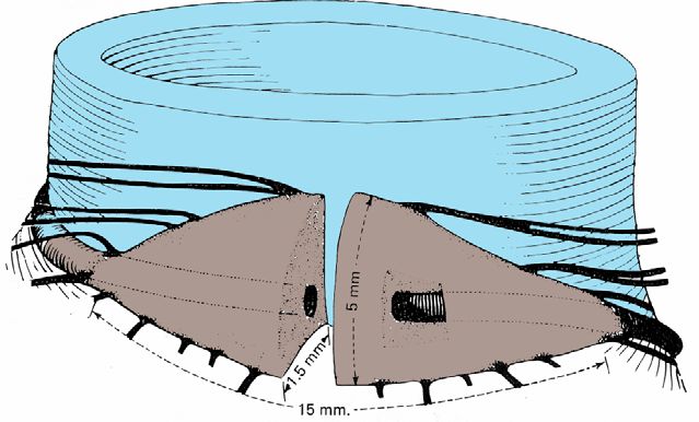
FINAL CONCLUSION AND
THEORETICAL CONSIDERATIONS
    CONCLUSÕES FINAIS E
  CONSIDERAÇÕES TEÓRICAS

     Andrés Ricardo Pérez-Riera MD
Coronariografia: TCE,DA e Cx normais; oclusão de CD no terço distal. Todos nós sabemos que uma oclusão proximal de CD com envolvimento do VD pode simular um IAM anteroseptal. Infelizmente não houve sucesso na tentativa de angioplastia primária; FLUXO TIMI 0. -------------------------------------------------------------------------------------------------------------------------------- Coronariography: LMCA, LAD and LCX: normal. RCA with distal obstruction. We all know that a proximal occlusion of the RCA with right ventricular involvement may simulate an acute anteroseptal MI. Unfortunately there was no success in the attempt of primary angioplasty, TIMI flow 0. ---------------------------------------------------------------------------------------------------------------------------------- Nem sempre é posivel identificar pelo ECG a artéria relacionada ao infarto (ARI). Traçados de ECGs com 18-derivações foram comparados com os respectivos resultados das angiográfias de 1024 pacientes consecutivos. Foram realizados em todos os pacientes mais de dois ECGs de 18 derivações dentro das 12 horas de iniciados os sintomas. Foram critérios de exclusão: Pacientes com IM prévio, com cirurgia de revascularização miocárdica, com implante de marcapassos ou com padrão BCRE e angiografia realizada com mais de 12 horas do início dos sintomas ---------------------------------------------------------------------------------------------------------------------------------- The infarct-related artery (IRA) could not always be identified by ECG. the reason for failed IRA identification by ECG based on the comparison between ECG records and coronary angiographic findings. All 18-lead ECG records were compared with respective angiographic findings in 1024 consecutive patients with STEM. More than two continous18-lead ECG records were performed within 12 hours of the symptom onset in all patients. Exclusion criteria: Patients with previous MI, coronary artery bypass surgery, pacemaker implantation or ECG evidence of LBBB and angiography was performed more than 12 hours time from symptom onset.
Of all 1024 patients enrolled, the IRA were correctly identified in 854 cases and identified wrong
in 96 cases and could not be identified in 74 cases by ECG. Of the failed identification in these
170 cases, IRA was LCX in 76 (44.7%)cases, RCA in 66 (38.8%) cases, LAD in 20 (11.8%)
cases, ramus medianus branch in 7 (4.1%) cases, and LMCA in 1(0.6%) case. Double-vessel
and triple-vessel diseases were recorded in 27(15.9%) patients and 47(27.6%) patients
respectively. Early repolarization syndrome occurred in 8 (4.7%) patients, and dextrocardia in 1
patient (0.6%). Angiographic study showed acute occlusion of a small branch in 6 (3.5%)
patients. The authors concluded that coronary collateral vessel can mislead judgments of the IRA
by ECG. When the IRA can not be determined by ECG, LCX is most likely to be the culprit
vessel. Occasionally, early repolarization syndrome and anatomic variation of the coronary artery
or heart and a small branch occlusion could be causes of misjudgments of IRA by ECG.
------------------------------------------------------------------------------------------------------------------------------
De todos os 1024 pacientes incluídos, foram corretamente identificados pelo ECG 854 casos.
Identificação equivocada ocorreu em 96 casos e não pôde ser identificada em 74. Dos 170 casos
com falha na identificação 76 (44,7%) foram da circunflexa esquerda, casos 66 (38,8%), da
coronária direita e em 20 pacientes (11,8%) a DA. O ramo mediano em 7 (4,1%) casos, e O
Tronco da CE em 1 (0,6%) caso. Doença de dois vasos foi observado em 27 (15,9%) e de três
vasos em 47 (27,6%), respectivamente. Síndrome de repolarização precoce ocorreu em 8 (4,7%)
e dextrocardia em um paciente (0,6%). O estudo angiográfico mostrou oclusão aguda de um
pequeno ramo em 6 (3,5%). Os autores concluíram que a presença de circulação colateral
coronária é o fator principal de falsa identificação da artéria culpada pelo ECG. Quando a
artéria relacionada a obstrução não pode ser determinado pelo ECG a circunflexa é o vaso mais
provávelmente culpado. Ocasionalmente, a síndrome de repolarização precoce e variação
anatômica da artéria coronariana ou cardíaca e oclusão de ramos pequenos poderiam ser
causas de incorreções de identificação da artéria culpada pelo ECG.
  1.   Zhang XJ, Yan HB, Zheng B, Song L, Wang J, Chi YP. Reasons for failed electrocardiographic identification of the
       infarct-related artery in patients with ST-elevation acute myocardial infarction. Zhonghua Xin Xue Guan Bing Za Zhi.
       2010 Oct;38:914-917.
Anterior STE is the classic ECG feature of anterior LV myocardial infarction due to occlusion of the LAD. However, anterior ST-segment elevation has rarely also been described in patients with RCA occlusion at several levels (Mainly proximal) without inferior ST-segment elevation. It is hypothesized that the inferior LV wall is protected by left-to-right collaterals, as seen on coronary angiography, with resultant isolated RV infarction RCA occlusions. Isolated RV infarction resulting in an ECG pattern mimicking anterior-wall LV infarction. STE na parede anterior é a característica clássico do ECG de infarto do miocárdio do VE anterior, devido à oclusão da artéria descendente anterior. No entanto, a elevação do segmento ST anterior raramente tem sido descrita também em pacientes com oclusão RCA em diversos níveis (principalmente proximal) sem elevação concomitante do segmento ST em parede inferior. A hipótese que tenta explicar esta ausência de isquemia na parede inferior do VE é a proteção oferecida por colaterais da esquerda para a direita, como visto na angiografia coronária, com conseqüente infarto isolado do VD oclusão da coronária direita. O infarto isolado do VD,pode resultar em um padrão de ECG que imita o infarto de parede anterior do VE.
Right ventricular infarction (RVI) during inferior MI is readily diagnosed when STE is recorded in
lead V4R. RVI may also yield precordial STE and such an ECG pattern may be misinterpreted
as a sign of anterior MI. Inferior-RVMI due to occlusion of a dominant RCA manifesting STE in
precordial and right chest leads1. RV dilation due to acute ischemic insult facilitated STE in leads
V1-V4 despite the dominant opponent inferior and posterolateral LV injury current. Dilation of an
infarcted RV should be considered when such an ECG pattern is encountered during inferior MI,
specifically a dominant one. Awareness of the circumstances under which this ECG pattern
develops facilitates avoidance of misinterpretation as a sign of anterior MI and proper
management.

1.   Andreou AY, Georgiou GM. Dominant right coronary artery occlusion entailing diffuse ST-segment elevation in the
     precordial leads. J Cardiovasc Med (Hagerstown). 2010 Nov;11:843-847.
ECG VALUE FOR LOCALIZATION OF “CULPRIT”
ARTERY IN ACUTE CORONARY SYNDROMES
(ACS) WITH ST SEGMENT ELEVATION (STEMI)
Andrés Ricardo Pérez Riera, MD
Chief of Electovectorcardiography Sector - Cardiology Discipline - ABC
Faculty – ABC Foundation – Santo André – São Paulo – Brazil
riera@uol.com.brTHE ECG IN ACUTE CORONARY SYNDROME(ACS)
Patients with ACS include those whose clinical presentations cover the following range of
diagnoses:
I) Unstable angina: New-onset exertion angina, angina increasing frequency or
duration or refractory to nitroglycerin, or angina at rest.
II) Non–ST-Elevation Myocardial Infarction (NSTEMI)
III) ST-elevation Myocardial Infarction (STEMI).
ST-segment Elevation Myocardial Infarction (STEMI)
New or presumably recent J point and ST segment elevation in 2 or more adjacent leads
> 2 mm in V1, V2 or V3 or > 1 mm in other leads
Non-ST segment Elevation Myocardial Infarction (NSTEMI)
ST segment depression
Isolated alterations of the T wave
This ACS spectrum concept is a useful framework for developing therapeutic strategies
1. Alpert JS, Thygesen K, Antman E, Bassand JP. Myocardial infarction redefined--a consensus document of The
Joint European Society of Cardiology/American College of Cardiology Committee for the redefinition of
myocardial infarction. J Am Coll Cardiol. 2000 Sep;36:959-969. Erratum in: J Am Coll Cardiol 2001 Mar 1;37:973.ACS
ACS
ELECTROCARDIOGRAM
STEMI NSTEMI
NSTEMI
STEMI
>>22mm
mmininVV1, ,VV2or
1 2
orVV3or
3
or>>11mm
mmininother
otherleads
leads
NEGATIVE POSITIVE
Q-WAVE
Q- WAVEMI
MI NON-QMI
NON-Q MI NEWLBBB
LBBB TRUE BIOMARKERS BIOMARKERS
NEW TRUE
POSTERIOR *
POSTERIOR UNSTABLE
UNSTABLE NON-QMI
NON-Q MI
MIPATTERN
MI PATTERN
ANGINA
ANGINA
THROMBOLYTICS OR PRIMARY
CORONARY ANGIOPLASTY
* Actual basal inferior.1 2 CORONARY CIRCULATION
Anterior Septal
6 Perforator Branches
3 S2 S1: First Septal Perforator branch
S1 S2: Second Septal Perforator
S3: Third Septal Perforator
4
S3
7 S’: Posterior Septal Perforators
S’
S’
S’ S’
S’
5
1. Left Main Coronary Artery (LMCA)
2. Left Anterior Descending Artery (LAD)
3. Left Circumflex Coronary Artery (LCX)
4. Right Coronary Artery (RCA)
5. Posterior Descending Artery (PDA). In this case is supplied by the RCA, then the coronary
circulation can be classified as "right-dominant“
6. First Diagonal (Dg)
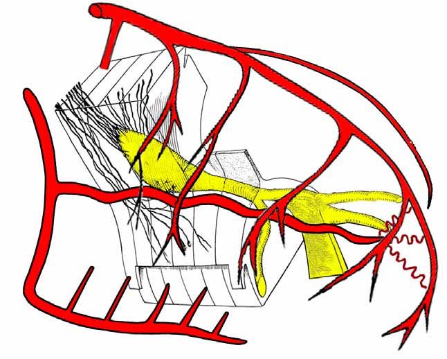
7. Acute Marginal (A. Mg)BLOOD SUPPLY OF SA-NODE
In the majority of individuals (≈ 59% of cases), the SA-node receives blood from a SA node
artery. This is the second branch of the RCA (the first one is the conus artery) and in 38% of
cases from the LCX and from both arteries in 3%1.
SVC
SA node
RCA OCCLUSION
SUPERIOR VENA CAVA
CONUS ARTERY
SANA AV-N
PROXIMAL
RCA
DRA
RV SA NODE
SA NO
DISTAL
DE A R
MIDDLE TERY
RCA RCA
Ac Mg
PDA LVB
RA – RIGHT ATRIUM
1. Kyriakidis MK, Kourouklis CB, Papaioannou JT, Christakos SG, Spanos GP, Avgoustakis DG. Sinus node coronary
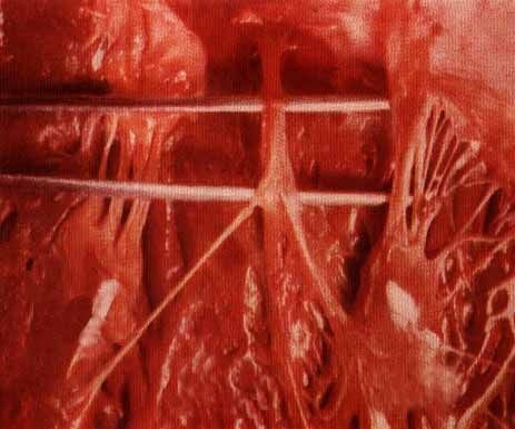
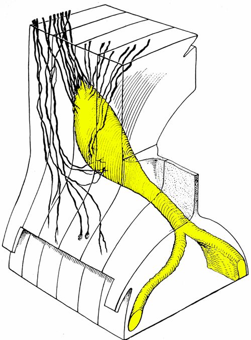
arteries studied with angiography. Am J Cardiol. 1983 Mar 1;51:749-750.THE JUNCTIONAL OR ATRIOVENTRICULAR AREA
The AV junction can be divided into 3 regions as follows:
1. Transitional cell zone = AN: Atrionodal Region
2. Compact AV node = N: Nodal Region
3. Penetrating portion of the AV bundle = NH
4. Penetrating portion of His bundle = H
POSTERIOR INTERNODAL BUNDLE OR OF THOREL
P MIDDLE INTERNODAL BUNDLE OF WENCKEBACH
M A ANTERIOR INTERNODAL BUNDLE
ATRIO-NODAL REGION (TRANSITIONAL CELL ZONE)
NODAL REGION (THE COMPACT PORTION)
TRACT OF AN
JAMES N HIS NODE REGION
NH (THE PENETRATING PART OF THE AV BUNDLE)
H HIS BUNDLE(NON BRANCHING PORTION)
LBB LEFT BUNDLE BRANCH
RBB RIGHT BUNDLE BRANCHBLOOD SUPPLY OF THE COMPACT AV NODE
In 85% of cases AV node receives its blood supply from the RCA. In the remaining 13% by the
LCX and in 2% by both arteries1.
The AV node becomes the AV His bundle at the point where the overall axis for conduction
penetrates into the central fibrous body2.
BLOOD SUPPLY OF THE HIS BUNDLE
This structure has double blood supply: from branches of the LAD and PDA3.
BLOOD SUPPLY OF THE LEFT BUNDLE BRANCH
(LBB)
• Branches of the PDA (90% of the RCA): AV node artery: ramus septi fibrosi, ramus
septi ventriculorum superior and ramus cristae.
• Branches of LAD: Ramus limbi sinistri (equivalent to ramus limbi dextri of the LDA).
1. Hadziselimović H. Vascularization of the conducting system in the human heart. Acta Anat (Basel). 1978;102:105-110.
2. Anderson RH, Ho SY, Becker AE. Anatomy of the human atrioventricular junctions revisited. Anat Rec. 2000 Sep
1;260:81-91.
3. Lumg G, Singletary HP. Blood Supply to the Atrioventricular Node and Bundle of His: A Comparative Study in Pig,
Dog, and Man Am J Pathol. 1962 Jul;41:65-75.BLOOD SUPPLY OF THE RIGHT BUNDLE BRANCH
(RBB)
AV- N
PROXIMAL PORTION is irrigated by the AV node artery of
the RCA and the first septal perforator artery
(S1) of the LAD.
MIDDLE PORTION is irrigated by:
Posterior Septal perforators of the PDA LBB
Second septal perforator artery(S2) of the LAD
Kugel’s artery, branch of the LCX.
F
LP
MIDDLE AND DISTAL PORTION: are irrigated by the LA
“Ramus limbi dextri”, branch of the S2 of the LAD. F
LSF
1) His Right Penetrating Portion.
2) His Right Branching Portion
3) Proximal or membranous of RBB
4) Middle, intramyocardial or mimetic
5) Inferior, distal or intra-moderator Band.BLOOD SUPPLY OF THE LEFT FASCICLES
1. Left Anterior Fascicle (LAF) Is supplied either by septal branches of the LAD or by the
AV nodal artery
2. Left Posterior Fascicle (LPF): The proximal part of LPF is supplied by the artery to the
AV Node and, at times, by septal branches of the LAD artery. The distal portion has a dual
blood supply from both anterior (S) and posterior (S`) Septal Perforator Arteries.
3. Left Septal Fascicle (LSF) or Left Median Fascicle: It is supplied exclusively by
septal branches of the LAD. Critical lesions of the LAD before the first septal perforator,
constitute the main cause of LSFB in the first world.
RESPONSIBLE LAF LPF LSF
SYSTEM
Branches of the LAD 40 % 10 % 100 %
Double irrigation 50 % 40 % 0%
(LAD & RCA)
RCA branches 10 % 50 % 0%LBB
LBB
LP
F
F
A
L
LSF
PMPM
ALPM 100% LAD
ALPM: ANTEROLATERAL PAPILLARY MUSCLE
PMPM: POSTERO MEDIAL PAPILLARY MUSCLELSF DISTRIBUTION AND TRAJECTORY
RB
LB
LAF
central
apical LSF
2 LA
3 LPF
1
1) Left Septal Fascicle: exclusively by septal branches of the LAD. Critical lesions of the
LAD before the S1, constitute the main cause of LSFB in the first world.
2) Left Anterior Fascicle
3) Left Posterior Fascicle.KILLIP SCORING SYSTEM - KILLIP CLASS OR THE
KILLIP-KIMBALL CLASSIFICATION1
The Killip classification is a system used in individuals with an acute myocardial infarction (AMI) in
order to risk stratify them. Individuals with a low Killip class are less likely to die within the first 30
days after their AMI than individuals with a high Killip class. Mortality rises dramatically through the
classes form I to IV. Patients were ranked by Killip class in the following way:
Killip class I: includes individuals with no clinical signs of heart failure: Absence of rales over the
lung fields and absence of a third heart sound(S3).Forrester: wedge ≤18mm Hg. Normal perfusion.
Killip class II: includes individuals with rales or crackles over 50% of the lung fields in the lungs,
an S3, and elevated jugular venous pressure. Forrester: wedge ≤18mm Hg. Poor perfusion.
Hipovolemic.
Killip class III: describes individuals with frank acute pulmonary edema: Rales over > 50% of
the lung fields and S3. Forrester: almost normal perfusion, increased pulmonary capillary pressure
and pulmonary congestion.
Killip class IV: describes individuals in cardiogenic shock or hypotension (measured as systolic
blood pressure < 90 mmHg), and evidence of peripheral vasoconstriction (oliguria, cyanosis or
sweating). Patients with or without lung congestion can be placed in class IV if they are in
cardiogenic shock.
1. Killip T 3rd, Kimball JT.Treatment of myocardial infarction in a coronary care unit : A Two year experience with 250
patients. Am J Cardiol. 1967 Oct; 20:457-464.Name: AR.; Date: 02/01/2009.; Age: 72 yo.; Gender: Male.; Ethnic Group: Caucasian Weight:
72 Kg.; Height: 1.74 m; Biotype: Mesomorphic; Management: Coronary Artery Bypass Graft
(CABG) 72 hours ago.
LMCA
LAFB
PAF: LSFB
Clinical features: ACS: 72-year-old male patient, admitted in the emergency room with typical
precordial pain that yielded after the administration of IV nitroglycerin.
ECG diagnosis: 1) LAFB + 2) LSFB: PAF + Lesion block + aVR lead with ST segment elevation
suggestive of obstruction in the LMCA. Laboratory: There was no increase of necrosis markers
(CK-MB/troponin).The coronary angiography revealed LMCA spasm + proximal critical lesion of
the LAD. Management: The patient was urgently revascularized, successfully. (Coronary Artery
Bypass Graft ).Name: AR; Date: 05/01/2009; Age: 72 yo;Gender: Male
Ethnic Group: Caucasian; Weight: 72 Kg; Height: 1.74 m; Biotype: Mesomorphic;
Management: Coronary Artery Bypass Graft (CABG) 72 hours ago.
Electrocardiogram conducted on the third day after successful surgery.
Both divisional blocks have disappeared: the extreme shift of QRS electric axis to the left in the
frontal plane (LAFB) is not seen, and prominent anterior forces (LSFB) has disappeared.Date: Date: Date: Date:
02/01/2009 05/01/2009 02/01/2009 05/01/2009
I V1
II V2
PAF
III
V3
WITH WITHOUT WITH PAF: WITHOUT
LAFB LAFB LSFB PAF: LSFB"TOMBSTONING" OF ST SEGMENT IN ACUTE MYOCARDIAL INFARCTION.1
Tombstoning (TOMB-ST) is manifested by a particular changing the shape of repolarization:
monophasic action potential-like patter consequence of proximal occlusion of LAD.
TOMB-ST has been associated with a poor prognosis ever since Wimalaratna's first description1;2
of this clinical entity, and the reasons for this are not fully understood.
Reperfusion injury reflected as TOMB-ST in patient following successful AMI PTCA3.
TOMB-ST can be seen also in an agonal ECG
TOMB-ST pattern suggest large infarction, low LVEF, increased mortality rate, HF, VF and higher
initial N-terminus pro-brain natriuretic peptide (NT-pro-BNP) level4.
In the population with TOMB-ST, increased mortality was independent of the total amplitude of ST
segment displacement; this relation was, however, observed in patients with STEMI without
TOMB-ST. The sum of amplitudes of ST segment deviations (SigmaST) >20 mm Is indicative for
the subgroup of patients with TOMB-ST and trend towards higher mortality. However, in patients
without TOMB-ST, SigmaST >20 mm identified two subgroups with significantly different mortality
rates (20% vs 4%, p=0.001) 5.
Rarely TOM-ST is secondary to acute pericarditis. In these rare cases the role of two-dimensional
echocardiogram is important6.
1. Wimalaratna HS. "Tombstoning" of ST segment in acute myocardial infarction. Lancet. 1993 Aug 21;342(8869):496.
2. Birnbaum Y, Sclarovsky S. "Tombstoning" of ST segment in acute myocardial infarction. Lancet. 1993 Dec
11;342(8885):1494.
3. Dalal J, Chambers CE.Marked ST elevation after successful PTCA for acute myocardial infarction. J Invasive Cardiol.
1994 Oct;6:263-266.
4. Tomcsányi J, Marosi A, Bózsik B, Somlói M, Zsoldos A, Vecsey T, et al. N-terminal pro-brain natriuretic peptide and
tombstoning ST-segment elevation in patients with anterior wall acute myocardial infarction. Am J Cardiol. 2005 Nov
1;96:1197-1199.
5. Kukla P, Dudek D, Szczuka K. "Tombstoning" of ST segment in acute myocardial infarction -- effect on clinical course.
Kardiol Pol. 2006 Mar;64:275-80.
6. Jain A. "Tombstone" anterior ST-segment elevations secondary to acute pericarditis: the role of two-dimensional
echocardiogram. Clin Cardiol. 1997 Apr;20:404-406.TOMB-ST ECG CHARACTERISTICS
An ST-segment elevation with a specific pattern is the principal element of TOMB-STEMI.
ST-segment elevation is often the earliest detected sign of acute MI. Initially, the ST segment
may straighten, with loss of the ST-T wave angle. Then the T wave becomes broader and the
ST segment elevates, losing its normal concavity. As further elevation occurs, the ST
segment tends to become convex upwards. As ST-segment elevation can be minimal, in
some cases, it may surpass the peak level of the R wave.
Thus, ST-segment elevation surpassing the R wave exhibits such a morphological
appearance that it reminds a tombstone.
---------------------------------------------------------------------------------------------------------------------------------
Tombstone appearance ST segment convex to the top
Ápex R wave
Here lie my desire
to study
Two electrophysiological mechanisms play a role in the formation of a tombstone
appearance: delayed transmural conduction and intramyocardial conduction block.TYPICAL ECG PATTERN OF LMCA OCCLUSION
Diffuse ST segment depression in the inferolateral leads
Minimal ST segment elevation
ST segment depression in II
ST segment elevation in aVR > V1 Depression of the ST segment from V4 to V5.
ST segment I and aVL
Why this pattern is observed? ST segment depression in V6 > ST
segment elevation in V1.The ST vector
lesion
pointing to aVR
LCx
ST segment LMCA
elevation
LAD
LV
RV
ST segment
depression in II
I and aVLaVR STSE > V1 STSE
Minimal ST segment elevation
ST segment elevation in aVR > V1. Why?
Because ST segment lesion vector is directed to upward and rightward, pointing to aVR
lead( RVOT)Ischemic evidences on
inferobasal wall:
ST segment depression
from V4 to V5
Z
ST SEGMENT
LESION VECTOR
POINTED TO RIGHT
AND BACKWARD
X
Minimal ST segment
elevation in V1
Depression of the ST
segment from V2 to V6LMCA OCCLUSION ECG CRITERIA
• ST segment elevation in aVR, and V1
• ST segment elevation in aVR > V1
• Ischemic evidences in inferobasal* wall: depression of the ST segment in II
and from V4 to V5
• ST segment depression in II or in inferior leads II>III
• Depression of ST segment in V6 > ST segment elevation in V1
• Diffuse ST segment depression in the inferolateral leads
• Eventually observation of RBBB, LAFB and/or LSFB.
4
10
15 4
* Formally called inferodorsal wallST segment elevation in aVR > V1 ST segment depression in II or in inferior leads II>III Depression of the ST ST depression segment segment in inferior leads from V3 to V6 Clinical Picture: Acute Coronary Syndrome associated with cardiogenic shock (Killip class IV) consequence of total occlusion of LMCA. Primary Angioplasty was performed, with immediately hemodynamic stabilization.
LMCA Occlusion complicated with Complete RBBB.

         ST depression II>III ST

    ST segment elevation in aVR

ST segment elevation in aVR and V1 (aVR > V1.). ST depression II>III ST depression
segment from V2 to V6.
ST segment elevation in aVR > V1 ST segment elevation = 6mm ST segment elevation = 5,5mm
QRS AXIS LOCATED IN RIGHT SUPERIOR QUADRANT

                                   LV
                        RV

                                                       ST segment
                                                      depression in
                                                      inferior leads
                     ST segment depression II > III        II>III
Z
ST SEGMENT DEVIATION
POINTING TO RIGHTWARD
AND BACKWARD BUT IN
POSITIVE QUADRANT OF V1
X
Ischemic evidences in
basal region: depression
of the ST segment from
V4 to V5.
= Positive quadrant of V1. The ST segment vector is inside of positive quadrant of V1LEFT ANTERIOR DESCENDING ARTERY (LAD)
OCCLUSION BEFORE FIRST SEPTAL
PERFORATOR(S1): PROXIMAL LAD OCCLUSIONAMI consequence of occlusion of LAD before the first septal
perforator and the first diagonal branch
ST segment elevation ≥ 2mm from V1 to V3
ST segment depression in inferior leads
ST segment elevation in aVL and aVR
ST segment depression in V5 and V6
Why we observe this pattern?ST SEGMENT LESION VECTOR DEVIATION
Dg
S1
LV
LAD X
RV
Y
ST SEGMENT VECTOR LESION POINTING TO UP, CAUSING ST SEGMENT
ELEVATION IN aVL AND aVR AND ST SEGMENT DEPRESSION IN INFERIOR
LEADS.ST segment elevation ≥ 2mm from V1 to V3 or V4 (ST segment lesion vector
directed to front). ST segment depression in V5 and V6 or isoelectric. Eventually
CRBBB and/or LAFB and/or LSFB.
ST segment
depression in V5
Z and V6 or
isoelectric.
X
ST SEGMENT LESION
VECTOR DIRECTED TO
FRONT AND MINIMAL
RIGHTWARD
ST segment elevation ≥ 2mm from V1 to V3 or V4AMI consequence of proximal LAD occlusion before S1
complicated with RBBB.
qR pattern
ST segment depression in inferior leads.
ST segment elevation ≥ 2mm from V1 to V4
ST segment elevation in aVL and aVRWELLENS' SYNDROME, LAD CORONARY T-WAVE
SYNDROME OR ACUTE CORONARY T-WAVE
SYNDROME.
Wellens' syndrome is a clinical-electrocardiographic entity.
It is a complex of symptoms and signals indicating the existence of an undesirable
condition secondary to critical high-grade proximal stenosis of the LAD coronary
artery characterized by the association of:
1) Prior history of ACS
2) Little or no elevation of markers of myocardial damage (unstable angina)
3) Characteristic ECG changes consistent with subepicardial anterior ischemic pattern in
the LAD territory (V1 through V5 or V6):
¾ Plus-minus T waves with inversion of the terminal portion: Type 1
¾ Persistently symmetrical, deep negative and broad-based T-waves: Type 2
4) Sensitivity and specificity for significant (≥ 70%) stenosis of the LAD artery was found
to be 69% and 89% respectively with positive predictive value 86%.²
1. de Zwaan C, Bär FW, Wellens HJ. Characteristic electrocardiographic pattern indicating a critical stenosis high in
left anterior descending coronary artery in patients admitted because of impending myocardial infarction. Am Heart
J. 1982 Apr;103:730-736.
2. Haines DE, Raabe DS, Gundel WD, Wackers FJ. Anatomic and prognostic significance of new T-wave inversion in
unstable angina. Am J Cardiol. 1983 Jul;52:14-18.ECG performed upon arrival to the Emergency Department (04/29/2008), and while having chest
pain. Deep negative and broad-based T-wave inversions in precordial leads from V2 through V6,
with high voltage R wave in V2 (R=18mm). Initial small q waves were observed in V2-V3. Left
septal initial q waves in left leads are absent. R/S ratio in V2 > 2. S wave depth in V2 < 5 mm.
Conclusion: Type 2 Wellens' pattern associated with prominent anterior forces: several Left
Septal Fascicular Block criteria are present.
1. Riera AR, Ferreira C, Ferreira Filho C, Dubner S, Schapachnik E, Uchida AH, Moffa P, Zhang L, de Luna AB. Wellens
syndrome associated with prominent anterior QRS forces: an expression of left septal fascicular block? J
Electrocardiol. 2008 Nov-Dec;41:671-674.ECGs performed one year before clinical manifestation (07/04/2007). There are not subepicardial T wave ischemic pattern and QRS complexes of the rS type in V2. Initial q wave are observed in left leads I, aVL, V5 and V6.
A) Basal anteroseptal precordial leads performed approximately one year before onset of clinical
picture.
B) The same leads performed during the clinical manifestation.
ECG performed ten days after (05/09/2008) the successful placement of the
stents in LAD. The ischemic pattern had disappeared, the lead V2 returned
to rS, the initial q wave in V3 disappeared, and small septal q waves
appeared in the left leads I, aVL, V5 and V6.
You can also read