Feasibility study in the combination of non-contrast computed tomography and transcranial Doppler for large vessel occlusion detection
←
→
Page content transcription
If your browser does not render page correctly, please read the page content below
Neurology and Neurosurgery

  Research Article                                                                                                                      ISSN: 2631-4339

Feasibility study in the combination of non-contrast
computed tomography and transcranial Doppler for large
vessel occlusion detection
Amber Y Dorn*, Samuel G Thorpe, Kian Jalaleddini, Seth J Wilk, Robert B Hamilton
Novasignal Corporation, Los Angeles, CA, 90064, USA

Abstract
Background and purpose: Accurate detection of Large Vessel Occlusion (LVO), and subsequent assessment of candidacy for endovascular intervention, present
challenging clinical obstacles to efficient stroke patient triage, especially when computed-tomography angiogram (CTA) is not readily available especially in non-
primary or comprehensive stroke centers. The Alberta Stroke Program Early CT Score (ASPECTS) is used to evaluate a patient’s non-contrast CT (NCCT) exam
for evidence of cerebral tissue damage and provides information about the tissue affected by stroke to determine course of treatment. Velocity curvature index (VCI)
is a biomarker derived from the morphological analysis of transcranial Doppler (TCD) waveforms and has shown utility in TCD assessment of LVO. The purpose
of this study was to evaluate the relationship between ASPECTS and VCI and to explore their combined diagnostic capabilities. We hypothesized that ASPECTS’
assessment of LVO can be augmented with TCD imaging modality for the purpose of faster door-to-intervention workflow and triage of LVO patients to stroke
centers.
Methods: NIHSS, standard of care, and TCD imaging were collected from two clinical populations; 33 LVO (16 female) and 33 In-Hospital Controls (13 female)
enrolled consecutively at a regional stroke center from October 2016 through September 2017. ASPECTS and VCI were derived from NCCT and TCD exams
respectively, and retrospectively evaluated in their performance against gold standard CTA.
Results: ASPECTS was not significantly correlated with VCI and predicted LVO with accuracy, sensitivity, and specificity of 82%, 97%, and 66% while VCI
performed with 91%, 91%, and 88%. Combining ASPECTS and VCI resulted in an accuracy of 91%, sensitivity of 97%, and specificity of 85%.
Conclusions: VCI and ASPECTS’ lack of correlation but independent strengths in LVO assessment indicate that they may shed light on differing mechanisms of
stroke pathology.

Introduction                                                                        trials underlines the critical importance of accurate and minimally
                                                                                    invasive diagnostics. Now more than ever, there is a need for rapid
     Acute ischemic strokes often result from a large vessel occlusion              and conclusive stroke triage and diagnosis to help facilitate rapid and
(LVO) of one of the major feeding arteries of the brain. Because of the             effective treatment [7].
brain’s reliance upon these major vessels for perfusion, LVO strokes
have high morbidity and mortality due to the difficulty of treatments in                 Currently, non-contrast computed tomography (NCCT), magnetic
achieving recanalization [1]. As such, these LVO strokes carry generally            resonance imaging (MRI), CT angiography (CTA), and magnetic
worse prognoses, sometimes even despite intravenous thrombolysis [2].               resonance angiography (MRA) are the standard minimally-invasive
When intravenous thrombolysis has been initiated but no recanalization              imaging protocols trusted by physicians in acute stroke assessment.
or clinical improvement has been observed, endovascular treatment                   When there are no limits of machine or expert availability, or patient
(EVT) is the next course of action and is only possible at comprehensive            contraindications, CTA is the preferred method of imaging. NCCT
stroke centers (CSC). Recent randomized control trials evaluating the               is a standard of stroke imaging and is relied upon heavily when CTA
efficacy of endovascular treatment have shown the importance of                     is unavailable. Once a NCCT exam has been performed, images can
timely initiation of this surgical intervention. The results of the SEER            be analyzed to derive a patient’s tissue status using the Alberta Stroke
collaboration demonstrate that a shorter time from stroke onset to                  Program Early CT Score (ASPECTS). A patient’s ASPECTS is based
reperfusion indicates potential for better outcome, which declines                  on assessment of the tissue in the MCA watershed regions such that
greatly for every minute of delay [3]. The STRATIS trial showed that                one point is deducted for each of 10 regions that appear hypoperfused
an increased time from EMS arrival to puncture (onset of endovascular
treatment) showed a reduced likelihood of good functional recovery
[4]. This important detail in successful stroke intervention was also
                                                                                    *Correspondence to: Amber Yvonne Eddy Dorn, Novasignal Corporation, Los
noted in the SWIFT-PRIME trial and HERMES meta-analysis, with                       Angeles, CA, 90064, USA, E-mail: amber.dorn@neuralanalytics.com
significant negative effects of prolonged periods from symptom onset
to mechanical thrombectomy negatively impacting outcome [5,6]. As                   Key words: transcranial doppler, TCD, ischemic stroke, large vessel occlusion,
recanalization becomes more attainable with increased availability                  ASPECTS
of endovascular intervention, the time-sensitivity observed in these                Received: April 02, 2020; Accepted: April 13, 2020; Published: April 16, 2020

Neuro Neurosurg, 2020       doi: 10.15761/NNS.1000130                                                                                              Volume 3: 1-6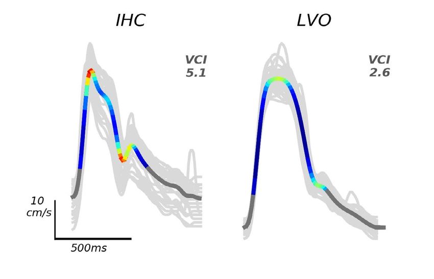
Dorn AY (2020) Feasibility study in the combination of non-contrast computed tomography and transcranial Doppler for large vessel occlusion detection

and can be used to asses a patient’s candidacy for surgical intervention;        ASPECTS’ assessment of LVO can be augmented with TCD imaging
a threshold of 7 is used in the prediction of both functional outcome            modality for the purpose of faster door-to-intervention workflow and
and final infarct volume in ischemic stroke [4,8–10]. In its prediction of       triage of LVO patients to stroke centers.
poor outcome, an ASPECTS threshold of 7 has been shown to reflect an
overall accuracy of 65.8% [11]. Though these studies have shown that             Methods
ASPECTS can provide relevant information once the presence of an                 Subject data collection
LVO has been established, its ability to initially indicate LVO alone has
not yet been investigated.                                                           TCD data for VCI derivation and NCCT ASPECTS scores were
                                                                                 collected from two clinical populations (In-Hospital Control and
     Although CTA is the gold standard of LVO imaging, limitations
                                                                                 LVO groups) enrolled consecutively at Erlanger Health Systems
include requirement of large-bore IV access, severe allergies to iodinated
                                                                                 Southeast Regional Stroke Center in Chattanooga, TN from October
contrast, and an available CTA scanner [12]. The availability of CTA at
all times is recommended for both primary and comprehensive stroke               2016 through September 2017. Upon admission to the emergency
centers per the Society of NeuroInterventional Surgery (SNIS), and               department (ED), all suspected stroke patients were evaluated by a
should only be substituted with MRA when absolutely necessary [12].              vascular neurologist using the National Institutes of Health Stroke Scale
Although the efficacy of CTA in stroke diagnosis is unquestionable as            (NIHSS) and received NCCT/CT Perfusion imaging to determine the
a gold standard and strongly endorsed, its clinical utilization remains          type of vascular pathology before undergoing CTA and TCD exams.
lower than desirable. In the IMS-III trial, investigators found that only        All TCD exam data were collected by a single expert technician who
about 46% of the initial study population underwent admission baseline           was blinded to the CTA results. CT/A/P examinations were performed
CTA, resulting in their amendment to the original study protocol to              using a GE Lightspeed VCT 64-section multidetector scanner (GE
require its use [13]. A 2015 study of these centers found that roughly 9%        Healthcare, Milwaukee, WI) with a slice thickness of 0.625 mm, and
of all primary stroke centers with CTA availability also met the criteria of     bolus injection of 70-150 mL of Omnipaque 350 (GE Healthcare,
comprehensive stroke centers with additional EVT capability on-site [14].        Milwaukee, WI) contrast material at a rate of 4.0 mL/s. CTA images
    Transcranial Doppler (TCD) ultrasound is a noninvasive ultrasonic            were reformatted in the coronal and sagittal planes, and 10-mm. The
imaging modality that directly measures Cerebral Blood Flow Velocity             LVO group was made up of patients with CTA-confirmed occlusion of
(CBFV) in major arteries of the head and neck which may be used                  the M1/M2 branches of the MCA and/or ICA (proximal extracranial
to gather information about a patients’ vascular status when CTA is              or terminal intracranial segments); only these occlusion locations were
unavailable. Morphological characteristics of the CBFV waveform                  selected because these larger cerebral arteries are most amenable to
are associated with various pathologies, including LVO [15-17]. TCD              endovascular intervention. The In-Hospital Control (IHC) group were
is currently indicated for use in acute stroke management, having                any stroke patients that were indicated as non-LVO by admission CTA.
demonstrated utility in detection and localization of occlusion in               We enrolled 66 total subjects, 33 LVO (16 female), and 33 IHC (13
numerous publications [18–21]. However, the current utilization of               female), further patient information can be found in table 1. Patients
TCD for stroke diagnosis is limited by its user-dependence in both               were eligible for enrollment if a complete TCD exam was acquired
administration and interpretation of a given exam.                               within 4 hours of CTA, and were excluded from enrollment if any of
    Recent research has focused on developing methods to make TCD                the following applied: 1) Head CT findings were consistent with acute
evaluation more objective, including CBFV waveform morphological                 primary intracranial hemorrhage; 2) Patient was hemodynamically
analysis and development of LVO decision criteria using TCD-only                 unstable and required pharmacological support for hypotension;
derived metrics, both of which agree with CTA with high accuracy                 3) Patient underwent partial or full craniotomy; 4) Additional
[17,18,22]. Thrombolysis in Brain Ischemia (TIBI) flow grades provide            intracranial pathologies present, including tumor or hydrocephalus;
a systematic method of classifying pathological waveforms as observed            5) Insufficient time was anticipated for scan as specified in study
on a TCD exam, and comparison of pre- and post-thrombolysis TIBI                 protocol; 6) Significant hemodynamic pharmacological agent present
scores can be used to indicate status of recanalization [17,23]. This            (cocaine, methamphetamine, etc.); 7) Subject under arrest for felony.
method supplements the diagnostic process; however, the subjective               Data collection and analysis of exams were approved by the University
nature of the assignment of these grades may be prohibitive of its               of Tennessee School of Medicine Institutional Review Board (ID: 16-
accuracy and consistency. The most recent of these morphological                 097). In examining differences between groups, we define statistical
analyses presented velocity curvature index (VCI) as a clinically-useful         significance as p < 0.05.
morphological biomarker in LVO assessment [22]. VCI is derived
from the mathematical principal of space curvature as applied to the             VCI Derivation from TCD CBFV waveforms
CBFV waveform, providing a quantifiable way of classifying ‘blunted’                 The mathematical curvature for each TCD beat waveform was
waveforms that have previously been determined subjectively and                  computed from the exams by a program written in Python according
shown to be associated with LVO [17,22,24].                                      to the methods described in Thorpe et al. [22]. This computation was
    Both NCCT and TCD have objective characteristics that have
been shown to contribute valuable information to the diagnosis                   Table 1. Average and standard deviation of each group’s assessment information
of LVO, but little consideration has been given to the advantage of                                         In-Hospital Control    Large-Vessel Occlusion
using these techniques in conjunction with one another. Evaluating               Variable                    Mean       Std Dev      Mean       Std Dev           P
the relationship between these two variables may shed light on their             Age (years)                  56.4        16.3         66.8        15.7      P < 0.001
individual and combined diagnostic ability. Here, we sought to evaluate          ASPECTS                      9.84        0.57         5.85        3.11      p < 0.001
the relationship between NCCT and TCD exams and explore their                    VCI                          4.95        1.24         2.66        0.88      P < 0.001
potential utility when combined into a simple and functionally realistic         NIHSS                        1.97        2.09        16.82        6.71      P < 0.001
decision criteria for use in acute stroke diagnosis. We hypothesized that        Time to TCD (minutes)         43          44          33           20       P = 0.26

Neuro Neurosurg, 2020     doi: 10.15761/NNS.1000130                                                                                                   Volume 3: 2-6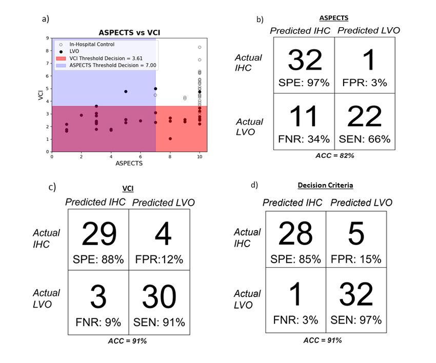
Dorn AY (2020) Feasibility study in the combination of non-contrast computed tomography and transcranial Doppler for large vessel occlusion detection
performed by an expert researcher who was blinded to the results of Results
each patient’s NCCT and CTP results. Multiple MCA velocities across
different depths between 45 and 60 mm were recorded trans temporally, Subject information and demographics
and the VCI values were extracted by choosing the minimum of the Of the possible subjects with adequate initial screenings, 50 LVO
bilateral curvature values associated with the maximal mean CBFV value and 38 IHC were initially enrolled. In the LVO group, three were
from a subject’s exam [22]. VCI is the summed curvature possessed by a discontinued (either expressed desire to discontinue or were transferred
single CBFV pulse waveform, thus assigning a lower numerical value to or died). An additional 14 LVO and five IHC subjects were excluded
a waveform that may appear ‘blunted’ to an interpreter (Figure 1). This due to the presence of disqualifying criteria not known upon initial
value provides a diagnostic biomarker that is representative of more enrollment. Within the LVO group, there were 20 subjects with M1
subjectively-classified morphology parameters at its useful threshold of occlusions, three with M2 occlusions, and eight ICA occlusions. One
3.61, chosen to maximize Youden’s J statistic described in Thorpe et al. [22]. additional subject had tandem occlusions in the M1 and ICA of the
Individual metric performance analysis same hemisphere, and one other subject had bilateral ICA occlusions in
addition to an M2 occlusion.
We correlated ASPECTS and VCI in each group using Pearson’s
correlation coefficient to understand what, if any, relationship exists For subject group demographics and attributes, see table 1 in the
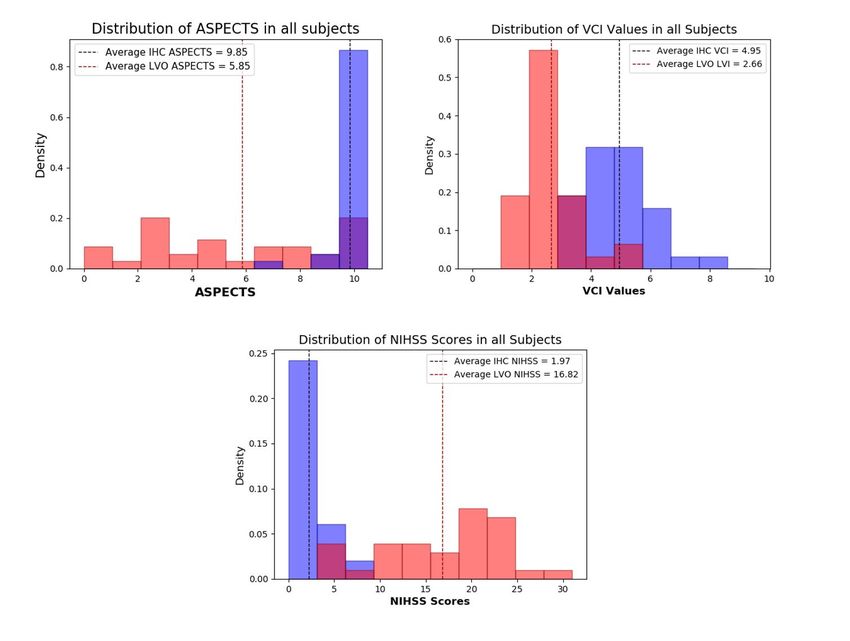
between these two stroke assessment techniques. In addition, differences previous section. Significant differences in ASPECTS, VCI, and NIHSS
between groups in ASPECTS and VCI were tested for significance using between the IHC and LVO groups were observed upon comparison.
The ranges of these metrics in each group, shown in figure 2.
the Mann-Whitney U Test. We evaluated the strength of agreement
between these two measures by finding the Cohen Kappa Statistic based ASPECTS and VCI are not Correlated
upon the incidence of agreement in designation. From these results, the
sensitivity (SEN), specificity (SPE) and accuracy (ACC) values for each In terms of Pearson’s correlation, we found no statistically
metric were computed to serve as a comparison for the performance significant correlation between these metrics in either experimental
of their combination. Additionally, we generated receiver operating group; in LVO, r = 0.249 (P=0.14); in IHC, r = 0.127 (P=0.48) (figure
characteristic (ROC) curves for the selection of the optimal thresholds 3a). ASPECTS and VCI agreed in the correct label of 29 of 33 IHC, and
of VCI and ASPECTS. Distribution analyses, correlation, and ROC in 22 of 33 LVO subjects. We evaluated the Cohen Kappa Statistic for
the agreement of VCI and ASPECTS and found it to be 0.46.
curves were computed using standard python libraries; SciPy Stats
version 1.0 and Scikit-learn version 0.19.1. Performance of ASPECTS and VCI in LVO Classification
Decision criteria formation Applied to our subject population, a threshold of 7 (in which
ASPECTS > 7 suggests non-LVO stroke) was used. In this study, the
We used a threshold of 3.61 for VCI and 7 for ASPECTS, as have
performance of ASPECTS when used at this threshold has ACC, SEN,
been previously published [8,22]. The optimal VCI threshold of 3.61
SPE of 82%, 66%, and 97%, respectively. ASPECTS alone predicted 1
was derived from the same population data used in this analysis [22]. IHC subject as LVO, and 11 LVO subjects as control (figure 3b). The
An ROC curve was generated to determine the optimal ASPECTS ROC area-under-the-curve (AUC) value for ASPECTS was 0.88.
threshold for use on this specific dataset, which was in line with
published clinical studies [8]. For subjects whose VCI or ASPECTS fell Across all subjects, the mean VCI value was 3.80 ± 1.56. Using the
below either of these thresholds, they were appended to the predicted same threshold value as the initial validation of VCI (3.61), VCI has
ACC, SEN, SPE of 91%, 91%, and 88%, respectively (Figure 3c). The
LVO group. Any subjects who did not fall below these thresholds were
ROC AUC value for VCI was 0.94.
designated as IHC. The decisions were presented as a binary set, and
the sensitivity, specificity, and accuracy values were generated based on Combination of ASPECTS and VCI in LVO Classification
comparison to the CTA ground-truth labels.
When we combined these two metrics and their thresholds into a
conditionally two-part decision process, we saw an increase from either
metric in sensitivity without major detriment to specificity, with our
criteria correctly identifying 32 of 33 LVO strokes (Figure 3d). This is
the most clinically imperative feature to improve, as the identification
of LVO can determine the method of intervention to follow. From these
results, it is evident that the inclusion of VCI has the capacity to strengthen
the diagnostic capacity of non-invasive LVO imaging protocols.
Using this diagnostic decision criteria in prediction of LVO and
IHC labels, it performed with ACC, SEN, and SPE values of 91%, 97%,
and 85%, respectively.
Discussion
The uncorrelated relationship between ASPECTS and VCI was
interesting, as these two metrics are useful in LVO assessment. Their
independent accuracies and their abilities to be used together, suggest
that ASPECTS and VCI may be sensitive to differing aspects of LVO
Figure 1. Example of IHC and LVO averaged CBFV waveforms, and their corresponding
VCI values. LVO is associated with a lower VCI value, as described in this figure from pathology; their combined use may allow for a more comprehensive
Thorpe et al. 2018, used with permission. diagnostic assessment of these patients.
Neuro Neurosurg, 2020 doi: 10.15761/NNS.1000130 Volume 3: 3-6Dorn AY (2020) Feasibility study in the combination of non-contrast computed tomography and transcranial Doppler for large vessel occlusion detection Figure 2. Distributions of (a) Alberta Stroke Program Early CT Scores (ASPECTS), (b) Velocity curvature index (VCI) values, and (c) National Institutes of Health Stroke Scale (NIHSS) Scores in both large vessel occlusion (LVO) and in-hospital control (IHC) groups Figure 3. a) Scatter plot of CTA-confirmed IHC or LVO subjects against the decision thresholds of both ASPECTS and VCI; false-negative rate (FNR) and false-positive rates (FPR) also included. b) Independent performance of ASPECTS in LVO prediction as compared to CTA. c) Performance of VCI alone in LVO prediction against CTA. d) Performance of ASPECTS and VCI together in the separation of IHC versus LVO stroke patients. Neuro Neurosurg, 2020 doi: 10.15761/NNS.1000130 Volume 3: 4-6
Dorn AY (2020) Feasibility study in the combination of non-contrast computed tomography and transcranial Doppler for large vessel occlusion detection

ASPECTS provides value in noninvasive LVO diagnosis                              Combining these noninvasive techniques shows promise for
                                                                                 centers lacking CTA
    ASPECTS has unquestionable value in the assessment of suspected
LVO patients and their candidacy for intervention, especially given its              Although CTA is required to image intracranial vasculature and
minimal impact on door-to-intervention workflow. However, its low                will remain the gold standard for centers with access, the results of this
sensitivity in this analysis could mean missed opportunity for optimal           analysis indicate that ASPECTS and VCI may indicate differing but
treatment of LVO patients if vascular imaging is not available. The              complimentary facets of stroke pathology. When utilized together, these
moderately accurate performance of ASPECTS using this threshold                  metrics could provide a more comprehensive and sensitive method
                                                                                 of LVO diagnosis in the absence of CTA. Based on the improved
for LVO prediction is markedly higher than previously published
                                                                                 performance of the combination of these metrics, VCI may be able to
performance of ASPECTS for other aspects of assessment at this                   contribute contextual information regarding a patient’s vascular status
threshold, such as outcome prediction or infarct volume estimation               that allows ASPECTS to provide more clarity.
[25,26]. A short time from symptom onset or good collateral flow are
potential confounds of ASPECTS, and future studies should examine                    The single LVO subject on which VCI and ASPECTS agreed on
                                                                                 predicting as a false negative (IHC; 4.73 and 10, respectively) had an
the relationship between ASPECTS and these variables.
                                                                                 acute NIHSS score of 13. CTA identified a right terminal ICA occlusion
    The increase in sensitivity observed when ASPECTS and VCI                    and displayed very good collateral flow. Radiology notes from this
are combined could bring clarity to cases which have conflicting or              subject’s NCCT noted evidence of subacute or chronic frontal lobe
uncertain results from preliminary assessment tools. For example,                infarction. The presence of good collateral flow, if strong enough, could
one of the CTA confirmed LVO subjects that ASPECTS missed had                    act as a confound for both VCI and ASPECTS due to the absence of
                                                                                 perfusion differences that are typically evident on both TCD and
presented to the ED four hours after symptom onset after transfer
                                                                                 NCCT exams.
from an outside facility. The patient had received an unknown dosage
of tPA in transfer, and CTA confirmed occlusion of the right distal              Study limitations
M1 segment of the MCA with distal reconstitution. Upon admission,
                                                                                      The performance of this decision criteria may be limited by the
their NIHSS was 12 and their ASPECTS was 10. Presence and flow
                                                                                 small sample size, and abnormally distributed sample populations.
in collateral arteries were also noted. The subject was administered             The largely-separate distributions of NIHSS and ASPECTS in the IHC
additional tPA, and mechanical thrombectomy achieved recanalization              and LVO groups may undermine the true differential ability of our
of TICI 2B. At 24-hour follow up, their NIHSS score increased to 15,             decision criteria; for this reason, the analysis should next be applied to
although this could also have been due to interrater variability. VCI of         a larger, more variable, study population. In a sample population where
2.6 was later derived from their acute TCD exam which his below the              more overlap between experimental group occurs for each evaluated
adopted threshold of 3.61.                                                       metric, the performance of each threshold may be better understood to
                                                                                 maximize their utility in this criterion.
TCD has a long history in LVO assessment
                                                                                     Although even CBFV morphological analyses are influenced by
     TCD has been present in stroke assessment since its adaptation              the angle of insonation, methods including VCI seek to mitigate this
for intracranial vessels; however, its user-dependence in both the               limitation by using the maximal velocity, which is assumed to be the
administration and interpretation of exams has prevented it from                 most-true value, for its computation. Additionally, the novelty of
evolving beyond its current perceived utility. Although progress has             VCI limits the experimental possibilities without a well-established
been made in the device’s size and bulk, these new technologies still            threshold to refer to, so subsequent evaluations of it are difficult to
require expert insonation or supervision [27,28]. Although automated             draw conclusions from. As VCI is used in a larger variety of diagnostic
TCD manipulation and analysis is a relatively new development,                   circumstances, its optimal threshold for LVO detection can be tested
there is potential for TCD to be simplified and made less operator               and verified.
independent such that if successful it could be more easily used in
                                                                                 Acknowledgements
a prehospital or centers without CTA. To mitigate the problem of
subjective interpretations of exams, signal processing techniques such              The authors are very grateful for the advice and expertise of Dr.
as VCI are being developed and provide promising and reliable options            Corey M. Thibeault, Ph.D. for his contributions to the concept design
[22]. Factors such as prior infarcts may be missed by TCD but would              and data analysis.
likely present in an NIHSS evaluation or NCCT exam.
                                                                                 Funding Information
     Although VCI is an independently strong biomarker of LVO,
                                                                                     The data collected and analyzed in this study was part of a feasibility
its potential utility lies in the information it can add to the standard
                                                                                 study designed and funded by Neural Analytics, Inc.
diagnostic battery of non-invasive stroke assessments; here the VCI
metric alone did not suggest LVO in three CTA confirmed subjects,                References
however, referencing NIHSS and ASPECTS only missed one. As an                    1. Hill MD, Barber PA, Demchuk AM, Newcommon NJ, Cole-Haskayne A, et al. (2002)
example, one presented to the ED with NIHSS of 24 and ASPECTS of 7,                 Acute Intravenous-Intra-Arterial Revascularization Therapy for Severe Ischemic
                                                                                    Stroke. Stroke 33: 279-82. [Crossref]
both of which suggested LVO diagnosis. CTA confirmed right proximal
M1 MCA occlusion. CTA also noted presence and patent bilateral                   2. Newell DW, Grady SM, Eskridge JM, Winn RH (1990) Distribution of Angiographic
                                                                                    Vasospasm after Subarachnoid Hemorrhage: Implications for Diagnosis by Transcranial
posterior communicating arteries. This subject received IV-tPA and                  Doppler Ultrasonography. Neurosurgery 27: 574-577. [Crossref]
mechanical thrombectomy. After 5 passes, recanalization of TICI 2B
                                                                                 3. Campbell BC, Hill MD, Rubiera M, Menon BK, Demchuk A, et al. (2016) Safety and
was achieved, and their 24-hour follow-up NIHSS score was 8. VCI                    Efficacy of Solitaire Stent Thrombectomy Individual Patient Data Meta-Analysis of
was found to be 4.98.                                                               Randomized Trials. Stroke 47: 798-806. [Crossref]

Neuro Neurosurg, 2020     doi: 10.15761/NNS.1000130                                                                                                  Volume 3: 5-6
Dorn AY (2020) Feasibility study in the combination of non-contrast computed tomography and transcranial Doppler for large vessel occlusion detection
4. Mueller-Kronast NH, Zaidat OO, Froehler MT, Jahan R, Aziz-Sultan MA, et al. 16. Demchuk AM, Christou I, Wein TH, Felberg RA, Malkoff M, et al. (2000) Accuracy
Systematic Evaluation of Patients Treated With Neurothrombectomy Devices for and criteria for localizing arterial occlusion with transcranial Doppler. J Neuroimaging
Acute Ischemic Stroke Primary Results of the STRATIS Registry. Stroke 48: 2760- 10: 1-12. [Crossref]
2768. [Crossref]
17. Demchuk AM, Burgin WS, Christou I, Felberg RA, Barber PA, et al. (2001)
5. Saver JL, Goyal M, van der Lugt A, Menon BK, Majoie CB, et al. (2016) Time to Thrombolysis in Brain Ischemia (TIBI) Transcranial Doppler Flow Grades Predict
Treatment With Endovascular Thrombectomy and Outcomes From Ischemic Stroke: A Clinical Severity, Early Recovery, and Mortality in Patients Treated With Intravenous
Meta-analysis. JAMA 316: 1279. [Crossref] Tissue Plasminogen Activator. Stroke 32: 89-93. [Crossref]
18. Thorpe SG, Thibeault CM, Canac N, Wilk SJ, Devlin T, et al. (2018) Decision Criteria
6. Saver JL, Goyal M, Bonafe A, Diener H-C, Levy I, et al. (2015) Stent-Retriever
for Large Vessel Occlusion Using Transcranial Doppler Waveform Morphology. Front
Thrombectomy after Intravenous t-PA vs. t-PA Alone in Stroke. N Engl J Med
Neurol 9: 847. [Crossref]
24:2285-2295. [Crossref]
19. Demchuk AM, Christou I, Wein TH, Felberg RA, Malkoff M, et al. (2000) Specific
7. Saver JL (2006) Time Is Brain-Quantified. Stroke 37: 263-266. [Crossref] transcranial Doppler flow findings related to the presence and site of arterial occlusion.
Stroke 31: 140-146. [Crossref]
8. Coutts SB, Lev MH, Eliasziw M, Roccatagliata L, Hill MD, et al. (2004) ASPECTS on
CTA Source Images Versus Unenhanced CT Added Value in Predicting Final Infarct 20. Tsivgoulis G, Sharma VK, Lao AY, Malkoff MD, Alexandrov AV (2007) Validation
Extent and Clinical Outcome. Stroke 35: 2472-6. [Crossref] of Transcranial Doppler With Computed Tomography Angiography in Acute Cerebral
Ischemia. Stroke 38: 1245-9. [Crossref]
9. Demeestere J, Garcia-Esperon C, Garcia-Bermejo P, Ombelet F, McElduff P, et al.
(2017) Evaluation of hyperacute infarct volume using ASPECTS and brain CT 21. Burgin WS, Malkoff M, Felberg RA, Demchuk AM, Christou I, et al. (2000)
perfusion core volume. Neurology 88: 2248-2253. [Crossref] Transcranial Doppler Ultrasound Criteria for Recanalization After Thrombolysis for
Middle Cerebral Artery Stroke. Stroke 31: 1128-32. [Crossref]
10. Naylor J, Churilov L, Chen Z, Koome M, Rane N, et al. (2017) Reliability,
Reproducibility and Prognostic Accuracy of the Alberta Stroke Program Early CT 22. Thorpe SG, Thibeault CM, Wilk SJ, O’Brien M, Canac N, et al. (2018) Velocity
Score on CT Perfusion and Non-Contrast CT in Hyperacute Stroke. Cerebrovasc Dis Curvature Index: a Novel Diagnostic Biomarker for Large Vessel Occlusion. Transl
44: 195-202. [Crossref] Stroke Res 10: 475-484. [Crossref]
11. González RG, Lev MH, Goldmacher G V., Smith WS, Payabvash S, et al. (2012) 23. Rubiera M, Ribo M, Pagola J, Coscojuela P, Rodriguez-Luna D, et al. (2011) Bridging
Intravenous-Intra-Arterial Rescue Strategy Increases Recanalization and the Likelihood
Improved Outcome Prediction Using CT Angiography in Addition to Standard
of a Good Outcome in Nonresponder Intravenous Tissue Plasminogen Activator-
Ischemic Stroke Assessment: Results from the STOPStroke Study. Kiechl S, ed. PLoS
Treated Patients A Case-Control Study. Stroke 42: 993-997. [Crossref]
One 7: e30352. [Crossref]
24. Thorpe SG, Thibeault CM, Canac N, Jalaleddini K, Dorn A, et al. (2020) Toward
12. McTaggart RA, Ansari SA, Goyal M, Abruzzo TA, Albani B, et al. (2015) Initial automated classification of pathological transcranial Doppler waveform morphology
hospital management of patients with emergent large vessel occlusion (ELVO): report via spectral clustering. PLoS One 15: e0228642. [Crossref]
of the standards and guidelines committee of the Society of NeuroInterventional
Surgery. J Neurointerv Surg 9: 316-323. [Crossref] 25. van Seeters T, Biessels GJ, Kappelle LJ, van der Schaaf IC, Dankbaar JW, et al. (2015)
The Prognostic Value of CT Angiography and CT Perfusion in Acute Ischemic Stroke.
13. Broderick JP, Palesch YY, Demchuk AM, Yeatts SD, Khatri P, et al. (2013) Cerebrovasc Dis 40: 258-269. [Crossref]
Endovascular Therapy after Intravenous t-PA versus t-PA Alone for Stroke. N Engl J
Med 368: 893-903. [Crossref] 26. Lin K, Lee SA, Zink WE (2011) What ASPECTS Value Best Predicts the 100-mL
Threshold on Diffusion Weighted Imaging? Study of 150 Patients with Middle
14. Schieb LJ, Casper ML, George MG (2015) Mapping Primary and Comprehensive Cerebral Artery Stroke. J Neuroimaging 21: 229-231. [Crossref]
Stroke Centers by Certification Organization. Circ Cardiovasc Qual Outcomes 8:
27. TCD Monitoring - Delica Transcranial Ultrasound Doppler - Products - SMT medical
S193-4. [Crossref] GmbH & Co. Available from: https://www.smt-medical.com/en/products/delica-
15. Christou I, Felberg RA, Demchuk AM, Grotta JC, Burgin WS, et al. (2001) A Broad transcranial-ultrasound-doppler/tcd-monitoring.html. Accessed September 30, 2019.
Diagnostic Battery for Bedside Transcranial Doppler to Detect Flow Changes With 28. Pietrangelo SJ, Lee HS, Sodini CG (2018) A Wearable Transcranial Doppler Ultrasound
Internal Carotid Artery Stenosis or Occlusion. J Neuroimaging 11: 236-242. [Crossref] Phased Array System. Acta Neurochir Suppl 126: 111-114. [Crossref]
Copyright: ©2020 Dorn AY. This is an open-access article distributed under the terms of the Creative Commons Attribution License, which permits unrestricted
use, distribution, and reproduction in any medium, provided the original author and source are credited.
Neuro Neurosurg, 2020 doi: 10.15761/NNS.1000130 Volume 3: 6-6You can also read