Altered Dynamic Amplitude of Low-Frequency Fluctuations in Patients With Migraine Without Aura - Frontiers
←
→
Page content transcription
If your browser does not render page correctly, please read the page content below
ORIGINAL RESEARCH
                                                                                                                                           published: 10 February 2021
                                                                                                                                     doi: 10.3389/fnhum.2021.636472

                                           Altered Dynamic Amplitude of
                                           Low-Frequency Fluctuations in
                                           Patients With Migraine Without Aura
                                           Hong Chen, Guiqiang Qi, Yingxia Zhang, Ying Huang, Shaojin Zhang, Dongjun Yang,
                                           Junwei He, Lan Mu, Lin Zhou and Min Zeng*

                                           Department of Radiology, The Third Affiliated Hospital of Chengdu Medical College, Pidu District People’s Hospital, Chengdu,
                                           China

                                           Migraine is a chronic and idiopathic disorder leading to cognitive and affective problems.
                                           However, the neural basis of migraine without aura is still unclear. In this study, dynamic
                                           amplitude of low-frequency fluctuations (dALFF) analyses were performed in 21 patients
                                           with migraine without aura and 21 gender- and age-matched healthy controls to identify
                                           the voxel-level abnormal functional dynamics. Significantly decreased dALFF in the
                                           bilateral anterior insula, bilateral lateral orbitofrontal cortex, bilateral medial prefrontal
                                           cortex, bilateral anterior cingulate cortex, and left middle frontal cortex were found
                                           in patients with migraine without aura. The dALFF values in the anterior cingulate
                         Edited by:
                    Bochao Cheng,          cortex were negatively correlated with pain intensity, i.e., visual analog scale. Finally,
            Sichuan University, China      support vector machine was used to classify patients with migraine without aura from
                        Reviewed by:       healthy controls and achieved an accuracy of 83.33%, sensitivity of 90.48%, and
                            Lijie Wang,
                                           specificity of 76.19%. Our findings provide the evidence that migraine influences the
  University of Electronic Science and
           Technology of China, China      brain functional activity dynamics and reveal the neural basis for migraine, which could
                          Jingjing Gao,    facilitate understanding the neuropathology of migraine and future treatment.
  University of Electronic Science and
           Technology of China, China      Keywords: migraine without aura, dynamic amplitude of low-frequency fluctuations, resting-state, functional MRI,
                 *Correspondence:          classification 3
                          Min Zeng
             minzengnihao@163.com
                                           INTRODUCTION
                   Specialty section:
         This article was submitted to     Migraine is an idiopathic and chronic disorder influencing the life quality of patients (Kruit et al.,
       Brain Imaging and Stimulation,      2004). The frequent migraine attacks also affect patients’ mental and physical health (Tietjen, 2004;
                a section of the journal   Borsook et al., 2012). The previous studies have demonstrated that long-term chronic pain leads to
    Frontiers in Human Neuroscience        functional damages in sensory, cognition, and affective processing (Montero-Homs, 2009; Linton,
       Received: 01 December 2020          2013; Denkinger et al., 2014). A lot of previous studies have revealed that migraine induced brain
        Accepted: 07 January 2021          gray matter volume and white matter integrity changes using structural and diffusion magnetic
       Published: 10 February 2021         resonance imaging (MRI) (DaSilva et al., 2007; Kim et al., 2008; Schmidt-Wilcke et al., 2008;
                             Citation:     Valfrè et al., 2008). With the use of resting-state functional MRI (fMRI), altered local regional
    Chen H, Qi G, Zhang Y, Huang Y,        homogeneity and whole-brain functional connectivity homogeneity were found in patients with
Zhang S, Yang D, He J, Mu L, Zhou L        migraine (Yu et al., 2012; Zhang et al., 2020). All these findings suggest that migraine could change
 and Zeng M (2021) Altered Dynamic
                                           the brain structure and functions.
         Amplitude of Low-Frequency
Fluctuations in Patients With Migraine
                                              The low-frequency oscillation of the brain is mainly characterized using resting-state fMRI with
                         Without Aura.     blood oxygen level-dependent (BOLD) signals (Biswal et al., 1995; Fox and Raichle, 2007). Resting-
   Front. Hum. Neurosci. 15:636472.        state fMRI has been widely applied to study the brain functional organization (Greicius et al., 2003;
   doi: 10.3389/fnhum.2021.636472          Fox et al., 2006; Wang et al., 2015, 2017c; Xu et al., 2019; Gao et al., 2020), to identify the functional

Frontiers in Human Neuroscience | www.frontiersin.org                             1                                        February 2021 | Volume 15 | Article 636472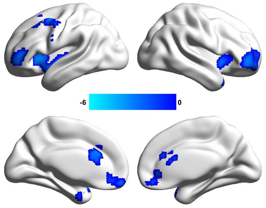
Chen et al.                                                                                                                  Altered dALFF in Migraine

abnormalities in patients with brain disorders (Greicius et al.,             Resting-State Functional MRI Data
2007; Brier et al., 2012; Muller et al., 2013; Wu et al., 2016; Sun          Acquisition
et al., 2018), and to reveal the neural basis of treatment response          MRI data were acquired on a 3-Tesla Siemens MRI scanner
(Guo et al., 2012; Mulders et al., 2016; Wang et al., 2017b,                 in the Department of Radiology, the Third Affiliated Hospital
2018; Xu et al., 2020). To quantitatively measure low-frequency              of Chengdu Medical College, Pidu District People’s Hospital of
oscillation, the amplitude of low-frequency fluctuations (ALFF)              Chengdu, China. The participants were instructed to close their
is proposed (Zang et al., 2007). Using ALFF, Xue et al. (2013)               eyes and not fall asleep, and earplugs and foam padding were used
found decreased ALFF values in the left rostral anterior cingulate           to reduce scanner noise and head motion. Resting-state fMRI
cortex and bilateral prefrontal cortex as well as increased ALFF             data were acquired using a gradient-echo echo-planar imaging
values in the right thalamus. Using ALFF to reveal the early                 (GRE-EPI) sequence with the following parameters: repetition
abnormal functional activity is promising to explore early marker            time (TR) = 2,000 ms, echo time (TE) = 30 ms, flip angle (FA)
for migraine. Recently, using sliding-window approach, the                   = 90◦ , matrix = 64 × 64, field of view (FOV) = 220 × 220 mm,
dynamic ALFF (dALFF) method was developed by calculating                     slice thickness = 4 mm with inter-slice gap = 0.6 mm, 32 axial
the variance of ALFF over time (Fu et al., 2018). The dALFF                  slices, and 250 time points. The information can be found in our
has been applied to study the functional activity abnormalities              previous study (Zhang et al., 2020).
in subjects under disease state and provides some new evidence
(Fu et al., 2018; Ma et al., 2020; Pang et al., 2020). Thus, to reveal       Resting-State Functional MRI Data
the dynamic changes of the low-frequency oscillation in migraine
patients may provide supplementary information to understand                 Preprocessing
its neural basis.                                                            The resting-state fMRI data were preprocessed using the
    In this study, with 21 patients with migraine without                    toolkit of DPARSF version 2.3 (Chao-Gan and Yu-Feng,
aura and 21 gender- and age-matched healthy controls, a                      2010) (www.restfmri.net/forum/DPARSF). To exclude unstable
voxel-wise dALFF method was used to reveal the dynamic                       magnetization effect, the first 10 volumes were discarded.
changes of low-frequency oscillation in migraine patients.                   Then, all the remaining volumes were realigned to the first
Moreover, using the dALFF values in brain regions showing                    volume to correct head motion. Next, all the images were
differences between patients and controls as features,                       normalized to standard EPI template in Montreal Neurological
support vector machine (SVM) was used to classify                            Institute (MNI) space and resampled to 3-mm voxel resolution.
migraine patients from healthy controls to further validate                  Subsequently, 6-mm Gaussian kernel was used to smooth
the results.                                                                 the fMRI data; and Friston 24-parameter of head motion,
                                                                             white matter, cerebrospinal fluid, and global mean signals were
                                                                             regressed out. Finally, the resting-state data were filtered with a
                                                                             frequency band of 0.01–0.08 Hz for dALFF analyses. To remove
                                                                             head-motion effects, the subjects were excluded if the head
MATERIALS AND METHODS                                                        movement exceeds 3 mm or 3◦ . Additionally, “scrubbing” was
Participants                                                                 used to delete the bad images before 2 time points and after 1 time
In this study, we recruited 21 right-handed patients with                    point exceeding the preset criteria [frame displacement (FD) <
migraine without aura (female/male = 16/5; age = 31.19 ± 6.38                0.5] (Power et al., 2012). We also calculated the mean FD values
years) and 21 gender- and age-matched right-handed healthy                   of healthy controls and migraine patients, and the two-sample
controls (female/male = 13/8; age = 30.19 ± 6.3 years) at the                t-test was used and did not find the significant difference (FD
Third Affiliated Hospital of Chengdu Medical College, Pidu                   values of patients = 0.16 ± 0.29; FD values of controls = 0.15
District People’s Hospital. The diagnosis of migraine without                ± 0.027; p = 0.49).
aura was based on the International Headache Society criteria.
The inclusion criteria for patients with migraine without aura               Dynamic Amplitude of Low-Frequency
were follows: (1) no migraine precipitated during or on the                  Fluctuations Calculation
day after the scan; (2) did not suffer from a migraine attack                The ALFF was proposed to characterize the resting-state
at least 72 h before the experiment; (3) for migraine patients               functional activity of each voxel in brain (Zou et al., 2008).
and healthy controls, no lifetime history of seizures, head                  To calculate ALFF, the time series was first transformed to
trauma, serious medical or surgical illness, substance dependence            frequency domain, and the ALFF is computed at the power
or abuse, and contraindications for MRI. The participants                    within the low-frequency range of 0.01–0.8 Hz. To calculate
were excluded if structural abnormalities were detected on                   dALFF, a sliding window method was used. The length of sliding
MRI examination, and no subject with structural deficits was                 window is determined based on the criterion that minimum
found. Written informed consent was provided and obtained                    window length should be larger than 1/f min , where f min is the
from all the subjects. This study was approved by the local                  minimum frequency of time series (Leonardi and Van De Ville,
ethics committees of the Third Affiliated Hospital of Chengdu                2015; Du et al., 2017; Li et al., 2019). Finally, a window length
Medical College, Pidu District People’s Hospital. The detailed               of 50 TR with step size of 5 TR was applied in this study. The
information for the subjects can be found in our previous study              ALFF map was computed in each window, and the variance
(Zhang et al., 2020).                                                        of the ALFF maps across all the windows was computed to

Frontiers in Human Neuroscience | www.frontiersin.org                    2                                  February 2021 | Volume 15 | Article 636472
Chen et al. Altered dALFF in Migraine
TABLE 1 | Demographics and clinical characteristics of the subjects used in
present study.
Migraine Controls p
(n = 21) (n = 21)
Gender (male/female) 16/5 13/8 0.51
Age (mean ± SD) 31.19 ± 6.38 30.19 ± 6.3 0.61
HAMA (mean ± SD) 7.95 ± 6.77
HAMD (mean ± SD) 6.71 ± 6.25
Duration of illness (months) 44.69 ± 61.13
VAS (mean ± SD) 4.33 ± 1.46
A Pearson chi-squared test was used for gender comparison. Two-sample t-tests were
used for age comparisons.
VAS, visual analog scale; HAMD, Hamilton Depression Rating Scale; HAMA, Hamilton
Anxiety Scale.
measure the dynamic. The dALFF maps were transformed to z- FIGURE 1 | Changed dynamic amplitude of low-frequency fluctuations (ALFF)
scores for statistical analyses. Two-sample t-test was performed in window length of 50 repetition time (TR) in patients with migraine without
to compare the dALFF maps between healthy controls and aura. Significantly decreased dynamic ALFF in the bilateral anterior insula,
patients with migraine without aura. The significant level was bilateral anterior cingulate cortex, bilateral lateral orbitofrontal cortex, and left
middle frontal cortex were found in patients with migraine without aura.
set at p < 0.05 using a Gaussian random field (GRF) correction
method. To further validate the results obtained with the window
length of 50 TR, the window length of 30 and 70 TR were
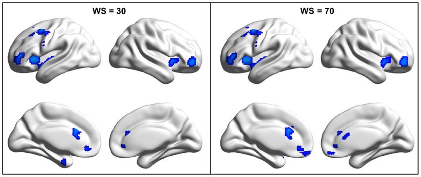
further applied. patients with migraine without aura compared with healthy
controls (Figure 1 and Table 2) was found. We also analyzed
Correlation Analyses dALFF in window length of 30 and 70 TR, and similar results
To explore the relationship between dALFF and clinical
were found (Figure 2).
measures, correlation analyses were performed between the
dALFF values of the areas showing differences in patients and
Correlation Analyses
visual analog scale (VAS), and disease duration. The significance
Correlation analyses identified significantly negative correlations
was set at p < 0.05 with Bonferroni correction.
between dALFF values in the anterior cingulate cortex and VAS
Support Vector Machine Classification scores (r = −0.5, p = 0.022) in patients with migraine without
A linear SVM classifier was performed using LIBSVM software aura after multiple comparisons correction (Figure 3).
(Chang and Lin, 2011). For classification, the mean dALFF
values of the areas showing differences between patients and Classification Results
controls were used as the features. To estimate the performance With the use of the changed dALFF values as features,
of our classifier, a leave-one-out cross-validation (LOOCV) test experimental results showed a correct classification rate of
was used to assess the generalization ability because of our 83.33%, sensitivity of 90.4%, and specificity of 76.19% using a
limited number of samples in the present study. The classification leave-one-out cross-validation method (Figure 4).
result was assessed using the classification accuracy, sensitivity,
and specificity. DISCUSSION
RESULTS In our study, dALFF method was applied and identified
decreased dALFF in the bilateral anterior insula, bilateral
Clinical Characteristics orbitofrontal cortex, bilateral anterior cingulate cortex, bilateral
There were no significant differences in age (p = 0.61) and sex medial prefrontal cortex, and left middle frontal cortex; and the
(p = 0.51) between healthy controls and patients with migraine changed dALFF in the anterior cingulate cortex was negatively
without aura (Table 1). correlated with VAS scores. Moreover, the changed dALFF can
serve as an effective neuromarker to differentiate patients with
Changed Dynamic Amplitude of migraine without aura from controls. Our findings revealed
Low-Frequency Fluctuations dynamic changes of brain functional activities in migraine
By the analysis of dALFF in window length of 50 TR, significantly patients and provide new evidence of neurophysiological
decreased dALFF in the bilateral anterior insula, bilateral lateral abnormalities in migraine.
orbitofrontal cortex, bilateral medial prefrontal cortex, bilateral We found decreased dALFF in the anterior cingulate
anterior cingulate cortex, and left middle frontal cortex in cortex and anterior insula in migraine patients without aura.
Frontiers in Human Neuroscience | www.frontiersin.org 3 February 2021 | Volume 15 | Article 636472Chen et al. Altered dALFF in Migraine
TABLE 2 | Regions with decreased dynamical amplitude of low-frequency fluctuations in patients with migraine without aura vs. healthy controls.
Brain regions Peak MNI coordinates t values Cluster size
X Y Z
Anterior insula −39 18 0 −4.71 383
Anterior insula 42 15 −9 −4.41 136
Lateral orbitofrontal cortex −39 39 6 −4.53 185
Lateral orbitofrontal cortex 42 45 −3 −3.89 266
Middle frontal cortex −46 0 51 −4.55 301
Medial prefrontal cortex −3 54 −15 −3.74 118
Anterior cingulate cortex −6 24 27 −3.87 102
FIGURE 2 | Changed dynamic amplitude of low-frequency fluctuations (ALFF) in migraine patients was validated in window length of 30 and 70 repetition times (TRs).
To validate the findings obtained with window length of 50 TR, the same procedures were performed in window length of 30 and 70 TRs and found similar results.
FIGURE 3 | Correlation analyses between dynamic amplitude of
low-frequency fluctuations (ALFF) and clinical measures. Significant correlation FIGURE 4 | Support vector machine (SVM) was applied to classify migraine
was found between the dynamic ALFF in the anterior cingulate cortex and patients and healthy controls with the dynamic amplitude of low-frequency
visual analog scale (VAS) scores in migraine patients. fluctuations (ALFF) values in brain areas showing differences between patients
and controls as features. SVM achieves a classification accuracy of 83.33%,
sensitivity of 90.48%, and specificity of 76.19%.
The anterior insula and anterior cingulate cortex are two
important nodes of salience network, which is mainly involved 2010; Menon, 2011; Wang et al., 2017a, 2019b). The functional
in coordinating the dynamic interaction between the external abnormality of anterior cingulate cortex and anterior insula
orient stimulus and internal self-perception (Menon and Uddin, found in our study is supported by previous functional and
Frontiers in Human Neuroscience | www.frontiersin.org 4 February 2021 | Volume 15 | Article 636472Chen et al. Altered dALFF in Migraine
structural studies (Peyron et al., 2000; Kim et al., 2008; Valfrè findings. Second, the samples of our study are small, and the
et al., 2008). Importantly, we found that decreased dALFF in findings need to be further validated in the future studies with
the anterior cingulate cortex is associated with clinical pain a larger sample.
intensity. Thus, the abnormal dynamic functional activities in the
anterior cingulate cortex may be a neuromarker to predict the CONCLUSION
pain intensity.
The decreased dALFF in the medial prefrontal cortex, middle This study revealed the abnormal dynamic low-frequency
frontal cortex, and lateral orbitofrontal cortex was also observed. oscillation in default mode network, salience network, and
The medial prefrontal cortex is the core brain area of the default executive control network in migraine patients. The decreased
mode network that participates in social cognition, emotional, dALFF in these areas may be associated with disrupted emotion
and self-referential processing (Gusnard et al., 2001; Amodio and cognitive functions. Our findings provide new evidence
and Frith, 2006; Etkin et al., 2011; Wang et al., 2019a,c). The that migraine could influence the brain functions leading to
abnormal functional activities in medial prefrontal cortex have functional impairments of emotion and cognitions in patients.
been reported in previous studies by analyses of static ALFF, local
regional homogeneity, and whole-brain functional connectivity DATA AVAILABILITY STATEMENT
homogeneity (Yu et al., 2012; Xue et al., 2013). In addition, the
middle frontal cortex and lateral orbitofrontal cortex play an The raw data supporting the conclusions of this article will be
important role in attention and executive control of pain-related made available by the authors, without undue reservation.
stimuli to modulate the descending pain system (Casey, 1999;
Lorenz et al., 2003; Wager et al., 2004). Thus, decreased dALFF ETHICS STATEMENT
values in the medial prefrontal cortex and middle prefrontal
cortex may be related to impaired cognitive and emotion The studies involving human participants were reviewed and
processing of pain. Moreover, we found that the changed dALFF approved by the Third Affiliated Hospital of Chengdu Medical
in brain areas including the bilateral anterior insula, bilateral College, Pidu District People’s Hospital. The patients/participants
anterior cingulate cortex, bilateral medial prefrontal cortex, and provided their written informed consent to participate in
left middle frontal cortex can well distinguish the migraine this study. Written informed consent was obtained from the
patients from controls. This finding indicates that the abnormal individual(s) for the publication of any potentially identifiable
dynamic activities in default mode network, salience network, images or data included in this article.
and executive network may be the neuropathology of migraine.
There are some limitations of our study. First, how to AUTHOR CONTRIBUTIONS
determine the length of sliding window is a problem for dynamic
analysis. We used different window lengths to validate the All authors have made a substantial contribution to this work.
REFERENCES and periaqueductal gray matter in migraine. Neuroreport 18, 301–305.
doi: 10.1097/WNR.0b013e32801776bb
Amodio, D. M., and Frith, C. D. (2006). Meeting of minds: the medial Denkinger, M. D., Lukas, A., Nikolaus, T., Peter, R., and Franke, S. (2014).
frontal cortex and social cognition. Nat. Rev. Neurosci. 7, 268–277. Multisite pain, pain frequency and pain severity are associated with depression
doi: 10.1038/nrn1884 in older adults: results from the ActiFE Ulm study. Age Ageing 43, 510–514.
Biswal, B., Yetkin, F. Z., Haughton, V. M., and Hyde, J. S. (1995). Functional doi: 10.1093/ageing/afu013
connectivity in the motor cortex of resting human brain using echo-planar Du, Y., Pearlson, G. D., Lin, D., Sui, J., Chen, J., Salman, M., et al. (2017).
MRI. Magnet. Resonance Med. 34, 537–541. doi: 10.1002/mrm.1910340409 Identifying dynamic functional connectivity biomarkers using GIG-ICA:
Borsook, D., Maleki, N., Becerra, L., and McEwen, B. (2012). Understanding application to schizophrenia, schizoaffective disorder, and psychotic bipolar
migraine through the lens of maladaptive stress responses: a model disease of disorder. Human Brain Mapp. 38, 2683–2708. doi: 10.1002/hbm.23553
allostatic load. Neuron 73, 219–234. doi: 10.1016/j.neuron.2012.01.001 Etkin, A., Egner, T., and Kalisch, R. (2011). Emotional processing in anterior
Brier, M. R., Thomas, J. B., Snyder, A. Z., Benzinger, T. L., Zhang, D., Raichle, M. cingulate and medial prefrontal cortex. Trends Cognitive Sci. 15, 85–93.
E., et al. (2012). Loss of intranetwork and internetwork resting state functional doi: 10.1016/j.tics.2010.11.004
connections with Alzheimer’s disease progression. J. Neurosci. 3, 8890–8899. Fox, M. D., Corbetta, M., Snyder, A. Z., Vincent, J. L., and Raichle, M.
doi: 10.1523/JNEUROSCI.5698-11.2012 E. (2006). Spontaneous neuronal activity distinguishes human dorsal and
Casey, K. L. (1999). Forebrain mechanisms of nociception and pain: ventral attention systems. Proc. Natl. Acad. Sci. U.S.A. 103, 10046–10051.
analysis through imaging. Proc. Natl. Acad. Sci. U.S.A. 96, 7668–7674. doi: 10.1073/pnas.0604187103
doi: 10.1073/pnas.96.14.7668 Fox, M. D., and Raichle, M. E. (2007). Spontaneous fluctuations in brain activity
Chang, C.-C., and Lin, C.-J. (2011). LIBSVM: a library for support vector machines. observed with functional magnetic resonance imaging. Nat Rev. Neurosci. 8,
ACM Trans. Intell. Syst. Technol. 2, 1–27. doi: 10.1145/1961189.1961199 700–711. doi: 10.1038/nrn2201
Chao-Gan, Y., and Yu-Feng, Z. (2010). DPARSF: a MATLAB toolbox for Fu, Z., Tu, Y., Di, X., Du, Y., Pearlson, G. D., Turner, J. A.,
“Pipeline” data analysis of resting-state fMRI. Front. Systems Neurosci. 4:13. et al. (2018). Characterizing dynamic amplitude of low-frequency
doi: 10.3389/fnsys.2010.00013 fluctuation and its relationship with dynamic functional connectivity:
DaSilva, A. F., Granziera, C., Tuch, D. S., Snyder, J., Vincent, M., and Hadjikhani, an application to schizophrenia. NeuroImage 180, 619–631.
N. (2007). Interictal alterations of the trigeminal somatosensory pathway doi: 10.1016/j.neuroimage.2017.09.035
Frontiers in Human Neuroscience | www.frontiersin.org 5 February 2021 | Volume 15 | Article 636472Chen et al. Altered dALFF in Migraine
Gao, Z., Guo, X., Liu, C., Mo, Y., and Wang, J. (2020). Right inferior frontal gyrus: Schmidt-Wilcke, T., Gänssbauer, S., Neuner, T., Bogdahn, U., and May, A. (2008).
An integrative hub in tonal bilinguals. Human Brain Mapp. 41, 2152–2159. Subtle grey matter changes between migraine patients and healthy controls.
doi: 10.1002/hbm.24936 Cephalalgia 28, 1–4. doi: 10.1111/j.1468-2982.2007.01428.x
Greicius, M. D., Flores, B. H., Menon, V., Glover, G. H., Solvason, H. B., Kenna, Sun, H., Luo, L., Yuan, X., Zhang, L., He, Y., Yao, S., et al. (2018). Regional
H., et al. (2007). Resting-state functional connectivity in major depression: homogeneity and functional connectivity patterns in major depressive disorder,
abnormally increased contributions from subgenual cingulate cortex and cognitive vulnerability to depression and healthy subjects. J. Affective Disord.
thalamus. Biol. Psychiatr. 62, 429–437. doi: 10.1016/j.biopsych.2006.09.020 235, 229–235. doi: 10.1016/j.jad.2018.04.061
Greicius, M. D., Krasnow, B., Reiss, A. L., and Menon, V. (2003). Tietjen, G. E. (2004). Stroke and migraine linked by silent lesions. Lancet 3:267.
Functional connectivity in the resting brain: a network analysis of the doi: 10.1016/S1474-4422(04)00729-X
default mode hypothesis. Proc. Natl. Acad. Sci. U.S.A. 100, 253–258. Valfrè, W., Rainero, I., Bergui, M., and Pinessi, L. (2008). Voxel-based
doi: 10.1073/pnas.0135058100 morphometry reveals gray matter abnormalities in migraine. Headache 48,
Guo, W. B., Liu, F., Chen, J. D., Gao, K., Xue, Z. M., Xu, X. J., et al. (2012). 109–117. doi: 10.1111/j.1526-4610.2007.00723.x
Abnormal neural activity of brain regions in treatment-resistant and treatment- Wager, T. D., Rilling, J. K., Smith, E. E., Sokolik, A., Casey, K. L., Davidson, R.
sensitive major depressive disorder: a resting-state fMRI study. J. Psychiatric J., et al. (2004). Placebo-induced changes in FMRI in the anticipation and
Res. 46, 1366–1373. doi: 10.1016/j.jpsychires.2012.07.003 experience of pain. Science 303, 1162–1167. doi: 10.1126/science.1093065
Gusnard, D. A., Akbudak, E., Shulman, G. L., and Raichle, M. E. (2001). Wang, C., Wu, H., Chen, F., Xu, J., Li, H., Li, H., et al. (2017a). Disrupted functional
Medial prefrontal cortex and self-referential mental activity: relation to a connectivity patterns of the insula subregions in drug-free major depressive
default mode of brain function. Proc. Natl. Acad. Sci. U.S.A. 98. 4259–4264. disorder. J. Affect. Disord. 234, 297–304. doi: 10.1016/j.jad.2017.12.033
doi: 10.1073/pnas.071043098 Wang, J., Becker, B., Wang, L., Li, H., Zhao, X., and Jiang, T. (2019a).
Kim, J. H., Suh, S. I., Seol, H. Y., Oh, K., Seo, W. K., Yu, S. W., et al. (2008). Regional Corresponding anatomical and coactivation architecture of the human
grey matter changes in patients with migraine: a voxel-based morphometry precuneus showing similar connectivity patterns with macaques. NeuroImage
study. Cephalalgia 28, 598–604. doi: 10.1111/j.1468-2982.2008.01550.x 200, 562–574. doi: 10.1016/j.neuroimage.2019.07.001
Kruit, M. C., van Buchem, M. A., Hofman, P. A. M., Bakkers, J. T. N., Terwindt, G. Wang, J., Wei, Q., Bai, T., Zhou, X., Sun, H., Becker, B., et al. (2017b).
M., Ferrari, M. D., et al. (2004). Migraine as a risk factor for subclinical brain Electroconvulsive therapy selectively enhanced feedforward connectivity from
lesions. JAMA 291, 427–434. doi: 10.1001/jama.291.4.427 fusiform face area to amygdala in major depressive disorder. Soc. Cognit.
Leonardi, N., and Van De Ville, D. (2015). On spurious and real fluctuations Affective Neurosci. 12, 1983–1992. doi: 10.1093/scan/nsx100
of dynamic functional connectivity during rest. NeuroImage 104, 430–436. Wang, J., Wei, Q., Wang, L., Zhang, H., Bai, T., Cheng, L., et al. (2018). Functional
doi: 10.1016/j.neuroimage.2014.09.007 reorganization of intra- and internetwork connectivity in major depressive
Li, C., Xia, L., Ma, J., Li, S., Liang, S., Ma, X., et al. (2019). Dynamic functional disorder after electroconvulsive therapy. Human Brain Mapp. 39, 1403–1411.
abnormalities in generalized anxiety disorders and their increased network doi: 10.1002/hbm.23928
segregation of a hyperarousal brain state modulated by insomnia. J. Affect. Wang, J., Xie, S., Guo, X., Becker, B., Fox, P. T., Eickhoff, S. B., et al. (2017c).
Disord. 246, 338–345. doi: 10.1016/j.jad.2018.12.079 Correspondent functional topography of the human left inferior parietal lobule
Linton, S. J. (2013). A transdiagnostic approach to pain and emotion. J. Appl. at rest and under task revealed using resting-state fmri and coactivation based
Biobehav. Res. 18, 82–103. doi: 10.1111/jabr.12007 parcellation. Human Brain Mapp. 38, 1659–1675. doi: 10.1002/hbm.23488
Lorenz, J., Minoshima, S., and Casey, K. L. (2003). Keeping pain out of mind: Wang, J., Yang, Y., Fan, L., Xu, J., Li, C., Liu, Y., et al. (2015). Convergent
the role of the dorsolateral prefrontal cortex in pain modulation. Brain 126, functional architecture of the superior parietal lobule unraveled with
1079–1091. doi: 10.1093/brain/awg102 multimodal neuroimaging approaches. Human Brain Mapp. 36, 238–257.
Ma, M., Zhang, H., Liu, R., Liu, H., Yang, X., Yin, X., et al. (2020). Static doi: 10.1002/hbm.22626
and dynamic changes of amplitude of low-frequency fluctuations in cervical Wang, L., Wei, Q., Wang, C., Xu, J., Wang, K., Tian, Y., et al.
discogenic pain. Front. Neurosci. 14:733. doi: 10.3389/fnins.2020.00733 (2019b). Altered functional connectivity patterns of insular
Menon, V. (2011). Large-scale brain networks and psychopathology: subregions in major depressive disorder after electroconvulsive
a unifying triple network model. Trends cognit. Sci. 15, 483–506. therapy. Brain Imaging Behav. 14, 753–761. doi: 10.1007/s11682-01
doi: 10.1016/j.tics.2011.08.003 8-0013-z
Menon, V., and Uddin, L. Q. (2010). Saliency, switching, attention and control: Wang, L., Yu, L., Wu, F., Wu, H., and Wang, J. (2019c). Altered whole
a network model of insula function. Brain Struct. Funct.214, 655–667. brain functional connectivity pattern homogeneity in medication-free major
doi: 10.1007/s00429-010-0262-0 depressive disorder. J. Affect. Disord. 253, 18–25. doi: 10.1016/j.jad.2019.04.040
Montero-Homs, J. (2009). Nocioceptive pain, neuropathic pain and pain memory. Wu, Y., Zhang, Y., Liu, Y., Liu, J., Duan, Y., Wei, X.,
Neurologia 24, 419–422. et al. (2016). Distinct changes in functional connectivity in
Mulders, P. C., van Eijndhoven, P. F., Pluijmen, J., Schene, A. H., Tendolkar, I., posteromedial cortex subregions during the progress of alzheimer’s
and Beckmann, C. F. (2016). Default mode network coherence in treatment- disease. Front Neuroanat. 10:41. doi: 10.3389/fnana.2016.
resistant major depressive disorder during electroconvulsive therapy. J. Affect. 00041
Disord. 205, 130–137. doi: 10.1016/j.jad.2016.06.059 Xu, J., Lyu, H., Li, T., Xu, Z., Fu, X., Jia, F., et al. (2019). Delineating functional
Muller, V. I., Cieslik, E. C., Laird, A. R., Fox, P. T., and Eickhoff, S. B. (2013). segregations of the human middle temporal gyrus with resting-state functional
Dysregulated left inferior parietal activity in schizophrenia and depression: connectivity and coactivation patterns. Human Brain Mapp. 40, 5159–5171.
functional connectivity and characterization. Front. Human Neurosci. 7:268. doi: 10.1002/hbm.24763
doi: 10.3389/fnhum.2013.00268 Xu, J., Wei, Q., Bai, T., Wang, L., Li, X., He, Z., et al. (2020).
Pang, Y., Zhang, H., Cui, Q., Yang, Q., Lu, F., Chen, H., et al. (2020). Combined Electroconvulsive therapy modulates functional interactions between
static and dynamic functional connectivity signatures differentiating bipolar submodules of the emotion regulation network in major depressive
depression from major depressive disorder. Australian N. Zealand J. Psychiatr. disorder. Translational Psychiatr. 10:271. doi: 10.1038/s41398-020-0
54, 832–842. doi: 10.1177/0004867420924089 0961-9
Peyron, R., Laurent, B., and Garcia-Larrea, L. (2000). Functional imaging of brain Xue, T., Yuan, K., Cheng, P., Zhao, L., Zhao, L., Yu, D., et al. (2013). Alterations
responses to pain. A review and meta-analysis neurophysiologie clinique. Clin. of regional spontaneous neuronal activity and corresponding brain circuit
Neurophysiol. 30, 263–288. doi: 10.1016/S0987-7053(00)00227-6 changes during resting state in migraine without aura. NMR Biomed. 26,
Power, J. D., Barnes, K. A., Snyder, A. Z., Schlaggar, B. L., and Petersen, S. 1051–1058. doi: 10.1002/nbm.2917
E. (2012). Spurious but systematic correlations in functional connectivity Yu, D., Yuan, K., Zhao, L., Zhao, L., Dong, M., Liu, P., et al. (2012). Regional
MRI networks arise from subject motion. NeuroImage 59, 2142–2154. homogeneity abnormalities in patients with interictal migraine without aura:
doi: 10.1016/j.neuroimage.2011.10.018 a resting-state study. NMR Biomed. 25, 806–812. doi: 10.1002/nbm.1796
Frontiers in Human Neuroscience | www.frontiersin.org 6 February 2021 | Volume 15 | Article 636472Chen et al. Altered dALFF in Migraine Zang, Y. F., He, Y., Zhu, C. Z., Cao, Q. J., Sui, M. Q., Liang, M., et al. (2007). Conflict of Interest: The authors declare that the research was conducted in the Altered baseline brain activity in children with ADHD revealed by resting- absence of any commercial or financial relationships that could be construed as a state functional MRI. Brain Dev. 29, 83–91. doi: 10.1016/j.braindev.2006. potential conflict of interest. 07.002 Zhang, Y., Chen, H., Zeng, M., He, J., Qi, G., Zhang, S., et al. (2020). Copyright © 2021 Chen, Qi, Zhang, Huang, Zhang, Yang, He, Mu, Zhou Abnormal whole brain functional connectivity pattern homogeneity and and Zeng. This is an open-access article distributed under the terms of the couplings in migraine without Aura. Front. Human Neurosci. 14:619839. Creative Commons Attribution License (CC BY). The use, distribution or doi: 10.3389/fnhum.2020.619839 reproduction in other forums is permitted, provided the original author(s) Zou, Q. H., Zhu, C. Z., Yang, Y., Zuo, X. N., Long, X. Y., Cao, Q. J., et al. and the copyright owner(s) are credited and that the original publication in (2008). An improved approach to detection of amplitude of low-frequency this journal is cited, in accordance with accepted academic practice. No use, fluctuation (ALFF) for resting-state fMRI: fractional ALFF. J. Neurosci. Methods distribution or reproduction is permitted which does not comply with these 172, 137–141. doi: 10.1016/j.jneumeth.2008.04.012 terms. Frontiers in Human Neuroscience | www.frontiersin.org 7 February 2021 | Volume 15 | Article 636472
You can also read