Deregulation of VEGFR-2 and PGFR expression and microvascular density in a triple-negative model of canine malignant mammary tumors with lymph ...
←
→
Page content transcription
If your browser does not render page correctly, please read the page content below
bioRxiv preprint first posted online Dec. 8, 2018; doi: http://dx.doi.org/10.1101/490144. The copyright holder for this preprint (which
       was not peer-reviewed) is the author/funder, who has granted bioRxiv a license to display the preprint in perpetuity.
                                 It is made available under a CC-BY-NC-ND 4.0 International license.

                                                                                                                                          1

                    Deregulation of VEGFR-2 and PGFR expression and microvascular density in a triple-
                       negative model of canine malignant mammary tumors with lymph node or lung
                                                        metastasis

                   Denner Santos dos Anjos1#, Aline Fernandes Vital1#, Patrícia de Faria Lainetti2, Antonio
                 Fernando Leis-Filho2, Fabiola Dalmolin3, Fabiana Elias3, Sabryna Gouveia Calazans1, Carlos
                                                 Eduardo Fonseca-Alves2*

                1. Veterinary Science Graduate Program, University of Franca (UNIFRAN), Franca, Brazil
                2. Department of Veterinary Surgery and Anesthesiology, School of Veterinary Medicine and
                Animal Science, São Paulo State University – UNESP, Botucatu, SP, Brazil.
                3. Federal University of the Southern Border, Realeza, PR, Brazil.

                *Correspondence: carlos.e.alves@unesp.br
                #
                 Both authors have contributed equally.

                Abstract
                          Canine mammary tumors (CMT) are the most common cancer in noncastrated female
                dogs. Interestingly, triple-negative tumors are the most common molecular subtype in female
                dogs. In this study, we proposed to evaluate the expression of VEGFR-2, PDGFR and
                microvascular density (MVD) in a group of metastatic and nonmetastatic triple-negative CMT
                and compare the expression based on clinical parameters. Twenty-six female dogs with triple-
                negative mammary tumors were divided into three groups: nonmetastatic tumors (NMT)
                (N=11), tumors with lymph node metastasis (LNM) (N=10) and tumors with lung metastasis
                (LM) (N=5). We observed increased VEGFR-2 expression in LNM compared with NMT and
                a positive correlation between tumor grade and VEGFR-2 expression. A positive correlation
                was noted between VEGFR-2 and PDGFR expression. Regarding microvascular density
                (MVD), we identified a higher number of vessels in primary tumors with lymph node
                metastasis and lung metastasis compared with tumors with no metastasis. The primary tumors
                with lung metastasis exhibited an increased MVD compared with carcinoma with lymph node
                metastasis. Overall, our results suggest a deregulation of VEGFR-2 and PDGFR and high
                MVD in metastatic tumors, indicating a role for angiogenesis in tumor progression.
                Key-words: angiogenesis. metastasis. mammary neoplasm.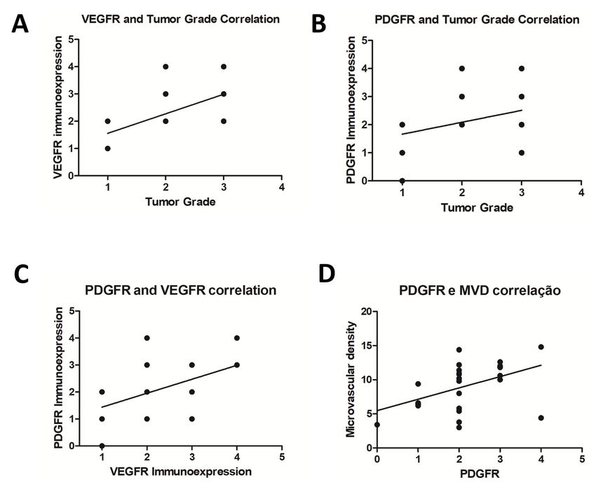
bioRxiv preprint first posted online Dec. 8, 2018; doi: http://dx.doi.org/10.1101/490144. The copyright holder for this preprint (which
       was not peer-reviewed) is the author/funder, who has granted bioRxiv a license to display the preprint in perpetuity.
                                 It is made available under a CC-BY-NC-ND 4.0 International license.

                                                                                                                                          2

                1. Introduction
                     Canine mammary tumors (CMT) are the most common tumor in noncastrated female dogs
                with a variable clinical behavior [1]. The incidence rates for CMT depends on the geographic
                origin given that it is a tumor with higher prevalence in countries where castration is not
                routinely performed [2]. In Brazil, the prevalence of CMT in intact female dogs is
                approximately 28% to 45% of all tumors in dogs [3, 4]. CMTs resemble human breast cancer
                (BC), and dogs represent an interesting model for comparative studies. The recent
                GLOBOCAN estimates of cancer presented an expectation of 2,0938,76 new cases of BC
                worldwide and 626, 679 deaths related to BC [5].
                     BC is the most important tumor in women as it is the most diagnosed cancer and the
                second cause of death related to cancer [6]. Human BC is subdivided into molecular subtypes,
                such as HER2 enriched, Luminal A, Luminal B and basal-like [6]. Triple-negative tumors are
                very important as these tumors represent a therapeutic challenging, and limited therapeutic
                options are available compared with other subtypes [7]. Recently, a study evaluating a large
                number of cases subdivided CMT into molecular subtypes and found an increased prevalence
                of triple-negative tumors in dogs [8]. These results indicate that female dogs serve as a natural
                model for human BC.
                     In humans, vascular endothelial growth factor-A (VEGF-A) expression increases based on
                tumor grade. Thus, a tumor with a higher histological grade presents higher VEGF-A levels.
                Moreover, increased VEGF-A expression correlates with tumor metastasis, indicating the role
                of the VEGF pathway in human tumors. Vascular endothelial growth factor receptor 2
                (VEGFR-2) is one of the principal mediators of VEGF-A activity [9]. VEGF-A and VEGFR-
                2 levels are associated with the worst outcome in patients with BC. Thus, the VEGF-
                A/VEGFR-2 signaling pathway exhibits prognostic and predictive value in female BC [9].
                VEGF expression was previously investigated in CMT. A correlation between VEGF
                expression and tumor angiogenesis was observed [10], and VEGF overexpression correlates
                with lymph node metastasis [11]. However, information regarding VEGFR-2 expression in
                CMT in the literature is lacking [12].
                     PDGFR and c-KIT expression is widely studied in human oncology, and both markers
                exhibit predictive value in human BC. Imatinib mesylate (Gleevec®) was previously
                evaluated in advanced/metastatic breast cancer expressing c-KIT or PDGFR [13, 14].
                However, both studies evaluated a low number of patients due to imatinib toxicity. Thus, it
                was concluded that imatinib mesylate as a monotherapy does not provide a clinical benefit for
                BC-affected patients and is associated with important side effects [13, 14]. However, PDGFR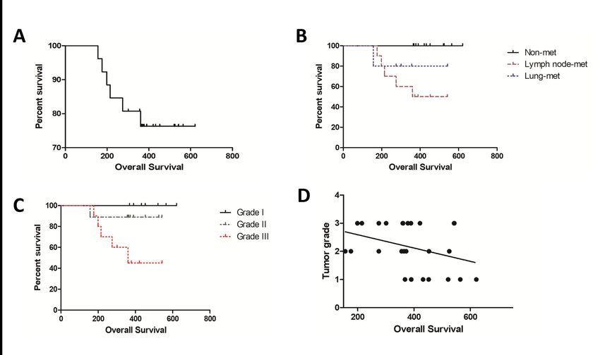
bioRxiv preprint first posted online Dec. 8, 2018; doi: http://dx.doi.org/10.1101/490144. The copyright holder for this preprint (which
       was not peer-reviewed) is the author/funder, who has granted bioRxiv a license to display the preprint in perpetuity.
                                 It is made available under a CC-BY-NC-ND 4.0 International license.

                                                                                                                                          3

                and c-KIT is overexpressed in human BC and still represent important predictive markers.
                New studies evaluating other PDGFR/c-KIT inhibitors represent a new therapeutic
                perspective.
                     In dogs with mammary tumors, c-KIT exhibits a controversial role in tumorigenesis [15,
                16, 17]. In general, c-KIT is expressed in normal mammary glands. During cancer
                progression, tumor cells lack c-KIT expression [15, 16, 17]. Regarding PDGFR, one previous
                study evaluated gene expression in CMT [17], and no previous study demonstrated PDGFR
                expression in CMT. The toxicity of different tyrosine kinase inhibitors has been studied in
                dogs [18, 19, 20]. Thus, dogs represent an important preclinical model for human cancers.
                     In humans and dogs, the development of metastasis is the major cause of cancer-related
                deaths [6, 15]. The PDGFR and PDGF signaling pathway is responsible for intratumoral
                lymphogenesis, promoting nodal metastasis [21], and the VEGF/VEGFR pathway induces
                neovasculogenesis [Shibuya, 2011]. Microvascular density (MVD) is very important for
                tumor progression and is induced by the production of proangiogenic factors by tumor cells
                [23]. MVD in BC is correlated with overall survival and disease-free interval in both humans
                [23, 24] and dogs [25]. Given the importance of dogs as a natural model for human BC, this
                research aimed to evaluate VEGF and PDGFR expression and assess MVD in metastatic and
                nonmetastatic CMT.

                2. Material and methods
                2.1 Study design
                          This was a prospective nonrandomized study including 26 female dogs from three
                institutions: Veterinary Teaching Hospital of University of Franca (UNIFRAN), the
                Veterinary Teaching Hospital of São Paulo State University (UNESP) and the Veterinary
                Teaching Hospital of the Federal University of the Southern Border (UFFS). All procedures
                were performed in accordance with the national and international guidelines for use of
                animals in research. This study was approved by each institutional Ethics Committee in the
                Use of Animals. The tissue samples were collected between May 2015 and September 2017.
                          We exclusively included patients with malignant tumors meeting the following
                criteria: a sufficient amount of tissue in the primary tumor and metastatic foci for
                immunohistochemical evaluation, received no previous systemic treatment, at least one year
                of clinical follow-up, only one tumor in the mammary gland and sentinel lymph node
                evaluation.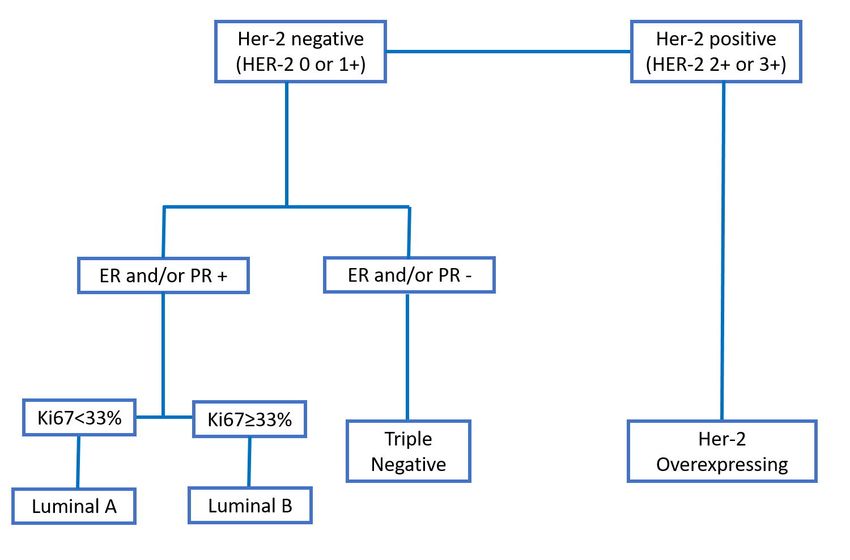
bioRxiv preprint first posted online Dec. 8, 2018; doi: http://dx.doi.org/10.1101/490144. The copyright holder for this preprint (which
       was not peer-reviewed) is the author/funder, who has granted bioRxiv a license to display the preprint in perpetuity.
                                 It is made available under a CC-BY-NC-ND 4.0 International license.

                                                                                                                                          4

                2.2 Patients
                          We included female dogs with only one mammary gland lesion, independent of the
                tumor size or location. All dogs underwent a previous cytological examination indicating a
                mammary gland tumor. The patients underwent sentinel lymph node assessment according to
                Beserra et al. [26]. Then, unilateral chain mastectomy was performed. The surgical specimens
                were stored in 10% buffered formalin for 24 hours. Then, histological processing was
                performed. Briefly, 4-μm tissue sections were processed for hematoxylin and eosin staining.
                The histological classification was performed according to Goldschmidt et al. [27], and tumor
                grade was evaluated according to Karayannopoulou et al. [28].

                2.3 Clinical evaluation
                          All patients underwent three-view thoracic radiographic examination, abdominal
                ultrasound and complete blood count. Then, we obtained the clinical stage from the staging
                system established by the World Health Organization for CMT and modified by Sorenmo et
                al. [2]. Patients were classified as stage I-V [2]. Patients with at least stage III disease and at
                least tumor grade II received adjuvant chemotherapy with four cycles of 300 mg/m2
                carboplatin and 5 mg/kg of firocoxib every 4 hours for six months Bonolo et al. [29]. Clinical
                follow-up was performed every three months in the first year with three-view thoracic
                radiographic examinations and complete blood counts.

                2.3 Molecular phenotype
                          We      exclusively       included        patients     with      triple-negative        mammary          tumors.
                Immunohistochemistry was performed according to Abadie [8]. Then, we used a combination
                of ERα, PR, HER-2, Ki67, CK5/6 and EGFR to classify the different molecular phenotypes
                (Figure 1).

                2.4 Tumor groups
                          Twenty-six patients met our inclusion criteria, and four samples were used. Our
                patients were divided into three groups: patients with malignant mammary tumor with no
                local or distant metastasis and at least one year of follow-up of evaluation (G1, nonmetastatic
                tumors), patients with malignant mammary tumor with metastasis to sentinel lymph nodes at
                diagnosis and no distant metastasis (G2, tumors with lymph node metastasis) and patients
                with malignant mammary tumors with negative sentinel lymph nodes at diagnosis and
                developed late lung metastasis (G3, tumors with distant metastasis).
bioRxiv preprint first posted online Dec. 8, 2018; doi: http://dx.doi.org/10.1101/490144. The copyright holder for this preprint (which
was not peer-reviewed) is the author/funder, who has granted bioRxiv a license to display the preprint in perpetuity.
It is made available under a CC-BY-NC-ND 4.0 International license.
5
2.4 Immunohistochemistry
Immunohistochemical evaluation was performed in 41 paraffin blocks: 11 primary
tumors from G1, 10 primary tumors and 10 lymph node metastasis from G2 and five primary
tumors and five lung metastasis from G3. Charged slides with 4-μm tissue sections were cut,
deparaffinized and submitted to antigen retrieval with citrate buffer pH 6.0 in a pressure
cooker (Pascal, Dako, Carpinteria, CA, USA). Endogenous peroxidase was blocked with 8%
hydrogen peroxide diluted in methanol for 10 minutes. Then, monoclonal VEGFR (Flk1,
Abcam, Cambridge, UK), rabbit monoclonal PGFR-β (clone 26E1, Cell signaling, Danvers,
MA, USA) and rabbit polyclonal CD31 (ThermoFisher Scientific, Waltham, MA, EUA)
antibodies at 1:300, 1:200 and 1:50, respectively, were applied for 18 hours. Afterward,
incubation with secondary antibody (Envision, Dako, Carpinteria, CA, USA) for 1 hour was
performed, and samples were incubated with 3,3'-diaminobenzidine (DAB, Dako, Carpinteria,
CA, USA) for 5 minutes. Counterstaining was performed with Harris hematoxylin for 1
minute. The positive controls were selected according to the Protein Atlas recommendations
(https://www.proteinatlas.org). For VEGFR, canine liver was used as a positive control. For
PGFR-β, normal testis was used as a positive control. For CD31, we used an internal control
(blood vessel in each tumor sample). Mouse (Negative Control Mouse, Dako, Carpinteria,
CA, USA) and rabbit immunoglobulin (Negative Control Rabbit, Dako, Carpinteria, CA,
USA) were used as negative controls. VEGFR, PGFR-β and CD31 cross-reactivities with
canine tissue were provided by the manufacturer.
For VEGFR and PGFR-β, the samples were evaluated by optical microscopy using a
semiquantitative score of 0 to 4 [30]. Briefly, 0: absence of labeling, 1: 1% up to 25% of
positive cells, 2: 26% up to 50% positive cells, 3: 51 up to 75% positive cells and 4: > 75%
positive cells. For Factor CD31, microvessel counts were performed in five fields using the
20x objective, and the mean of the sum of the five fields was used according to Weidner [23].
2.5 Statistical evaluation
The results were previously submitted to Shapiro-Wilk normality tests and analysis of
variance (ANOVA). If the variables presented a Gaussian distribution, Tukey’s test or
nonparametric Kruskal-Wallis test was used for microvascular density analysis. Spearman's
test was used to investigate correlations between variables. Regarding the VEGFR and
PDGFR immunoexpression, Chi-square or Fisher exact tests were performed. StatisticalbioRxiv preprint first posted online Dec. 8, 2018; doi: http://dx.doi.org/10.1101/490144. The copyright holder for this preprint (which
was not peer-reviewed) is the author/funder, who has granted bioRxiv a license to display the preprint in perpetuity.
It is made available under a CC-BY-NC-ND 4.0 International license.
6
analyses were performed using the GraphPad Prism® program (version 6.0 - GraphPad
Software, Inc. 2015) with a significance level of 0.05.
3. Results
3.1 Clinical and Pathological evaluation
Regarding the pathological parameters, tubulopapillary carcinoma was the most
commonly observed diagnosis (8/26) followed by solid carcinoma (7/26), complex carcinoma
(5/26), comedocarcinoma (4/26) and mixed carcinoma (2/26). Seven carcinomas were
classified as grade 1, eight as grade 2 and eleven as grade 3 (Table 1). The clinical parameters
are described in Table 1. The mean survival time for all patients independent of the metastatic
status was 384.96 days (±123.6) (Figure 2A). Patients with nonmetastatic disease at the
diagnosis experienced an increased survival time compared with patients with lymph node
metastasis (P=0.0359) (Figure 2B). Patients with grade III tumors experienced a shorter
survival time compared with grades I and II (P=0.534) (Figure 2C). A negative correlation
was observed between tumor grade and overall survival (P=0.0274; Spearman R= -0.4244).
Thus, patients with high tumor grade experienced a reduced survival time (Figure 2D).
2.2 Immunohistochemistry
We identified VEGFR-positive expression in all primary and metastatic samples.
Patients exhibiting lymph node metastasis at diagnosis exhibited increased VEGFR
expression compared with nonmetastatic carcinomas (P=0.0238). On the other hand, we did
not observe a significant difference when we compared primary carcinomas with lung
metastasis with nonmetastatic carcinomas (P=0.1239). We did not observe a significant
difference between lymph node metastasis and lung metastasis (P=0.7243). We also did not
observe a significant difference when comparing the primary carcinomas with its respective
metastasis. We identified a positive correlation between tumor grade and VEGFR expression
(P=0.001; Spearman R=0.6071). No correlation between VEGFR expression and overall
survival was observed (P=0.125; Spearman R=-0.3087). VEGFR immunoexpression results
are presented in Figure 3 and Table 2.
Regarding PDGFR immunoexpression, we observed positive expression in 25 out of
26 samples. No significant difference in PDGFR expression was noted among the different
groups. No correlation was observed between PDGFR expression and tumor grade
(P=0.0692; Spearman R=0.3620) or overall survival (P=0.2581; Spearman R= -0.2301).bioRxiv preprint first posted online Dec. 8, 2018; doi: http://dx.doi.org/10.1101/490144. The copyright holder for this preprint (which
was not peer-reviewed) is the author/funder, who has granted bioRxiv a license to display the preprint in perpetuity.
It is made available under a CC-BY-NC-ND 4.0 International license.
7
However, we identified a positive correlation between VEGFR and PDGFR expression. Thus,
samples exhibiting the highest scores for VEGFR also presented the highest PDGFR
expression (P=0.01; Spearman R=0.4959). PDGFR immunoexpression results are presented
in Figure 3 and Table 2.
Regarding MVD (Figure 3), we identified an increased number of vessels in primary
tumors with lymph node metastasis (P=0.0151) and lung metastasis (P=0.0046) compared
with tumors with no metastasis. Primary tumors with lung metastasis exhibited increased
MVD compared with carcinoma with lymph node metastasis (P=0.0496). Interestingly, we
did not find a correlation between MVD and VEGFR expression (P=0.0827; Spearman R=
0.3467); however, a positive correlation between MVD and PDGFR was observed (P=0.0102;
Spearman R= 0.4946) (Figure 4). Thus, samples with high PDGFR expression also exhibited
high MVD (Figure 4).
4. Discussion
Canine mammary gland tumors are one of the most important cancers in intact female
dogs and represents a therapeutic challenge. Although surgery and chemotherapy have been
used for CMT treatment, there is no standardized chemotherapy or target therapy. This
research evaluated VEGFR-2, PDGFR2 and MVD in canine mammary tumors, aiming to
associate different prognostic factors with these proteins. One interesting aspect of our
research is a very restricted criterion used in patient selection. Typically, lymph node
metastasis is evaluated after chain mastectomy in inguinal lymph nodes, and the inguinal
lymph node is not always draining the tumor. The sentinel lymph node technique allowed us
to identify the tumor-draining lymph node and increase the probability of identifying
metastasis.
Another interesting aspect was the inclusion of a group of patients with no metastatic
disease detected at diagnosis but with late lung metastasis. In clinical practice, it is relatively
common to find female dogs with late lung metastasis after months or even years post
surgery. However, given that metastatic disease takes a while to be detected on the X-ray, it
was not possible to achieve a high number of patients in this group. These particular criteria
can explain the highest survival time of patients with lung metastasis compared with patients
with lymph node metastasis. We considered overall survival between the diagnosis and the
time of current follow-up/death. Given that lung metastasis appeared in the patients from this
group, the overall survival was compared with no lymph metastasis patients.bioRxiv preprint first posted online Dec. 8, 2018; doi: http://dx.doi.org/10.1101/490144. The copyright holder for this preprint (which
was not peer-reviewed) is the author/funder, who has granted bioRxiv a license to display the preprint in perpetuity.
It is made available under a CC-BY-NC-ND 4.0 International license.
8
We did not include patients with more than one tumor in the mammary chain. This
criterion excluded many animals from our study. Fifteen dogs were excluded from this study
due to the presence of multiple mammary tumors (data not shown). In the context of multiple
mammary tumors, it is not possible to guarantee which nodule the metastasis originated from.
Regarding the nonmetastatic group, some patients with multiple tumors exhibited different
molecular subtypes (data not shown), making it difficult to establish a prognosis based on the
molecular subtype. Triple-negative tumors seem to be the most common molecular subtype in
dogs [8]. This finding highlight the utility of dogs as a model for human triple-negative BC.
VEGFR-2 expression is correlated with angiogenesis and modulation of the tumor
microenvironment [12]. In human [31] and canine [12] mammary tumors, VEGFR-2
expression is important in tumor growth and development and exhibits prognostic value [12].
Moreover, VEGFR-2 is a tyrosine kinase protein that can be inhibited by different target
therapies [32]. Our results strongly suggested that VEGFR-2 is overexpressed in tumors with
metastasis, indicating its predictive and prognostic value. Given that the VEGFR-2 inhibitor is
not routinely used in human and veterinary oncology, clinical studies in dogs can benefit both
species. We demonstrated a correlation between VEGFR-2 expression and tumor grade,
indicating that high-grade tumors may require increased angiogenesis to maintain cell
proliferation. Although we did not identify a correlation between VEGFR-2 expression and
MVD, we identified increased vascular density in metastatic carcinomas.
These results together demonstrate the dependency of high-grade/metastatic tumors on
angiogenic factors. Santos et al. [12] investigated VEGFR-2 expression in CMT, and
overexpression of this protein was associated with carcinosarcomas (very aggressive tumor
subtype). Although MVD did not correlate with VEGFR-2 expression, we identified a
correlation between PDGFR and MVD. PDGFR induced intratumoral lymphogenesis [21],
and we identified a correlation between intratumoral vasculogenesis and high levels of
PDGFR. In addition, PDGFR and VEGFR-2 exhibited a positive correlation. These results
collectively demonstrated the role of angiogenesis in the development and potential
aggressiveness of CMT. Interestingly, both primary tumors and respective metastases were
positive for VEGFR and PDGFR immunoexpression. Given that numerous VEGFR/PDGFR
inhibitors are available, these results indicate the use of target therapy patients with CMT.
Thus, our results support the idea of future clinical trials investigating the role of
VEGFR/PDGFR inhibitors for the treatment of metastatic CMT.
5. ConclusionsbioRxiv preprint first posted online Dec. 8, 2018; doi: http://dx.doi.org/10.1101/490144. The copyright holder for this preprint (which
was not peer-reviewed) is the author/funder, who has granted bioRxiv a license to display the preprint in perpetuity.
It is made available under a CC-BY-NC-ND 4.0 International license.
9
Metastatic mammary carcinomas present VEGFR-2 overexpression and high
microvascular density, indicating the role angiogenesis in tumor progression. PDGFR may
induce vasculogenesis in metastatic mammary carcinomas. Overall, our results suggest the
use of antiangiogenic and specific target therapies in a subset of patients with mammary
tumors.
Acknowledgments
We would like to thank the São Paulo Research Foundation (FAPESP) for their financial
support (grant number: 2015/02798-5).
6. References
1. Misdorp, W. Tumors of the mammary gland. In Tumors in Domestic Animals, 4th ed.;
Meuten, D.J., Eds.; Iowa State Press: Ames, IA, USA, 2002, pp. 575–606.
2. Sorenmo, K.U.; Worley, D.R.; Goldschmidt, M.H. Tumors of the Mammary Gland. In
Withrow & MacEwen's Small Animal Clinical Oncology, 5th ed.; Withrow S.J., Vail D.M.,
Page R.L., Eds.; Elsevier: St Louis, MO, USA, 2013, pp. 538-556.
3. DE NARDI, A.B.; RODASKI, S.; SOUSA, R.S.; COSTA, T.A.; MACEDO, T.R.;
RODIGHERI, S.M.; RIOS, A.; PIEKARZ, C.H. Prevalência de neoplasias e modalidades de
tratamentos em cães, atendidos no hospital veterinário da Universidade Federal do Paraná.
Arch Vet Sci. 2002, 7, 15-26; http://dx.doi.org/10.5380/avs.v7i2.3977
4. Santos, I.F.C.; Cardoso, J.M.M.; Oliveira, K.C.; Laisse, C. J. M.; Bessa, S.A.T. Prevalência
de neoplasias diagnosticadas em cães no Hospital Veterinário da Universidade Eduardo
Mondlane, Moçambique. Arq Bras Med Vet Zootec. 2013, 65, 773-782;
http://dx.doi.org/10.1590/S0102-09352013000300025
5. Bray, F.; Ferlay, J.; Soerjomataram, I.; Siegel, R.L.; Torre, L.A.; Jemal, A. Global cancer
statistics 2018: GLOBOCAN estimates of incidence and mortality worldwide for 36 cancers
in 185 countries. CA Cancer J Clin. 2018, 68, 394-424; https://doi.org/10.3322/caac.21492bioRxiv preprint first posted online Dec. 8, 2018; doi: http://dx.doi.org/10.1101/490144. The copyright holder for this preprint (which
was not peer-reviewed) is the author/funder, who has granted bioRxiv a license to display the preprint in perpetuity.
It is made available under a CC-BY-NC-ND 4.0 International license.
10
6. Cancer Genome Atlas Network. Comprehensive molecular portraits of human breast
tumours. Nature. 2012, 490, 61-70.
7. McCart Reed, A.E.; Kalita-de Croft, P.; Kutasovic, J.; Saunus, J.M.; Lakhani, S.R. Recent
advances in breast cancer research impacting clinical diagnostic practice. J Pathol. (accepted);
https://doi.org/10.1002/path.5199
8. Abadie, J.; Nguyen, F.; Loussouarn, D.; Peña, L.; Gama, A.; Rieder, N.; Belousov, A.;
Bemelmans, I.; Jaillardon, L.; Ibisch, C.; Campone, M. Canine invasive mammary carcinomas
as models of human breast cancer. Part 2: immunophenotypes and prognostic significance.
Breast Cancer Res Treat. 2018, 167, 459-468; https://doi.org/10.1007/s10549-017-4542-8
9. Ghosh, S.; Sullivan, C.A.; Zerkowski, M.P.; Molinaro, A.M.; Rimm, D.L.; Camp, R.L.;
Chung, G.G. High levels of vascular endothelial growth factor and its receptors (VEGFR-
1,VEGFR-2, neuropilin-1) are associated with worse outcome in breast cancer. Hum Pathol.
2008, 39, 1835-43; https://doi.org/10.1016/j.humpath.2008.06.004
10. Queiroga, F.L.; Pires, I.; Parente, M.; Gregório, H.; Lopes, C.S. COX-2 over-expression
correlates with VEGF and tumour angiogenesis in canine mammary cancer. Vet J. 2011, 189,
77-82; https://doi.org/10.1016/j.tvjl.2010.06.022
11. Qiu, C.W.; Lin, D.G.; Wang, J.Q.; Li, C.Y.; Deng, G.Z. Expression and significance of
PTEN and VEGF in canine mammary gland tumours. Vet Res Commun. 2008, 32, 463-72;
https://doi.org/10.1007/s11259-008-9049-7
12. Santos, A.; Lopes, C.; Gärtner, F.; Matos, A.J. VEGFR-2 expression in malignant tumours
of the canine mammary gland: a prospective survival study. Vet Comp Oncol. 2016, 14, 83-
92; https://doi.org/10.1111/vco.12107
13. Cristofanilli, M.; Morandi, P.; Krishnamurthy, S.; Reuben, J.M.; Lee, B.N.; Francis, D.;
Booser, D.J.; Green, M.C.; Arun, B.K.; Pusztai, L.; Lopez, A.; Islam, R.; Valero, V.;
Hortobagyi, G.N. Imatinib mesylate (Gleevec) in advanced breast cancer-expressing C-Kit or
PDGFR-beta: clinical activity and biological correlations. Ann Oncol. 2008, 19, 1713-9;
https://doi.org/10.1093/annonc/mdn352bioRxiv preprint first posted online Dec. 8, 2018; doi: http://dx.doi.org/10.1101/490144. The copyright holder for this preprint (which
was not peer-reviewed) is the author/funder, who has granted bioRxiv a license to display the preprint in perpetuity.
It is made available under a CC-BY-NC-ND 4.0 International license.
11
14. Modi, S.; Seidman, A.D.; Dickler, M.; Moasser, M.; D'Andrea, G.; Moynahan, M.E.;
Menell, J.; Panageas, K.S.; Tan, L.K.; Norton, L.; Hudis, C.A. A phase II trial of imatinib
mesylate monotherapy in patients with metastatic breast cancer. Breast Cancer Res Treat.
2005, 90, 157-6; https://doi.org/10.1007/s10549-004-3974-0
15. Salvador, R.C.L.; Raposo, T.M.M.; Fonseca-Alves, C.E.; Terra, E.M.; Varallo, G.R.;
Laufer-Amorim, R. Evaluation of c-KIT protein expression in canine mammary tumors. In
São Paulo Advanced School of Comparative Oncology: Abstracts, Águas de São Pedro, SP,
Brasil, 30/09/2012; Rainho , C.A.; dos Reis, P.P; Rogatto, S.R., Eds.; BMC Proceedings, 7,
63-63; https://doi.org/10.1186/1753-6561-7-S2-P63
16. Brunetti, B.; Beha, G.; Benazzi, C.; Bondin, V.; De Tolla, L.; Sarli, G. CD117 expression
influences proliferation but not survival in canine mammary tumours. J Comp Pathol. 2014,
151, 202–206; https://doi.org/10.1016/j.jcpa.2014.04.018
17. Koltai, Z.; Szabó, B.; Jakus, J.; Vajdovich, P. Tyrosine kinase expression analyses in
canine mammary gland tumours - A pilot study. Acta Vet Hung. 2018, 66, 294-308;
https://doi.org/10.1556/004.2018.027
18. London, C.A.; Hannah, A.L.; Zadovoskaya, R.; Chien, M.B.; Kollias-Baker, C.;
Rosenberg, M,; Downing, S.; Post, G.; Boucher, J.; Shenoy, N.; Mendel, D.B.; McMahon, G.;
Cherrington, J.M. Phase I dose-escalating study of SU11654, a small molecule receptor
tyrosine kinase inhibitor, in dogs with spontaneous malignancies. Clin Cancer Res. 2003, 9,
2755-68.
19. Nakano, Y.; Kobayashi, T.; Oshima, F.; Fukazawa, E.; Yamagami, T.; Shiraishi, Y.;
Takanosu, M. Imatinib responsiveness in canine mast cell tumors carrying novel mutations of
c-KIT exon 11. J Vet Med Sci. 2014, 76, 545-8; https://doi.org/10.1292/jvms.13-0156
20. Foskett, A.; Manley, C.; Naramore, R.; Gordon, I.K.; Stewart, B.M.; Khanna, C.
Tolerability of oral sorafenib in pet dogs with a diagnosis of cancer. Vet Med (Auckl). 2017, 8,
97-102; DOI: 10.2147/VMRR.S149678bioRxiv preprint first posted online Dec. 8, 2018; doi: http://dx.doi.org/10.1101/490144. The copyright holder for this preprint (which
was not peer-reviewed) is the author/funder, who has granted bioRxiv a license to display the preprint in perpetuity.
It is made available under a CC-BY-NC-ND 4.0 International license.
12
21. Cao, R.; Björndahl, M.A.; Religa, P.; Clasper, S.; Garvin, S.; Galter, D.; Meister, B.;
Ikomi, F.; Tritsaris, K.; Dissing, S.; Ohhashi, T.; Jackson, D.G.; Cao, Y. PDGF-BB induces
intratumoral lymphangiogenesis and promotes lymphatic metastasis. Cancer Cell. 2004, 6,
333-45. Erratum in Cancer Cell. 2006, 9, 239; https://doi.org/10.1016/j.ccr.2004.08.034
22. Shibuya, M. Vascular Endothelial Growth Factor (VEGF) and Its Receptor (VEGFR)
Signaling in Angiogenesis: A Crucial Target for Anti- and Pro-Angiogenic Therapies. Genes
Cancer. 2011, 2, 1097-105; https://doi.org/10.1177/1947601911423031
23. Weidner, N.; Folkman, J.; Pozza, F.; Bevilacqua, P.; Allred, E.N.; Moore, D.H.; Meli, S.;
Gasparini, G. Tumor angiogenesis: A new significant and independent prognostic indicator in
early-stage breast carcinoma. J Nat Cancer Inst. 1992, 84, 1875-1887.
24. Bosari, S.; Lee, A.K.C.; DeLellis, R.A.; Wiley, B.D.; Heatley, G.J.; Silverman, M.L.
Microvessel quantification and prognosis in invasive breast carcinoma. Human Pathol. 1992,
23, 755-761.
25. Graham, J.C.; Myers, R.K. The prognostic significance of angiogenesis in canine
mammary tumors. J Vet Intern Med. 1999, 13, 416–418; https://doi.org/10.1111/j.1939-
1676.1999.tb01456.x
26. Beserra, H.E.O.; Cavalcante, R.V.; de Pinho Pessoa, A.W.; Pinheiro, L.G.P. Technical of
sentinel lymph detection in canine mammary gland using Patent Blue V and technetium [sup.
99m] Tc/Tecnica de deteccao do linfonodo sentinela da glandula mamaria de cadelas
utilizando Azul Patente V e tecnecio [sup. 99m] Tc/Tecnica para la deteccion del ganglio
linfatico centinela en glandulas mamarias de perras con Azul Patente y tecnecio [sup. 99m]
Tc. Vet. Zootec . 2011, 18, 57-60.
27. Goldschimidt, M.; Peña, L.; Rasotto, R.; Zappuli, V. Classification and grading of canine
mammary tumors. Vet Pathol. 2011, 48, 117–131; DOI: 10.1177/0300985810393258
28. Karayannopoulou, M.; Kaldrymidou, E.; Constantinidis, T.C.; Dessiris, A. Histological
grading and prognosis in dogs with mammary carcinomas: application of a humangrading
method. J Comp Pathol. 2005, 133, 246-52; DOI: 10.1016/j.jcpa.2005.05.003bioRxiv preprint first posted online Dec. 8, 2018; doi: http://dx.doi.org/10.1101/490144. The copyright holder for this preprint (which
was not peer-reviewed) is the author/funder, who has granted bioRxiv a license to display the preprint in perpetuity.
It is made available under a CC-BY-NC-ND 4.0 International license.
13
29. Bonolo De Campos, C.; Lavalle, G.E.; Monteiro, L.N.; Pêgas, G.R.A.; Fialho, S.L.;
Balabram, D.; Cassali, G.D. Adjuvant Thalidomide and Metronomic Chemotherapy for the
Treatment of Canine Malignant Mammary Gland Neoplasms. In Vivo 2018, 32, 1659-1666;
DOI: 10.21873/invivo.11429
30. Fonseca-Alves, C.E.; Kobayashi, P.E.; Rivera Calderón, L.G.; Felisbino, S.L.; Rinaldi,
J.C.; Drigo, S.A.; Rogatto, S.R.; Laufer-Amorim, R. Immunohistochemical panel to
characterize canine prostate carcinomas according to aberrant p63 expression. PLoS One.
2018, 13, 1-16; https://doi.org/10.1186/1753-6561-7-S2-P63
31. Yan, J.D.; Liu, Y.; Zhang, Z.Y.; Liu, G.Y.; Xu, J.H.; Liu, L.Y.; Hu, Y.M. Expression and
prognostic significance of VEGFR-2 in breast cancer. Pathol Res Pract. 2015, 211, 539-43;
DOI: 10.1016/j.prp.2015.04.003
32. Sloan, B.; Scheinfeld, N.S. Pazopanib, a VEGF receptor tyrosine kinase inhibitor for
cancer therapy. Current opinion in investigational drug (London, England: 2000), 2008, 9,
1324-1335.
Figure 1. Classification of the molecular phenotypes of canine mammary carcinomas
according to each immunohistochemical marker.
Figure 2. Overall survival of dogs with mammary carcinomas based on clinical parameters. A:
Percent survival of all female dogs independent of metastasis status. B: Female dogs with
nonmetastatic tumors exhibited increased survival time followed by patients with lung
metastasis and lymph node metastasis. C: Overall survival independent of metastasis status
according to tumor grade. Patients with grade III experienced a reduced survival time. D:
Negative correlation between tumor grade and overall survival. Patients with low-grade
tumors exhibited increased survival time.
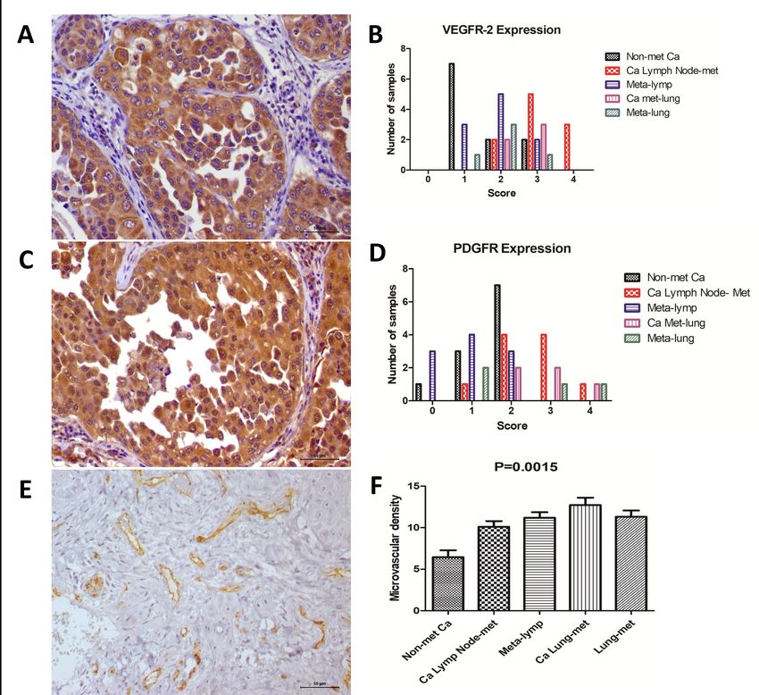
Figure 3. Immunoexpression of the different markers. A: VEGFR-2 (score 4) expression in a
mammary carcinoma with lymph node metastasis. B: Graphic representation of each
immunohistochemical score for VEGFR-2 expression in all tumor groups. C: PDGFRbioRxiv preprint first posted online Dec. 8, 2018; doi: http://dx.doi.org/10.1101/490144. The copyright holder for this preprint (which
was not peer-reviewed) is the author/funder, who has granted bioRxiv a license to display the preprint in perpetuity.
It is made available under a CC-BY-NC-ND 4.0 International license.
14
expression in a mammary carcinoma (score 2) with lymph node metastasis. D: Graphic
representation of each immunohistochemical score for PDGFR expression in all tumor
groups. E: Microvascular density (MVD) in a mammary carcinoma with lymph node
metastasis. F: Graphic representation of MVD, indicating an increased number of vessels in
metastatic tumors.
Figure 4. Correlation between immunohistochemical markers and different clinical
parameters. A: Positive correlation between VEGFR-2 immunoexpression and tumor grade
(P=0.001; Spearman R=0.6071). B: Absence of correlation between PDGFR expression and
tumor grade (P=0.0692; Spearman R=0.3620). C: Positive correlation between VEGFR-2 and
PDGFR expression (P=0.01; Spearman R=0.4959). D: Positive correlation between PDGFR
expression and microvascular density (P=0.0102; Spearman R= 0.4946).
Table 1. Clinical parameters of female dogs affected by mammary gland tumors.
Table 2. Immunohistochemical evaluation of VEGFR-2, PDGFR and microvascular density
in canine mammary gland tumor samples.bioRxiv preprint first posted online Dec. 8, 2018; doi: http://dx.doi.org/10.1101/490144. The copyright holder for this preprint (which
was not peer-reviewed) is the author/funder, who has granted bioRxiv a license to display the preprint in perpetuity.
It is made available under a CC-BY-NC-ND 4.0 International license.
Table 1. Clinical parameters of female dogs affected by mammary gland tumors.
Clinical parameters N %
Age
< 10.5 16 61.5
> 10.5 10 38.5
Neutering Status
Intact 20 100%
Neutered 0 0
Tumor size
< 5 cm 18 69.2%
> 5 cm 8 30.8%
Nodal stage*
N0 16 61.5%
N1 10 38.5%
Histological grade*
Grade I 7 26.9%
Grade II 8 30.8%
Grade III 11 42.3%
*Nodal stage based on the sentinel lymph node.
**According to Karayannopoulou et al., 2005.bioRxiv preprint first posted online Dec. 8, 2018; doi: http://dx.doi.org/10.1101/490144. The copyright holder for this preprint (which
was not peer-reviewed) is the author/funder, who has granted bioRxiv a license to display the preprint in perpetuity.
Table 2. Immunohistochemical evaluation of VEGFR-2, PDGFR and microvascular density in canine mammary gland tumor samples.
It is made available under a CC-BY-NC-ND 4.0 International license.
VEGFR-2 score PDGFR score Microvascular Density
0 1 2 3 4 0 1 2 3 4 Mean (SD*)
Non-metastatic 0% 45.5% 45.5% 9% 0% 9% 27.3% 63.7% 0% 0%
6.4 (± 2.7)
Carcinomas (N=11) (N=0) (N=5) (N=5) (N=1) (N=0) (N=1) (N=3) (N=7) (N=0) (N=0)
Carcinomas Lymph 0% 0% 20% 50% 30% 0% 40% 40% 10%
10% (N=1) 10.1 (±2.2)
Node Metastasis (N=10) (N=0) (N=0) (N=2) (N=5) (N=3) (N=0) (N=4) (N=4) (N=1)
Lymph Node Metastasis 0% 30% 50% 20% 0% 30% 30% 0% 0%
40% (N=4) 11.2 (±2.0)
(N=10) (N=0) (N=3) (N=5) (N=2) (N=0) (N=3) (N=3) (N=0) (N=0)
Carcinomas Lung 0% 0% 0% 60% 40% 0% 40% 40% 20%
0% (N=0) 12.7 (±2.0)
Metastasis (N=5) (N=0) (N=0) (N=0) (N=3) (N=2) (N=0) (N=2) (N=2) (N=1)
0% 20% 60% 20% 0% 20% 0% 20% 20%
Lung Metastasis (N=5) 40% (N=2) 11.3 (±1.7)
(N=0) (N=1) (N=3) (N=1) (N=0) (N=1) (N=0) (N=1) (N=1)
*SD: standard deviationYou can also read