A Cancer-Favoring, Engineered Vaccinia Virus for Cholangiocarcinoma - MDPI
←
→
Page content transcription
If your browser does not render page correctly, please read the page content below
cancers
Communication
A Cancer-Favoring, Engineered Vaccinia Virus
for Cholangiocarcinoma
So Young Yoo 1,2, * , Narayanasamy Badrinath 3 , Hye Lim Lee 2 , Jeong Heo 3 and
Dae-Hwan Kang 2,3
 1   BIO-IT Foundry Technology Institute, Pusan National University, Busan 46241, Korea
 2   Research Institute for Convergence of Biomedical Science and Technology, Pusan National University
     Yangsan Hospital, Yangsan 50612, Korea; roasua@hanmail.net (H.L.L.); sulsulpul@naver.com (D.-H.K.)
 3   Biomedical Sciences, School of Medicine, Pusan National University, Yangsan 50612, Korea;
     badrisamy@gmail.com (N.B.); sodium@korea.com (J.H.)
 *   Correspondence: yoosy2@gmail.com; Tel.: +82-51-510-3417

 Received: 14 October 2019; Accepted: 24 October 2019; Published: 27 October 2019

 Abstract: While oncolytic vaccinia virus-based therapy has shown promising results for uncured
 patients with cancer, its effects on cholangiocarcinoma (CCA) remain unclear. Here, we evaluated
 the anti-cancer activity of the cancer-favoring oncolytic vaccinia virus (CVV), which was recognized
 as a promising therapy for stem cell-like colon cancer cells (SCCs) and metastatic hepatocellular
 carcinoma (HCC) in previous studies. CCA presents major challenges, such as clinical complexity,
 stem cell cancer characteristics, a high refractory rate, resistance to conventional therapy, and a dismal
 prognosis. In the present study, we confirmed the oncolytic activity of the CVV in CCA with a slightly
 alkaline microenvironment (pH 7–8), in which the CVV was stable and highly effective at infecting
 CCA. Taken together, our findings suggest that CVV-based therapy is highly suitable for the treatment
 of CCA.

 Keywords:      cholangiocarcinoma (CCA); cancer-favoring vaccinia virus (CVV); alkaline
 extracellular microenvironment

1. Introduction
      DNA recombination technology provides unprecedented opportunities to utilize viruses for
biomedical applications, including tissue engineering, drug/gene delivery, targeting/imaging, etc. [1–6].
Virotherapy utilizes engineered viruses via the above technologies as therapeutics to cure diseases [4,7,8].
Oncolytic viruses (OVs) are unique nanomedicines with merits over conventional anti-cancer drugs
in terms of their tumor selectivity and killing mechanism [9]. Selectivity can be achieved by either
genetic engineering or evolutionary bioselection, which attenuates viral replication in normal cells [10].
Strategies depend on disrupting genes that are critical for the virus to replicate in host cells, but that
are limited in normal cells and redundant in cancer cells. For the vaccinia virus (VV), the most popular
gene for disruption encodes the vaccinia thymidine kinase enzyme (vTk); the disruption of this gene
results in the preferred replication of VV in cancer cells with high cellular TK expression [11,12].
      A refractory cancer one that is resistant to conventional therapy [13,14]; cholangiocarcinoma
(CCA) is curable only in its early stage by surgical removal of the tumor mass, but it becomes complex
and refractory in later stages, with a dismal prognosis. Therefore, therapeutics with a new mode of
action should be developed and employed to treat CCA. OVs have shown promising results for treating
various cancers [3,7,15,16]. In our previous study, clinically, JX-594 demonstrated tumor selectivity
via viral thymidine kinase (vTK) inactivation because cancer cells have high cellular TK levels due to
Epidermal growth factor receptor (EGFR) pathway activation, in which the VV was evolved to replicate

Cancers 2019, 11, 1667; doi:10.3390/cancers11111667                             www.mdpi.com/journal/cancers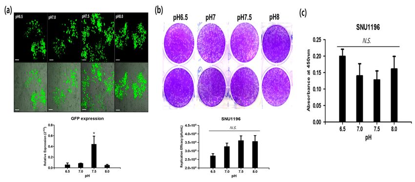
Cancers 2019, 11, x FOR PEER REVIEW 2 of 11
Cancers 2019, 11, 1667 2 of 11
levels due to Epidermal growth factor receptor (EGFR) pathway activation, in which the VV was
evolved toinreplicate
selectively tumors.selectively
Engineered in VVs
tumors.haveEngineered VVs have
also been shown also been shown
to successfully to successfully
kill even cancer stem kill
even cancer stem cell-like cancers using mechanisms different from conventional
cell-like cancers using mechanisms different from conventional anti-cancer drugs [15,16]. Specifically, anti-cancer drugs
we[15,16].
developedSpecifically, we developed
an evolutionary an evolutionary
cancer-favoring cancer-favoring
engineered vaccinia virusengineered
(CVV) and vaccinia virus (CVV)
demonstrated its
and demonstrated its ability to selectively and efficiently find and kill stem
ability to selectively and efficiently find and kill stem cell-like colon cancers [15]. CVV was evolved cell-like colon cancers
[15].Wyeth
from CVV was evolved
strain vacciniafrom Wyeth
virus. Thestrain vaccinia
evolution wasvirus. The evolution
achieved by isolation wasofachieved
Wyeth strainby isolation
vacciniaof
Wyeth
virus fromstrain vaccinia
the blood of virus fromvirus-injected
a vaccinia the blood of aVX2 vaccinia
tumorvirus-injected
animal model. VX2Thetumor animal
isolation of model. The
virus was
isolation of virus was done during the tumor volume reduction, which
done during the tumor volume reduction, which released viruses in into the serum. From that process, released viruses in into the
serum. From that process, we got evolved virus (called EVV), and subsequently,
we got evolved virus (called EVV), and subsequently, we engineered EVV by deleting the Tk gene to we engineered EVV
bythe
get deleting the Tk
virus with gene tocancer
enhanced get theselectivity
virus with enhanced
(called CVV).cancer selectivity
The higher (called CVV).
cancer-favoring The higher
characteristics
cancer-favoring characteristics of the CVV were successfully utilized as a
of the CVV were successfully utilized as a strategic therapeutic for metastatic hepatocellular carcinomastrategic therapeutic for
metastatic
(HCC) and hepatocellular
induced complete carcinoma (HCC)
regression of and
liverinduced completeand
tumorigenicity regression
metastasisof liver tumorigenicity
to the colon [16].
However, few studies have dealt with CCA in terms of utilizing an OV as a treatment.ofCCA
and metastasis to the colon [16]. However, few studies have dealt with CCA in terms utilizing
has aan
OV as a treatment. CCA has a different appearance and tumor localization
different appearance and tumor localization from other cancers. Furthermore, it is generally thought from other cancers.
Furthermore,
that cancers have it isa generally thought
slightly acidic that cancers have
microenvironment a slightly
[17,18] comparedacidictomicroenvironment
normal cells. However, [17,18]
compared to normal cells. However, the pH of CCA may be different from
the pH of CCA may be different from general cancers because it is associated with the bile duct, and bilegeneral cancers because
it isa associated
has with the
slightly alkaline pHbile
(i.e.,duct, and bile has a slightly alkaline pH (i.e., pH 7–8).
pH 7–8).
2.2.Results
Results
2.1.Cancer-Favoring,
2.1. Cancer-Favoring,Evolutionarily-Engineered
Evolutionarily-EngineeredVaccinia
VacciniaVirus
Virus
TheCVV
The CVVwaswas generated
generated through
through evolution
evolution ofWyeth
of the the Wyeth
strainstrain
of theof the
VV, VV, deletion
deletion of thymidine
of thymidine kinase,
kinase,
and and of insertion
insertion of the green
the green fluorescence protein fluorescence protein (GFP) and
(GFP) and guanine-hypoxanthine guanine-hypoxanthine
phosphoribosyltransferase
phosphoribosyltransferase
(GPT) (GPT)1a).
selection markers (Figure selection
Real-timemarkers
qPCR (Figure 1a). Real-time
results indicated a lowerqPCR results
expression of indicated
vTK and a a
lowerexpression
higher expressionofofGFP
vTKinand a higher expression
CVV-infected cancer celloflines
GFP(HeLa).
in CVV-infected cancer celloflines
A higher expression vTK(HeLa).
and theA
higher
lack expressionofofGFP
of expression vTKwere
and also
the lack of expression
confirmed of GFP
in wild-type were
(WT) also confirmed
VV-infected in wild-type
cell lines (WT)
(Figure 1b,c).
VV-infected
These cell lines (Figure
results confirmed 1b,c). These
GFP expression results
in the CVV. confirmed GFP expression in the CVV.
Figure1. 1.(a)(a)
Figure The cancer-favoring
The cancer-favoring oncolytic vaccinia
oncolytic virusvirus
vaccinia (CVV) was engineered
(CVV) from the
was engineered Wyeth
from the strain
Wyeth
ofstrain
the vaccinia
of the virus (VV).
vaccinia The (VV).
virus The vTk
viral gene was
viral deleted
gene and the
vTk was GFP and
deleted andGPT
the selection
GFP andmarkers were
GPT selection
inserted along the sides of the early/late promoters, and then CVV was evolved and
markers were inserted along the sides of the early/late promoters, and then CVV was evolved andselected after serial
culture in serum
selected of infected
after serial cultureand responded
in serum tumor model.
of infected (b) vTk expression
and responded in WT
tumor model. (b)and
vTkCVV-infected
expression in
HeLa cancer cell lines. Experiments were done in triplicate. (c) Confirmation of GFP
WT and CVV-infected HeLa cancer cell lines. Experiments were done in triplicate. (c) Confirmation expression in the
CVV-infected HeLa cancer cell line. Experiments were done in duplicate.
of GFP expression in the CVV-infected HeLa cancer cell line. Experiments were done in duplicate.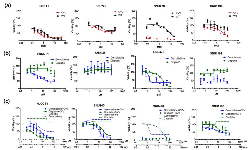
Cancers 2019, 11, 1667 3 of 11
Cancers 2019, 11, x FOR PEER REVIEW 3 of 11
2.2.High
2.2. HighReplication
ReplicationEfficacy
Efficacyofofthe
theCVV
CVVininCCA
CCACell
CellLines
Linesand
andTumors
Tumors
ToToevaluate
evaluate thethe replication
replication efficacy
efficacy of theofCVV
the in
CVV CCA, in HuCCT1
CCA, HuCCT1 and SNU1196
and SNU1196 cells werecells were
infected
infected
with a 0.1with a 0.1 multiplicity
multiplicity of infectionof (MOI)
infection (MOI)
of the WToforthe CVV.WTInfected
or CVV.cellInfected
lysatescell lysates
were usedwere used
to infect
to infect U2OS cells for replicated virus titration. The results indicated the
U2OS cells for replicated virus titration. The results indicated the superior replication efficacy of thesuperior replication
efficacy
CVV of the
relative CVV
to the WTrelative
in both to
cellthe WT
lines in both
(Figure cellFurthermore,
2a,b). lines (Figurethe 2a,b). Furthermore,
results confirmed the the robust
results
confirmedofthe
infectivity therobust
CVV infectivity
in the CCAofcell thelines.
CVV Thein the CCA efficacy
higher cell lines.ofThe
CVV higher efficacy
in cancer canof CVV
give itsin cancer
greater
can give its greater cancer selectivity. To test in vivo tumor selectivity and safety,
cancer selectivity. To test in vivo tumor selectivity and safety, tumor tissues and normal organ tissues tumor tissues and
normal
were organ tissues
harvested 2 weekswere
afterharvested 2 weeks
intraperitoneal after intraperitoneal
injection of the virus from injection of the virus from
HT29-xenografted nude HT29-
mice.
xenografted
They showednude mice.and
exclusive They showed
robust CVV exclusive
infection andin robust
tumorsCVV withinfection in tumors
negligible effects onwith
thenegligible
normal
effects
bile ductoncells
the (Figure
normal bile
2c). duct
These cells (Figure
results 2c). These
confirmed theresults confirmed infection
tumor-selective the tumor-selective
of the CVVinfection
in the
of the
CCA CVV in the CCA model.
model.
Figure2.2.Replication
Figure Replicationefficacy
efficacyofofthe
theCVV
CVVinincholangiocarcinoma
cholangiocarcinoma(CCA)
(CCA)cell
celllines
linesand
andtumors.
tumors.The
TheWT WT
ororCVV (0.1 multiplicity of infection (MOI)) was used to infect HuCCT1 and SNU1196
CVV (0.1 multiplicity of infection (MOI)) was used to infect HuCCT1 and SNU1196 cells. After cells. After24,
24,
48,48,
andand
7272 h of
h of infection,
infection, cells
cells were
were harvested.
harvested. Virus
Virus titration
titration was
was performed
performed using
using U2OSU2OS cells.
cells. (a)
(a) Replication efficacy of the WT and CVV in HuCCT1 cells. Experiments were
Replication efficacy of the WT and CVV in HuCCT1 cells. Experiments were done in duplicate. (b) done in duplicate.
(b) Replication
Replication efficacy
efficacy of of
thetheWT WT and
and CVV
CVV inin SNU1196
SNU1196 cells.
cells. Experiments
Experiments were were done
done in in duplicate.
duplicate. (c)
(c) The bio-distribution of the WT and CVV in HT29-xenograft nude mice confirms the
The bio-distribution of the WT and CVV in HT29-xenograft nude mice confirms the tumor selectivity tumor selectivity
ofofthe
theCVV
CVV(compared
(comparedtotothe theWT)
WT)ininthis
thisininvivo model(n(n==3).
vivomodel 3).
2.3. Oncolytic Efficacy of the CVV Enhances the Therapeutic Efficacy of Chemotherapeutic Drugs In Vitro
2.3. Oncolytic Efficacy of the CVV Enhances the Therapeutic Efficacy of Chemotherapeutic Drugs In Vitro
Next, to evaluate the cytotoxicity of the CVV in CCA cells, the HuCCT1, SNU245, SNU478,
Next, to evaluate
and SNU1196 cell linesthe
werecytotoxicity
used. Cellsof the
were CVV in CCA
seeded in cells,
96-welltheplates
HuCCT1,
and SNU245,
the CVV SNU478,
or WT was and
SNU1196
used cell linesat
for infections were
0.01,used.
0.1, Cells
1, 10, were
and 20seeded
MOIs.in 96-well
To monitorplates and theand
cisplatin CVV or WT was toxicity,
gemcitabine used for
these drugs were used at concentrations of 0.1–100 µM. The WST-1 assays indicated robustdrugs
infections at 0.01, 0.1, 1, 10, and 20 MOIs. To monitor cisplatin and gemcitabine toxicity, these and
were used at concentrations of 0.1–100 µM. The WST-1 assays indicated robust
dose-dependent cytotoxicity of the CVV in four different CCA cell lines (Figure 3a). Gemcitabine and dose-dependent
cytotoxicity
showed of the CVVinin
high cytotoxicity allfour different
cell lines exceptCCA cell (Figure
SNU245 lines (Figure
3b), and3a).
mostGemcitabine showed
CCA cells were high
resistant
cytotoxicity in all cell lines except SNU245 (Figure 3b), and most CCA cells were
to cisplatin (Figure 3b). The combined treatment of the CVV and cisplatin showed higher cellular resistant to cisplatin
(Figurein3b).
toxicity The combined
HuCCT1 and SNU478 treatment of while
cell lines, the CVV and cisplatin
the combined showed
treatment higher
of the CVVcellular toxicity in
and gemcitabine
HuCCT1 and SNU478 cell lines, while the combined treatment of the CVV and gemcitabine led to
higher cellular toxicity in SNU1196 and SNU245 (Figure 3c). These results indicated that CVV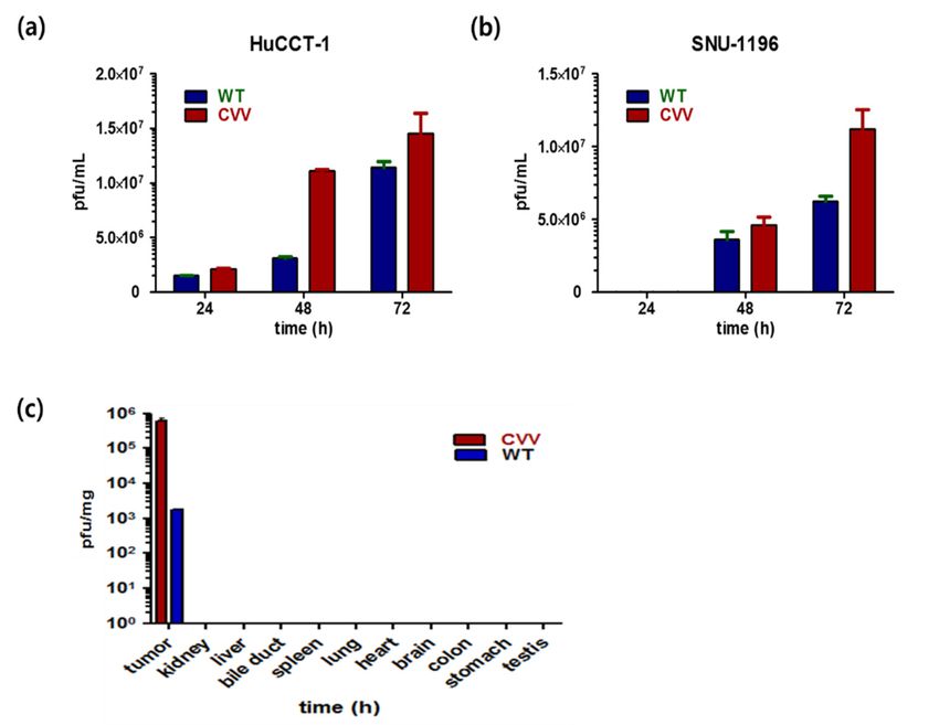
Cancers 2019, 11, 1667 4 of 11
Cancers 2019, 11, x FOR PEER REVIEW 4 of 11
led to higher cellular toxicity in SNU1196 and SNU245 (Figure 3c). These results indicated that
monotreatment
CVV monotreatment also works well inwell
also works CCA, inwhich
CCA, is very is
which resistant to conventional
very resistant anticancer
to conventional drugs,
anticancer
such
drugs, such as gemicitabine and cisplatin. The reason why CVV works well in CCA regardlessofofthe
as gemicitabine and cisplatin. The reason why CVV works well in CCA regardless the
resistance
resistancetoto gemicitabine
gemicitabine or cisplatin isis because
becauseCVV
CVVtakes
takescancer
cancerselective
selectiveinfection/replication
infection/replication
asas
its
its cancer
cancer killing
killing mechanism,
mechanism, whichwhich is different
is different from
from that that of conventional
of conventional anti-canceranti-cancer drugs.
drugs. Therefore,
Therefore, CVVininfection
CVV infection in CCA
CCA cells cells may
may enhance theenhance
efficacythe efficacy of chemotherapeutic
of chemotherapeutic agents by
agents by overcoming
overcoming the drug-resistance.
the drug-resistance.
Figure
Figure3.3.Oncolytic
Oncolyticactivity
activityofofthe
theCVV
CVVandanditsitscombinations
combinationswith
withcisplatin
cisplatinand
andgemcitabine.
gemcitabine.(a) (a)The
The
viability (%) of four different CCA cell lines against the CVV and WT at different MOIs.
viability (%) of four different CCA cell lines against the CVV and WT at different MOIs. (b) The viability(b) The
viability (%)
(%) of four of four
different CCAdifferent
cell linesCCA cellcisplatin
against lines against cisplatinatand
and gemcitabine gemcitabine
different at different
concentrations. (c) The
concentrations.
combination of (c)CVVThe+ cisplatin/CVV
combination of + gemcitabine
CVV + cisplatin/CVV + in
viability (%) gemcitabine viability
four different (%)lines.
CCA cell in four
different CCA cell lines.
2.4. High Expression of Stemness Markers in CCA Cell Lines
2.4. High
TheExpression
difficulty of
ofStemness
optimal Markers in CCA Cell Lines
clinical management of CCA is mainly due to its clinical complexity
contributed by itsofmultiple
The difficulty optimalcell origins,
clinical stemness of
management features,
CCA is and biliary
mainly due tumor microenvironment
to its clinical complexity
(TME) [19]. To evaluate the stemness of CCA cell lines, mRNA expressions
contributed by its multiple cell origins, stemness features, and biliary tumor microenvironment of Nanog, Sox2, Oct4,
(TME)
andTo
[19]. c-Myc werethe
evaluate analyzed
stemness inofthe
CCAHuCCT1 andmRNA
cell lines, SNU1196 cell lines.
expressions Cells were
of Nanog, Sox2,harvested
Oct4, andatc-Myc
three
different time intervals (24, 48, and 72 h) for mRNA expression analysis. HuCCT1
were analyzed in the HuCCT1 and SNU1196 cell lines. Cells were harvested at three different time cells cultured for 72 h
showed the upregulation of all four genes (Figure 4a). Nanog and c-Myc expression
intervals (24, 48, and 72 h) for mRNA expression analysis. HuCCT1 cells cultured for 72 h showed were upregulated
after
the 72 h in SNU1196
upregulation of all cells, whereas
four genes Sox2 4a).
(Figure and Nanog
Oct4 genes and were
c-Mycupregulated
expression in cells
were cultured forafter
upregulated 48 h
(Figure 4b). mRNA expression of stemness markers confirmed the stemness characteristics
72 h in SNU1196 cells, whereas Sox2 and Oct4 genes were upregulated in cells cultured for 48 h (Figure of CCA cell
lines,
4b). whichexpression
mRNA may contribute to drug
of stemness resistance
markers [15]. the stemness characteristics of CCA cell lines,
confirmed
which may contribute to drug resistance [15].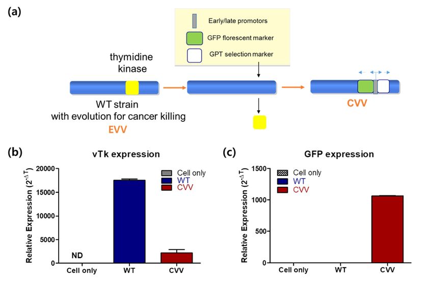
Cancers 2019, 11, 1667 5 of 11
Cancers 2019, 11, x FOR PEER REVIEW 5 of 11
Cancers 2019, 11, x FOR PEER REVIEW 5 of 11
Figure 4.4. Expression
Expressionofofstemness
stemness markers
markers in CCA
in CCA cell lines.
cell lines. HuCCT1HuCCT1 and SNU1196
and SNU1196 cells werecells were
cultured
Figure 4. Expression of stemness markers in CCA cell lines. HuCCT1 and SNU1196 cells were
cultured inplates.
in six-well six-well plates.
Cells wereCells were harvested
harvested and total and
RNAtotalwasRNA was extracted
extracted at three time
at three different different time
intervals:
cultured in six-well plates. Cells were harvested and total RNA was extracted at three different time
intervals:
24, 48, and24,
72 48, and 72 h. Complementary
h. Complementary DNA (cDNA) DNA (cDNA)
synthesis synthesis
was wasfrom
performed performed from
total RNA andtotal
usedRNA
for
intervals: 24, 48, and 72 h. Complementary DNA (cDNA) synthesis was performed from total RNA
and used for real-time qPCR expression analysis. (a) mRNA expression of stemness
real-time qPCR expression analysis. (a) mRNA expression of stemness markers (Nanog, Sox2, Oct4, markers (Nanog,
and used for real-time qPCR expression analysis. (a) mRNA expression of stemness markers (Nanog,
Sox2, Oct4, and
and c-Myc) c-Myc) in
in HuCCT1 HuCCT1
cells and (b)cells and (b) in
in SNU1196 SNU1196 cells.
cells.
Sox2, Oct4, and c-Myc) in HuCCT1 cells and (b) in SNU1196 cells.
2.5. The
2.5. The Effect
Effect of
of pH
pH onon CCA
CCA Cell
Cell Lines
Lines
2.5. The Effect of pH on CCA Cell Lines
To evaluate
To evaluatethe theeffect
effectofofpHpH ononCCA
CCAcells, thethe
cells, HuCCT1,
HuCCT1,SNU245,
SNU245,and and
SNU478 cell lines
SNU478 were were
cell lines used.
Cell To evaluate
culture mediathewith
effectthe
ofappropriate
pH on CCApH cells, the HuCCT1,
values were used SNU245,
for cell and SNU478
culture. To cellan
create lines were
acidic pH,
used. Cell culture media with the appropriate pH values were used for cell culture. To create an acidic
used.
1pH, Cell
N HCl culture
was used; media with
to create the appropriate
an alkaline pH
pH, 1 pH, values
N NaOH were used
was used. for cell
The cells culture. To create an acidic
1 N HCl was used; to create an alkaline 1 N NaOH was used. Thewere
cellsstable
were in media
stable with a
in media
pH,
pH 1ranging
N HCl wasfrom used;
7 to 9to(Figure
create an
5). alkaline
The pH, 1and
survival N NaOH
spreadwas
of used.cell
CCA Thelines
cellsatwere
pH stable
7–9 in media
indicated the
with a pH ranging from 7 to 9 (Figure 5). The survival and spread of CCA cell lines at pH 7–9
with a pHnature
alkaline rangingof fromcells
these 7 toand
9 (Figure
their 5). The survival and spread of CCA cell lines at pH 7–9
environments.
indicated the alkaline nature of these cells and their environments.
indicated the alkaline nature of these cells and their environments.
Figure 5. The effect of pH on CCA cells. The pH of the medium was adjusted using HCl and NaOH.
Figure 5.5.The
Theeffect of pH ononCCA cells. The pH ofof
the medium was adjusted using HCl and NaOH.
Figure
The images ofeffect
cells of
in pH CCA
different pHcells.
mediaThe pH
were the medium
captured after was
24 h adjusted
under anusing HCl
inverted and NaOH.
microscope
The images of cells in different pH media were captured after 24 h under an inverted microscope
The images of cells in different pH media were captured after 24 h under an inverted microscope (scale
(scale bar = 100 µm).
(scale
bar =bar
100= µm).
100 µm).
2.6. Alkaline
2.6.Alkaline pH
AlkalinepH Favors
pHFavors CVV
FavorsCVV
CVV Infection
Infection in the CCA Cell Line
2.6. Infection inin
thethe CCA
CCA Cell
Cell Line
Line
To assess
Toassess the
assessthe oncolytic
theoncolytic efficacy
oncolyticefficacy of
efficacyofofthethe
theCVVCVV under
CVVunder
underan an alkaline
an alkaline pH,
pH, HuCCT1
HuCCT1was was cultured
culturedatat
wascultured four
atfour
four
To alkaline pH, HuCCT1
pH conditions
pHconditions ranging
conditionsranging from
rangingfrom
from6.56.5 to 8.0.
6.5toto8.0. The
8.0.The CVV
TheCVV
CVVwaswas then
wasthen infected
theninfected (0.1
infected(0.1 MOI),
(0.1MOI), GFP
MOI),GFP expression
GFPexpression
expressionwas was
was
pH
captured,
captured, andand cells
and cells were harvested for qPCR GFP expression. Green fluorescence expression
captured, cells were
wereharvested
harvested for for
qPCR GFP GFP
qPCR expression. Green Green
expression. fluorescence expression
fluorescence indicated
expression
indicated robust infectivity of the CVV at alkaline pH values of 7.5 and 8.0 (Figure 6a, top). qPCR
indicated robust infectivity of the CVV at alkaline pH values of 7.5 and 8.0 (Figure 6a, top). qPCR
GFP expression was the highest at pH 7.5 (Figure 6a, bottom). These in vitro results support the robust
GFP expression was the highest at pH 7.5 (Figure 6a, bottom). These in vitro results support the robustCancers 2019, 11, 1667 6 of 11
robust infectivity of the CVV at alkaline pH values of 7.5 and 8.0 (Figure 6a, top). qPCR GFP expression
Cancers 2019, 11, x FOR PEER REVIEW 6 of 11
was the highest at pH 7.5 (Figure 6a, bottom). These in vitro results support the robust infectivity of the
CVV underofan
infectivity thealkaline
CVV underpH. Replication efficacy
an alkaline pH. in SNU1196
Replication in in
efficacy different
SNU1196 pHinmedia (6.5,pH
different 7, 7.5,
mediaand 8)
shows
(6.5, 7, 7.5, and 8) shows its preference to a slightly alkaline environment (pH 7.5–8) (Figure 6b), which cell
its preference to a slightly alkaline environment (pH 7.5–8) (Figure 6b), which corresponds
toxicity
corresponds(decrease of absorbance)
cell toxicity (decrease of at absorbance)
respective pH media (Figure
at respective pH media6c). (Figure
The differences in replication
6c). The differences
efficacy and cytotoxicity
in replication efficacy andin SNU1196 were
cytotoxicity not significant
in SNU1196 > 0.05), which
were not(psignificant is perhaps
(p > 0.05), which isbecause
perhaps CCA
is already an alkaline cancer. When we used HeLa (extracellular pH of
because CCA is already an alkaline cancer. When we used HeLa (extracellular pH of most cancer is most cancer is generally
considered to be mildly
generally considered to beacidic
mildlyin acidic
the rage of 6.4–7.0)
in the [20], the[20],
rage of 6.4–7.0) highest replication
the highest efficacyefficacy
replication of CVV ofwas
found
CVV was in pH 7.5 (*
found < 0.05,
inppH 7.5 (*Figure S1).Figure
p < 0.05, From theS1).results,
From theweresults,
proposewe that the oncolytic
propose that theactivity
oncolytic of the
activity
CVV of the and
is stable CVVhighly
is stable and highly
effective effective
at infecting at infecting
cancer cells in acancer
slightly cells in a slightly
alkaline alkaline
microenvironment
microenvironment
(pH 7–8), suggesting (pH 7–8),
that CVV suggesting
is highlythat CVV for
suitable is highly suitable for
the treatment the treatment of CCA.
of CCA.
Figure 6.
Figure CVVinfectivity
6. CVV infectivityininCCA.
CCA.(a)(a)Green
Green fluorescence
fluorescence expressions
expressions in SNU245
in SNU245 at different
at different pHspHs
(top), 48 h after CVV infection. GFP gene expression from cells cultured in different
(top), 48 h after CVV infection. GFP gene expression from cells cultured in different pHs of media pHs of media
(bottom) (scale
(scalebar
bar200 um);* *p p< <
200um); 0.05,
0.05, one-way
one-way ANOVA.
ANOVA. (b)(b) Replication
Replication efficacy
efficacy in SNU1196
in SNU1196 cultured
cultured
different pHs
in different pHsof ofmedia.
media.(c)(c)WST-1
WST-1 assay
assay results
results of of SNU1196
SNU1196 cells
cells cultured
cultured in different
in different pHspHs of media.
of media.
N.S., ** pp >>0.05,
0.05,one-way
one-wayANOVA.
ANOVA.
2.7. TherapeuticEfficacy
2.7. Therapeutic EfficacyofofCVV
CVVininthe
theXenograft
Xenograft Model
Model
To prove
To prove thethe oncolytic
oncolyticefficacy
efficacyofofthe
theCVV
CVVininCCA CCA inin vivo,
vivo, thethe SNU1196
SNU1196 xenograft
xenograft modelmodelwaswas
used. SNU1196 cellscells(2 7
(2××1010) were
) were injected subcutaneously into
used. SNU1196 7 injected subcutaneously into thethe
leftleft
flankflank of nude
of nude mice.
mice. Mice Mice
werewere
categorized intofive
categorized into fivegroups
groups(PBS, (PBS,cisplatin,
cisplatin,gemcitabine,
gemcitabine, WT,WT, and and CVV),
CVV), withwith
fivefive
micemice
per per
groupgroup
and were treated
treated asas per
per Section
Section2.6. 2.6.After
Afterthe
thetumor
tumorvolume
volume reached 300 mm 3 , 100 µL of PBS, 1 ×6 106
reached 300 mm , 100 µL of PBS, 1 × 10
3
plaque-forming
plaque-forming units unitsofofthetheCVV CVVororWT, WT,andand2525mg/kg
mg/kg ofof cisplatin
cisplatin or or gemcitabine
gemcitabine werewere injected
injected
intratumorally
intratumorally to to the
the corresponding
correspondingmice micegroups
groupsonceonceperperweek.
week.The The tumor
tumor volume
volume (mm (mm 3 ) was
3) was
twice per
measured twice per week
weekusing
usingthe theformula:
formula:LL××W2/2,W2/2,where
whereLLisisthe the tumor
tumor length
length andand
WW is the
is the tumor
tumor A
width. width.
slower A tumor
slowerburden
tumor was burden was observed
observed in CVV-treated
in CVV-treated mice thanmice mice (p < 0.05,
than in PBS-treated
in PBS-treated
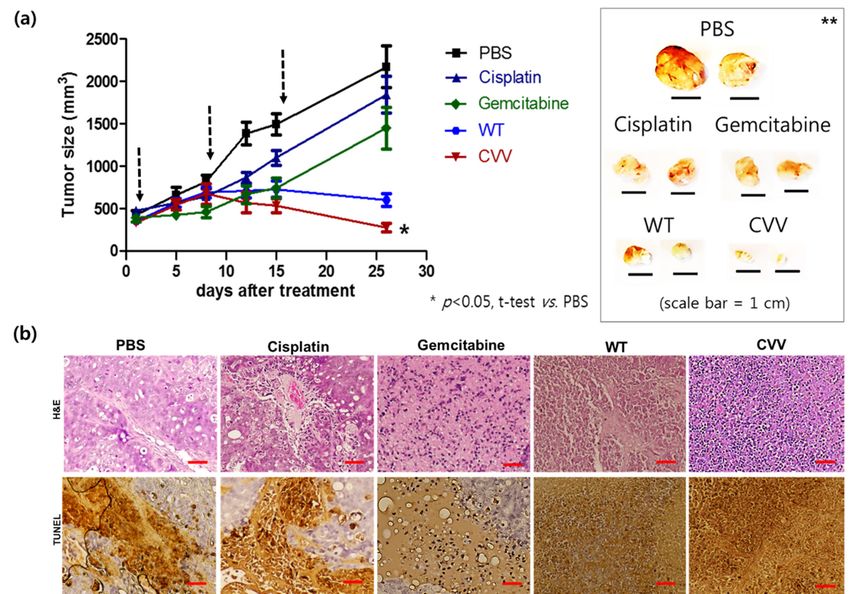
mice (p7a).
Figure < 0.05, Figure 7a). Gemcitabine
Gemcitabine may work more may work
rapidlymore rapidly (approximately
(approximately up to 10 up daysto after
10 days after
treatment),
treatment), but surviving, drug-resistant stem cells like CCA started to grow
but surviving, drug-resistant stem cells like CCA started to grow after that. In addition, regression ofafter that. In addition,
regression
tumor burdenof was
tumor burdenonly
observed wasinobserved onlyand
the viral (WT in CVV)
the viral (WT and
treatment group. CVV) treatment total
Additionally, group.
cancer
Additionally, total cancer cell lysis was easily found within the tumor mass
cell lysis was easily found within the tumor mass only in the viral treatment groups. As expected, only in the viral treatment
groups.
the As efficacy
highest expected, thefound
was highest efficacy
in the CVVwasgroupfound in the
(tumor CVV
size: PBS group (tumor>size:
> cisplatin PBS > cisplatin
gemcitabin > WT > >CVV
gemcitabin > WT > CVV group). Hematoxylin and eosin (H&E) staining
group). Hematoxylin and eosin (H&E) staining results of tumor mass and terminal deoxynucleotidyl results of tumor mass and
terminal deoxynucleotidyl transferase dUTP nick end labeling (TUNEL) staining results of tumor
transferase dUTP nick end labeling (TUNEL) staining results of tumor mass of each group indicated
mass of each group indicated that CVV most effectively induced apoptosis in the tumor mass (Figure
that CVV most effectively induced apoptosis in the tumor mass (Figure 7b). This result confirmed the
7b). This result confirmed the therapeutic efficacy of the CVV in the CCA xenograft model.
therapeutic efficacy of the CVV in the CCA xenograft model.Cancers 2019, 11, 1667 7 of 11
Cancers 2019, 11, x FOR PEER REVIEW 7 of 11
Figure
Figure 7.7. Therapeutic
Therapeutic efficacy
efficacy of
of the
the CVV
CVV in in the
the SNU1196
SNU1196 xenograft
xenograft model.
model. (a)
(a) Tumor
Tumor size
size measured
measured
in
in xenograft
xenograft models
models treated
treated with
with PBS,
PBS, cisplatin,
cisplatin, gemcitabine,
gemcitabine, and and WTWT oror CVV.
CVV. SNU1196
SNU1196 tumors
tumors were
were
induced in nude mice. When the tumor volume reached 300
3 mm 3, treatment was started. The
induced in nude mice. When the tumor volume reached 300 mm , treatment was started. The treatment
treatment
scheduled scheduled
days are noted days with
are noted with downwards
downwards arrows. Tumor arrows. Tumorinvolume
volume nude mice in nude
after mice after
treatment
treatment
(left). Tumor (left). Tumor
volume aftervolume
treatment after treatment
(left). Image of (left). Imagetumors
SNU1196 of SNU1196
4 weekstumors 4 weeks(right).
after treatment after
treatment
One-paired(right). One-paired
t-test were t-test the
used to detect were used to detect
significance in tumorthe volume
significance
growth in (* p < 0.05)
tumor volume growth
and one-way
(* p < 0.05)
ANOVA testand one-way
shows ANOVA
that tumor volumetestatshows
day 27that
are tumor volume
significantly at daybetween
different 27 are significantlyCancers 2019, 11, 1667 8 of 11
(Figure 3a–c). The therapeutic efficacy of the CVV against stem cell-like colon cancer was proven
in a previous study [15]. To prove the therapeutic efficacy of the CVV against stem cell-like CCA
tumors, upregulated mRNA expressions of stemness markers, such as Nanog, Sox2, Oct4, and c-Myc,
were shown in the HuCCT1 and SNU1196 cell lines (Figure 4a); hence, we used SNU1196 to evaluate
the therapeutic efficacy of the CVV in the xenograft model. We considered the alkaline environment of
CCA tumors in the bile duct to check whether extracellular pH modulation, in addition to enhanced
cancer selectivity of CVV, would be able to overcome the drug-resistance and complexity of the TME,
would enhance the therapeutic activity of CVV on these refractory cancers. Three different CCA cell
lines were cultured in media with four different pH values, and the normal proliferation of HuCCT1
cells in a slightly alkaline pH environment was observed (Figure 5). GFP expression of the CVV in
terms of green fluorescence and mRNA expression in HuCCT1 cells confirmed the replication and
oncolytic efficacy of the CVV in SNU1196 were highest at pH 7.5 (Figure 6). These results support
that the oncolytic efficacy of the CVV on CCA cell lines is enhanced with the alkaline pH in vitro.
In the xenograft model, the slower growth and total lysis (apoptosis) within SNU1196 tumor mass in
CVV-treated nude mice indicated the therapeutic efficacy of the CVV against CCA tumors (Figure 7).
It is widely accepted that the extracellular pH (pHe ) of cancer cells is more acidic than normal cells
and the intracellular pH (pHi ) of them is neutral or even more alkaline than that in normal. Recently,
some suggested that an approach to manipulating pHe could be achieved by the targeted delivery of
alkaline nanoparticles to the tumor tissue as a future clinical application [26]. As an aspect of the impact
of TME and stem-like plasticity in CCA, this pHe modulation approach in conjunction with CVV is
likely to be very effective in cancer therapy. This application is very applicable for other cancer cell
types as well. The promise of this therapeutic potential will be confirmed with a clinical assessment of
its efficacy and safety in further studies. Before the clinical evaluation, therapeutic efficacy tests of CVV
in syngeneic and orthotopic animal models should be considered. In clinical studies, administration
of the virus, how to modulate pHe in surrounding cancer tissue, and optimization in corresponding
cancer types should be also considered.
4. Materials and Methods
4.1. Cell Culture and Virus
Human CCA cell lines (HuCCT1, SNU245, SNU478, and SNU1196) were obtained from a
Korean cell line bank (KCLRF, Seoul, South Korea). All cell culture media and their related reagents
were obtained from Welgene (Gyeongsan, South Korea). These cells were thawed and cultured
in Roswell Park Memorial Institute (RPMI) 1640 medium (10% fetal bovine serum (FBS) and 1%
penicillin/streptomycin(P/S)) in the presence of 5% CO2 at 37 ◦ C in an incubator. For HeLa and HT29
cells, they were thawed and cultured in Dulbecco Modified Eagle medium (DMEM) (10% FBS and 1%
P/S). Cells were subcultured when they achieved 70% confluence. The CVV was engineered as per
Figure 1a. Phase-contrast images of the cells were taken by a microscope (EVOS Cell Imaging Systems,
Thermo Fisher Scientific, Waltham, MA, USA).
4.2. Replication Efficacy Assay
To assess the replication efficacy of the CVV in CCA (HuCCT1 and SNU1196), 2 × 10⁵ cells per
well were seeded in six-well plates. After 90% confluence, 0.1 MOI of the CVV was used to infect
cells. Cells were harvested after 72 h and frozen at −80 ◦ C for virus titration. For virus titration,
2 × 10⁵ U2OS (ATCC®HTB-96™, Manassas, VA, USA) cells per well were seeded in six-well plates.
Harvested CVV-infected cell lysate was used to infect U2OS cells by serial dilution. At 72 h post
infection, cells were stained with 0.1% crystal violet solution. After 2–4 h, destaining was performed
and the plates were dried to count the plaques.Cancers 2019, 11, 1667 9 of 11
4.3. WST-1 Assay
The WST-1 assay was used to evaluate the cellular toxicity of the CVV in various CCA cell lines.
First, 1 × 10⁴ cells per well were seeded in 96-well plates. After 24 h, chemotherapeutic drugs or the
CVV were used to infect cells at different concentrations (0.1, 1.0, 10, 100, and 1000 µM, and 0.01, 0.1,
1.0, 10, and 100 MOI, respectively) in serum-free media. Serum-free media were replaced with normal
media after 2 h. At 72 h post infection, cells were analyzed using the WST-1 assay (EzCytotox, ITSBIO,
Seoul, Korea). As per the manufacturer’s instructions, absorbance was measured at 450 and 680 nm
(reference wavelengths).
4.4. Cells Culturing in pH-Adjusted Media
To adjust pH of the media, 1N NaoH or 1 N HCl was used. HuCCT1, SNU245, SNU245,
and SNU1196 cells were seeded in six-well plates. After 24 h of seeding, the normal media (RPMI1640,
10%FBS, 1%P/S) was replaced with corresponding pH-adjusted media (RPMI1640; no FBS; pH: 2.0,
5.0, 6.0, 6.5, 7.0, 7.5, 8.0, 9.0, and 11.0). Cells were kept in 2 h in pH-adjusted media. After this,
pH-adjusted media was replaced with normal media. The proliferation of cells was noted after 24 h
using inverted microscope.
4.5. Real-Time PCR Assay
Total RNA was extracted from cell lines using Trizol reagent (Thermo Fisher Scientific Korea,
Seoul, South Korea). To confirm RNA purity, absorbance was measured at 260 and 280 nm. For cDNA
synthesis, 2 µg total RNA was used with the PrimeScript 1st Strand cDNA Synthesis Kit (Takara Korea,
Seoul, Korea). For the real-time polymerase chain reaction (PCR), 1 µg cDNA was used with the
SYBR-Green quantitative PCR (qPCR) mixture (Roche, Basel, Switzerland) reagent. A LightCycler 96
Real-Time PCR System (Roche) was used to perform the real-time PCR assay. The program consisted
of a 40-cycle amplification at 95 ◦ C for 20 s, 60 ◦ C for 20 s, and 72 ◦ C for 25 s. β-actin mRNA expression
was used to normalize the expression of the selected genes using the 2−∆∆Ct method.
4.6. Animal Study
All animal experiments were approved by the Institutional Animal Care and Use Committee of
Pusan National University (PNU-2017-1533). Nude mice were purchased from Orient (Gapyeong,
South Korea). SNU1196 (2 × 10⁷ cells/100 µL) was injected subcutaneously into the left flank of nude
mice. Mice were divided into five groups; namely, PBS, cisplatin, gemcitabine, WT, and CVV, with five
mice per group. The tumor volume (mm3 ) was measured twice per week using the formula: L × W2/2,
where L is the tumor length and W is the tumor width. After the tumor volume reached 300 mm3 ,
100 µL of PBS, 1 × 10⁶ plaque-forming units of the CVV or WT, cisplatin (25 mg/kg, Selleckchem,
Houston, TX, USA), and gemcitabine (Merck, Darmstadt, Germany) were injected intratumorally to
the corresponding mice groups once per week. We used 106 plaque-forming units (pfu) virus/mouse
because CVVs have a high replication rate [15,16] and the infectious dose of the WT or JX594 viruses
used in a previous in vivo study was more than 107 pfu [27]. After four weeks of injection, mice were
euthanized and the tumors were harvested for imaging. For the bio-distribution of viruses, organs and
tumors were collected from HT29-xenograft nude mice, which received 1 × 10⁶ plaque-forming units
of the CVV or WT intraperitoneally and were sacrificed after 2 weeks.
4.7. H&E staining and TUNEL Assay
To assay tomor generation and morphological characterics, H&E staining and TUNEL assays were
performed on paraffin-embedded tumor sections. Images were acquired using EVOS Cell Imaging
Systems (Thermo Fisher Scientific).Cancers 2019, 11, 1667 10 of 11
4.8. Statistical Analysis
Student’s unpaired t-tests were used to analyze the statistical significance of the SNU1196 tumor
volumes in nude mice. The level of statistical significance was set at p < 0.05.
5. Conclusions
In conclusion, the CCA cell lines and xenograft-based results demonstrated the oncolytic efficacy
of the CVV and confirmed its therapeutic efficacy in different circumstances in relation to the stem
cell-like characteristics and the alkaline nature of CCA tumors. The findings of this study support the
fact that CVV-based virotherapy is also suitable for the treatment of drug-resistant CCA.
Supplementary Materials: The following are available online at http://www.mdpi.com/2072-6694/11/11/1667/s1,
Figure S1: Replication efficacy in HeLa cultured in different pHs of media. * p < 0.05, one-way ANOVA.
Author Contributions: Conceptualization, methodology, and investigation, S.Y.Y., N.B., and H.L.L.; resources,
S.Y.Y. and D.-H.K.; writing the manuscript: S.Y.Y. and N.B.; project administration, S.Y.Y.; funding acquisition,
S.Y.Y. and J.H.
Funding: This research was supported by the Basic Science Research Program through the National Research
Foundation of Korea (NRF) funded by the Ministry of Education (grant numbers NRF-2016R1D1A1B03935221,
NRF-2017R1C1B5015034, and NRF-2018R1D1A1B07050358).
Conflicts of Interest: The authors declare no conflict of interest.
References
1. Lee, H.-S.; Kang, J.-I.; Chung, W.-J.; Lee, D.H.; Lee, B.Y.; Lee, S.-W.; Yoo, S.Y. Engineered Phage Matrix
Stiffness-Modulating Osteogenic Differentiation. ACS Appl. Mater. Interfaces 2018, 10, 4349–4358. [CrossRef]
[PubMed]
2. Moon, J.-S.; Kim, W.-G.; Shin, D.-M.; Lee, S.-Y.; Kim, C.; Lee, Y.; Han, J.; Kim, K.; Yoo, S.Y.; Oh, J.-W.
Bioinspired M-13 bacteriophage-based photonic nose for differential cell recognition. Chem. Sci. 2017, 8,
921–927. [CrossRef] [PubMed]
3. Yoo, S.Y.; Badrinath, N.; Woo, H.Y.; Heo, J. Oncolytic Virus-Based Immunotherapies for Hepatocellular
Carcinoma. Mediat. Inflamm. 2017, 2017, 5198798. [CrossRef] [PubMed]
4. Yoo, S.Y.; Jeong, S.N.; Kang, J.I.; Lee, S.W. Chimeric Adeno-Associated Virus-Mediated Cardiovascular
Reprogramming for Ischemic Heart Disease. ACS Omega 2018, 3, 5918–5925. [CrossRef]
5. Yoo, S.Y.; Jin, H.-E.; Choi, D.S.; Kobayashi, M.; Farouz, Y.; Wang, S.; Lee, S.-W. M13 Bacteriophage and
Adeno-Associated Virus Hybrid for Novel Tissue Engineering Material with Gene Delivery Functions.
Adv. Healthc. Mater. 2016, 5, 88–93. [CrossRef]
6. Yoo, S.Y.; Shrestha, K.R.; Jeong, S.N.; Kang, J.I.; Lee, S.W. Engineered phage nanofibers induce angiogenesis.
Nanoscale 2017, 9, 17109–17117. [CrossRef]
7. Heo, J.; Reid, T.; Ruo, L.; Breitbach, C.J.; Rose, S.; Bloomston, M.; Cho, M.; Lim, H.Y.; Chung, H.C.; Kim, C.W.;
et al. Randomized dose-finding clinical trial of oncolytic immunotherapeutic vaccinia JX-594 in liver cancer.
Nat. Med. 2013, 19, 329–336. [CrossRef]
8. Varela-Guruceaga, M.; Tejada-Solís, S.; García-Moure, M.; Fueyo, J.; Gomez-Manzano, C.; Patiño-García, A.;
Alonso, M.M. Oncolytic Viruses as Therapeutic Tools for Pediatric Brain Tumors. Cancers 2018, 10, 226.
[CrossRef]
9. Badrinath, N.; Heo, J.; Yoo, S.Y. Viruses as nanomedicine for cancer. Int. J. Nanomed. 2016, 11, 4835–4847.
[CrossRef]
10. Ricordel, M.; Foloppe, J.; Antoine, D.; Findeli, A.; Kempf, J.; Cordier, P.; Gerbaud, A.; Grellier, B.; Lusky, M.;
Quemeneur, E.; et al. Vaccinia Virus Shuffling: deVV5, a Novel Chimeric Poxvirus with Improved Oncolytic
Potency. Cancers 2018, 10, 231. [CrossRef]
11. Guse, K.; Cerullo, V.; Hemminki, A. Oncolytic vaccinia virus for the treatment of cancer. Expert Opin.
Biol. Ther. 2011, 11, 595–608. [CrossRef] [PubMed]Cancers 2019, 11, 1667 11 of 11
12. Parato, K.A.; Breitbach, C.J.; Le Boeuf, F.; Wang, J.; Storbeck, C.; Ilkow, C.; Diallo, J.S.; Falls, T.; Burns, J.;
Garcia, V.; et al. The oncolytic poxvirus JX-594 selectively replicates in and destroys cancer cells driven
by genetic pathways commonly activated in cancers. Mol. Ther. J. Am. Soc. Gene Ther. 2012, 20, 749–758.
[CrossRef] [PubMed]
13. Furuse, J.; Okusaka, T. Targeted Therapy for Biliary Tract Cancer. Cancers 2011, 3, 2243–2254. [CrossRef]
[PubMed]
14. Kokuryo, T.; Yokoyama, Y.; Nagino, M. Recent advances in cancer stem cell research for cholangiocarcinoma.
J. Hepato Biliary Pancreat. Sci. 2012, 19, 606–613. [CrossRef] [PubMed]
15. Yoo, S.Y.; Bang, S.Y.; Jeong, S.N.; Kang, D.H.; Heo, J. A cancer-favoring oncolytic vaccinia virus shows
enhanced suppression of stem-cell like colon cancer. Oncotarget 2016, 7, 16479–16489. [CrossRef]
16. Yoo, S.Y.; Jeong, S.N.; Kang, D.H.; Heo, J. Evolutionary cancer-favoring engineered vaccinia virus for
metastatic hepatocellular carcinoma. Oncotarget 2017, 8, 71489–71499. [CrossRef]
17. Gillies, R.J.; Liu, Z.; Bhujwalla, Z. 31P-MRS measurements of extracellular pH of tumors using
3-aminopropylphosphonate. Am. J. Physiol. 1994, 267, C195–C203. [CrossRef]
18. Vaupel, P.; Kallinowski, F.; Okunieff, P. Blood flow, oxygen and nutrient supply, and metabolic
microenvironment of human tumors: A review. Cancer Res. 1989, 49, 6449–6465.
19. Raggi, C.; Invernizzi, P.; Andersen, J.B. Impact of microenvironment and stem-like plasticity in
cholangiocarcinoma: Molecular networks and biological concepts. J. Hepatol. 2015, 62, 198–207. [CrossRef]
20. Griffiths, J.R. Are cancer cells acidic? Br. J. Cancer 1991, 64, 425–427. [CrossRef]
21. Moeini, A.; Sia, D.; Bardeesy, N.; Mazzaferro, V.; Llovet, J.M. Molecular Pathogenesis and Targeted Therapies
for Intrahepatic Cholangiocarcinoma. Clin. Cancer Res. Off. J. Am. Assoc. Cancer Res. 2016, 22, 291–300.
[CrossRef] [PubMed]
22. Badrinath, N.; Yoo, S.Y. Recent Advances in Cancer Stem Cell-Targeted Immunotherapy. Cancers 2019, 11, 310.
[CrossRef] [PubMed]
23. Coelen, R.J.S.; de Keijzer, M.J.; Weijer, R.; Loukachov, V.V.; Wiggers, J.K.; Mul, F.P.J.; van Wijk, A.;
Fong, Y.; Heger, M.; van Gulik, T.M. In vitro detection of cholangiocarcinoma cells using a fluorescent
protein-expressing oncolytic herpes virus. Cancer Gene Ther. 2017, 24, 227–232. [CrossRef] [PubMed]
24. Pugalenthi, A.; Mojica, K.; Ady, J.W.; Johnsen, C.; Love, D.; Chen, N.G.; Aguilar, R.J.; Szalay, A.A.; Fong, Y.
Recombinant vaccinia virus GLV-1h68 is a promising oncolytic vector in the treatment of cholangiocarcinoma.
Cancer Gene Ther. 2015, 22, 591–596. [CrossRef]
25. Lange, S.; Lampe, J.; Bossow, S.; Zimmermann, M.; Neubert, W.; Bitzer, M.; Lauer, U.M. A novel armed
oncolytic measles vaccine virus for the treatment of cholangiocarcinoma. Hum. Gene Ther. 2013, 24, 554–564.
[CrossRef]
26. Hao, G.; Xu, Z.P.; Li, L. Manipulating extracellular tumour pH: An effective target for cancer therapy.
RSC Adv. 2018, 8, 22182–22192. [CrossRef]
27. Breitbach, C.J.; Arulanandam, R.; De Silva, N.; Thorne, S.H.; Patt, R.; Daneshmand, M.; Moon, A.; Ilkow, C.;
Burke, J.; Hwang, T.H.; et al. Oncolytic vaccinia virus disrupts tumor-associated vasculature in humans.
Cancer Res. 2013, 73, 1265–1275. [CrossRef]
© 2019 by the authors. Licensee MDPI, Basel, Switzerland. This article is an open access
article distributed under the terms and conditions of the Creative Commons Attribution
(CC BY) license (http://creativecommons.org/licenses/by/4.0/).You can also read