Aberrant Expression of PIWIL1 and PIWIL2 and Their Clinical Significance in Ductal Breast Carcinoma
←
→
Page content transcription
If your browser does not render page correctly, please read the page content below
ANTICANCER RESEARCH 38: 2021-2030 (2018)
doi:10.21873/anticanres.12441

         Aberrant Expression of PIWIL1 and PIWIL2 and Their
            Clinical Significance in Ductal Breast Carcinoma
                MONIKA LITWIN1, ANNA SZCZEPAŃSKA-BUDA1, DAGMARA MICHAŁOWSKA1,
            JĘDRZEJ GRZEGRZÓŁKA2, ALEKSANDRA PIOTROWSKA2, AGNIESZKA GOMUŁKIEWICZ2,
                    ANDRZEJ WOJNAR3, PIOTR DZIĘGIEL2,4 and WOJCIECH WITKIEWICZ1

            1Regional Specialist Hospital in Wroclaw, Research and Development Centre, Wroclaw, Poland;
               2Department of Histology and Embryology, Wroclaw Medical University, Wroclaw, Poland;
                 3Department of Pathomorphology, Lower Silesian Oncology Centre, Wroclaw, Poland;
                4Department of Physiotherapy, University School of Physical Education, Wroclaw, Poland

Abstract. Background/Aim: P-Element-induced wimpy               Breast cancer is the most frequently diagnosed tumor in
testis (PIWI) proteins in complex with PIWI-interacting RNA     women and the leading cause of cancer-related death.
(piRNA) are involved in epigenetic regulation of gene           Currently recommended classification of breast cancer is
expression in germline cells. Aberrant expression of piRNA      based on expression of estrogen receptor (ER), progesterone
and PIWI proteins have been identified in various types of      receptor (PR), human epidermal growth factor receptor 2
tumour cells. The aim of this study was to evaluate the         (HER2) and the proliferation-associated nuclear protein Ki67
expression profiles of PIWI-like protein-1, -2 (PIWIL1 and      to identify clinical supbgroups for appropriate therapy.
PIWIL2), their immunohistochemical (IHC) characteristics        Nevertheless, triple-negative breast cancer characterized by
in ductal breast cancer, and determine their correlation with   a lack of ER, PR and HER2 expression has a worse clinical
clinicopathological parameters of this type of cancer.          outcome and an increased risk of recurrence due to lack of
Materials and Methods: Material for IHC studies comprised       targeted therapies (1). Furthermore, heterogeneity of the
of 101 invasive ductal carcinoma (IDC) cases and 31             tumour cells within the cancerous lesion has led to the
mastopathy tissues. Frozen fragments of paired tissue           concept of the existence of ‘cancer stem cells’ (CSCs or
specimens (tumour and adjacent non-malignant breast             tumour-initiating cells), which present characteristics of stem
tissue) taken from 55 patients with IDC and 18 samples of       cells, such as self-renewal capacity, fast proliferation and
mastopathy were used for molecular studies using real-time      multilineage differentiation (2, 3). These unique properties
polymerase chain reaction (RT-PCR). Results: A statistically    of CSCs explain the failure of many antitumor therapies,
significantly higher level of PIWIL1 and PIWIL2 was found       which in most cases target rapidly dividing cells. Further
in IDC compared to mastopathy samples (p≤0.0001).               extensive characterization of CSCs in individual tumour
Increased expression of PIWIL1 was correlated with              types is needed in order to identify specific markers for the
increased PIWIL2 expression in breast cancer tissue.            heterogeneous population of CSCs and consequently be able
Surprisingly, PIWIL1 mRNA was detected only in cancer and       to target CSCs selectively.
mastopathy, but was not found in most normal breast tissues,       P-Element-induced wimpy testis (PIWI) proteins bind to
although it is noteworthy that the PIWIL2 mRNA level was        a newly discovered class of non-coding small RNAs called
statistically significantly lower in mastopathy and IDC         PIWI-interacting RNAs (piRNA). Complexes of piRNA and
samples compared to normal breast tissues. Conclusion: Our      PIWI proteins are predominantly expressed in germline cells,
results affirm the hypothesis that reactivation of PIWI         where they have been demonstrated to be involved in
expression in various caner types is crucial for cancer         germline development, stem cell self-renewal and
development.                                                    gametogenesis (4). The human PIWI proteins form a
                                                                subfamily of the highly conserved Argonaute proteins and
                                                                consist of four members: PIWIL1, PIWIL2, PIWIL3 and
                                                                PIWIL4 (5). To date, a model of function for PIWI and
Correspondence to: Monika Litwin, Research and Development
                                                                piRNA in normal tissue and cancer has been proposed.
Centre Novasome Sp. z o.o., 51-423 Wroclaw, Poland. E-mail:
m.litwin@cbr.novasome.pl                                        piRNAs, along with abundant expression of PIWI, in
                                                                germline stem cells regulate transposone silencing through
Key Words: PIWIL1, PIWIL2, breast cancer.                       DNA methylation during spermatogenesis (6-8). In this way,

                                                                                                                          2021
ANTICANCER RESEARCH 38: 2021-2030 (2018)
germline cells (expressing PIWI at a high level) develop Table I. Clinical and pathological data of studied patients with breast
normally into somatic tissues in which PIWI protein cancer.
expression is absent. However, in cancer cells, high
Parameter IHC group Real-time
expression of PIWI and piRNAs results in aberrant DNA PCR group
methylation, silencing of tumour-suppressor genes and an
abnormal ‘stem-like’ state of cancer cells (6, 7). Number of patients 101 55
Numerous data have shown the aberrant expression of PIWI Median age (range) 55.9 (33-76) 58.5 (37-84)
≤50 Years 33 (32.7) 18 (32.7)
genes/proteins in various cancer types, suggesting that both
>50 Years 68 (67.3) 37 (67.3)
PIWI proteins and selected piRNAs are involved in Histopathological grading, n (%)
tumorigenesis (4, 9). Preliminary studies suggest that over- Grade 1 10 (9.9) 17 (30.8)
and ectopic expression of PIWIL1 is associated with several Grade 2 58 (57.4) 31 (56.6)
different types of tumours (10, 11). Based mainly on immuno- Grade 3 33 (32.7) 7 (12.6)
Tumour size, n (%)
histochemical (IHC) studies, the increased expression of
T1-2 91 (90.1) 43 (78.2)
PIWIL1 has been detected in colon, breast, oesophageal, T3-4 10 (9.9) 12 (21.8)
pancreatic, gastric and hepatocellular carcinoma and was Regional lymph node status, n (%)
correlated with histological grade of tumour, clinical stage N0 56 (55.4) 21 (38.2)
and poorer clinical outcome of patients (12-17). Our recent N1-N3 45 (44.6) 34 (61.8)
Distant metastasis, n (%)
study showed significantly higher mRNA level of PIWIL1
Absent 80 (79.2) 50 (90.9)
and decreased level of PIWIL2 mRNA in colorectal cancer Present 21 (20.8) 5 (9.1)
tissues compared to corresponding non-cancerous samples ER, n (%)
(18). Recent studies suggest that deregulated expression of Positive 76 (75.2) 33 (60)
PIWI genes/proteins is linked to deregulated cell Negative 25 (24.8) 22 (40)
PR, n (%)
proliferation, altered apoptosis, genomic instability and
Positive 65 (64.3) 26 (47.3)
invasion of tumours. PIWI proteins seem to be promising Negative 36 (35.7) 29 (52.7)
CSC markers due to the fact that their deregulated expression HER2, n (%)
was also correlated with clinicopathological features and Positive 58 (57.4) 47 (85.4)
survival of cancer patients (19, 20). Negative 43 (43.6) 8 (14.6)
Triple-negative, n (%) 17 (16.8) 3 (5.4)
The aim of our study was to evaluate the expression
profiles of the PIWIL1 and PIWIL2 in invasive ductal IHC: Immunohistochemistry, ER: estrogen receptor, PR: progesterone
carcinoma (IDC) samples in comparison to mastopathy receptor, HER2: human epidermal growth factor receptor 2, PCR:
tissues. We aimed to determine possible prognostic polymerase chain reaction.
significance of PIWIL1 and PIWIL2 in breast cancer
development and progression.
for real-time polymerase chain reaction (RT-PCR) studies. Fresh
Materials and Methods frozen, paired tissue specimens (tumour IDC and adjacent non-
malignant breast tissue samples) were obtained from patients with
Clinical samples. The study was conducted on two separate patient breast cancer operated at the Regional Specialist Hospital in
cohorts: one for IHC and one for real-time polymerase chain Wroclaw, Research and Development Centre, between 2011 and
reaction (PCR) studies. For IHC studies, paraffin blocks containing 2014. Mastopathy samples and non-malignant breast tissue were
diffuse cystic mastopathy tissue derived from 31 patients, and IDC used as control. The fresh tissue samples were collected in
from 101 patients, treated at the Lower Silesian Oncology Center RNAlater™ Stabilization Solution (Ambion by Life Technologies,
in Wrocław, Poland between years 1999-2005. All the tissue Carlsbad, CA, USA), frozen and stored at −80˚C until use. All
specimens utilized for the study were obtained before beginning tumour and normal tissue samples were submitted for
treatment. The patient’s clinicopathological data are listed in Table histopathological analysis. Detailed description of patients is
I. Paraffin sections stained with haematoxylin and eosin (H&E) presented in Table I.
were used to verify the diagnosis and malignancy grade of the The study protocol was approved by the Medical Ethics
tumors, according to World Health Organisation criteria (21). All Committee of Regional Specialized Hospital, Research and
patients with IDC were treated by mastectomy or quadrantectomy, Development Centre in Wroclaw (KB/ No. 10/2015). The study was
and subsequent axillary lymph node resection. The mean patient age conducted in accordance with the Helsinki Declaration of 1975 and
at diagnosis was 55.9 years. Adjuvant chemotherapy was applied to its later amendments or comparable ethical standards.
79 patients with IDC. Only seven patients received neoadjuvant
chemotherapy prior to primary tumor resection and 50 of the Quantitative real-time RT-PCR assay. Total RNA was extracted from
patients were treated with adjuvant radiotherapy. (30 mg) tumour, non-cancerous adjacent tissues and mastopathy
The second patient cohort comprised 55 tissue samples of tissues by a combination of TRI Reagent® (Ambion by Life
patients with IDC and 18 tissue mastopathy samples and were used Technologies) method with silica-based purification technology using
2022Litwin et al: Aberrant Expression of PIWIL1 and PIWIL2 in Ductal Breast Carcinoma NucleoSpin RNA isolation kit (Machery-Nagel GmbH & CO. Kg, using EnVision™ FLEX Hematoxylin for 5 min. After dehydration Düren, Germany) according to the manufacturers’ instructions. in graded ethanol concentrations (70%, 96%, absolute) and in Briefly, disruption and homogenization steps with MagNA Lyser xylene, all slides were coversliped in SUB-X Mounting Medium Green Beads were performed in 1 ml of TRI Reagent® (Ambion by (Agilent Technologies, Dako). Life Technologies) using the MagNA Lyser System (Roche Molecular Systems, Inc. Pleasanton, CA, USA) (3×30 s at 7000 rpm). The Histopathological examination and analysis of IHC reactions. All homogenized samples were incubated at room temperature for 5 min IHC preparations were examined using an Olympus BX-41 light and then 0.2 ml of chloroform was added. The mixture was microscope (Olympus, Shinjuku, Tokyo, Japan). The intensity of thoroughly shaken by vortexing for 15 s and incubated at room IHC reactions for PIWIL1 and PIWIL2 was estimated using the temperature for an additional 15 min. Phase separation was carried semi-quantitative immunoreactive (IRS) scale of Remmele and out by centrifugation at 4˚C and 12,000 × g for 15 min. The aqueous Stegner (23). IRS score represent the results of multiplication of the phase was collected and RNA precipitation was performed by adding percentage of positively stained cells and the intensity of staining an equal volume of 70% ethanol. The RNA samples were further (values from 0-12). IRS of 8 or lower were defined as negative, purified using spin columns according to the manufacturer’s values above 8 as positive. recommendation. The concentration and purity of isolated RNA was measured in Statistical analysis. Statistical analysis was performed with a NanoDrop2000 spectrophotometer (ThermoFischer Scientific, GraphPad Prism 5.0 (GraphPad Software, Inc., La Jolla, CA, USA). Wilmington, DE, USA). For the reverse transcription reaction, 250 Shapiro–Wilk test was used to evaluate the normality assumption of ng of RNA was used. The reverse transcription reaction was examined groups. To compare the differences between the performed using the High Capacity cDNA Reverse Transcription Kit expression of examined markers in the all patient pairs of groups (Applied Biosystem by Life Technologies) according to the and clinicopathological data the unpaired t-test and Mann–Whitney procedure of the manufacturer. Diluted cDNA (4- fold in nuclease- test were used. To compare the differences between more than two free water) was used for the subsequent real time PCR using groups, the Kruskal–Wallis and Dunn’s multiple comparison tests TaqMan Fast Universal PCR Master Mix and TaqMan Gene were performed. Additionally, the Spearman correlation test was Expression Assays (Applied Biosystem by Life Technologies): used to analyse the existing correlations. The Kaplan–Meier method Hs01041737_s1 specific for human PIWIL1, Hs01032720_m1 for was used to construct survival curves. In all analyses, the results PIWIL2 and Hs039299097_g1 for glyceraldehyde 3-phosphate were considered to be statistically significant at p2 and 8). IHC Santa-Cruz Biotechnology, Inc.) were used as the primary reaction for PIWIL2 was negative (IRS
ANTICANCER RESEARCH 38: 2021-2030 (2018)
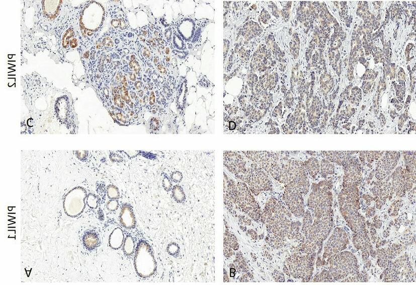
Figure 1. Immunohistochemical (IHC) identification of PIWI-like protein-1 (PIWIL1) (A, B) and PIWIL2 (C, D) in mastopathy (A,C) in comparison
to invasive ductal breast cancer cases (B, D). Polyclonal goat anti-human PIWIL1 (N-17, sc-22685), polyclonal goat anti-human PIWIL2 (K-18,
sc-67502) were used for PIWIL1 and PIWIL2 identification, respectively (Santa Cruz Biotechnology, Inc). Magnification, ×200.
correlation was found between PIWIL1 and PIWIL2 PCR analysis was performed. As presented in Table III, in
expression in IDC and the expression of ER, PR and HER2 normal breast tissues, expression of PIWIL1 was mostly not
receptors, the size of primary tumours (pT), metastasis to detected (in 52 samples out of 55), while in mastopathy and
lymph nodes (pN) and distant metastasis or the age of the IDC samples, the PIWIL1 expression was highly elevated.
patients. A positive association between PIWIL1 expression Significantly lower expression of PIWIL2 was detected in
and tumour size (T) as well as PIWIL2 and distant metastasis IDC tissues (RQ=0.33) and mastopathy (RQ=0.43) as
on the border of statistical significance was observed. The compared to adjacent noncancerous samples (RQ=1.33)
correlations between the presence of PIWIL1 and PIWIL2 (Figure 5B, Table III). The data showed a statistically
evaluated in IDC and clinicopathological parameters of the significant correlation between low PIWIL1 and PIWIL2
patients are summarized in Table II. mRNA expression and increased tumour grade for breast
In order to evaluate the prognostic value of PIWIL1 and IDC tissues (Figure 6A and B, respectively, Table IV).
PIWIL2 expression in IDC cases, the survival of patients
depending on the expression level was analysed. However, Discussion
no significant differences were noted in patient survival
regarding the intensity of PIWIL1 and PIWIL2 expression The complex of PIWI proteins and piRNAs regulate the gene
(Figure 4A and B). The variables which affected the duration expression at the epigenetic post-transcriptional level. In
of patient survival without statistical significance were tumor germline cells, PIWI proteins are essential for germline
size (T staging) (p=0.062) and tumor malignancy grade development, stem cell self-renewal and gametogenesis (24).
(p=0.65) (Figure 4C and D). Aberrant expression of different PIWI types on the mRNA and
protein levels as well as different piRNAs have been reported
PIWIL1 and PIWIL2 mRNA expression level in IDC, in several types of tumour (9, 25). Interestingly, overexpression
adjacent non-malignant and mastopathy samples. In order to and ectopic expression of PIWIL1 has been detected in many
determine mRNA level of PIWIL1 and PIWIL2 in IDC, types of cancer and in most cases an increased level of PIWIL1
mastopathy and normal tissue adjacent to tumor, real-time was correlated with clinical and pathological parameters of
2024Litwin et al: Aberrant Expression of PIWIL1 and PIWIL2 in Ductal Breast Carcinoma
Table II. Association of PIWI-like protein-1 (PIWIL1) and PIWIL2 expression by immunoreactive score (IRS) with clinical and pathological data
of the studied patients with breast cancer.
PIWIL1 expression, n (%) PIWIL2 expression, n (%)
Characteristic IRS≤8 IRS>8 p-Value IRS≤8 IRS>8 p-Value
Age
≤50 Years 23 (22.8) 10 (9.9) 0.476 29 (28.7) 4 (4.0) 0.746
>50 Years 52 (51.5) 16 (15.8) 61 (60.4) 7 (6.9)
Histopathological grading
Grade 1 6 (5.9) 4 (4.0) 0.566 8 (7.9) 2 (2.0) 0.177
Grade 2 41 (40.6) 17 (16.8) 50 (49.5) 8 (7.9)
Grade 3 28 (27.7) 5 (5.0) 32 (31.7) 1 (1.0)
Tumour size
T1-2 70 (69.3) 21 (20.8) 0.064 81 (80.2) 10 (9.9) 0.916
T3-4 5 (5.0) 5 (5.0) 9 (8.9) 1 (1.0)
Regional lymph node status
N0 45 (44.6) 11 (10.9) 0.118 49 (48.5) 7 (6.9) 0.5626
N1-N3 30 (29.7) 15 (14.9) 41 (40.6) 4 (4.0)
Distant metastasis
Absent 58 (57.4) 22 (21.8) 0.430 70 (69.3) 10 (9.9) 0.066
Present 17 (16.8) 4 (4.0) 20 (19.8) 1 (1.0)
ER
Positive 55 (54.5) 21 (20.8) 0.449 66 (65.3) 10 (9.9) 0.202
Negative 20 (19.8) 5 (5.0) 24 (23.8) 1 (1.0)
PR
Positive 48 (47.5) 17 (16.8) 0.899 58 (57.4) 7 (6.9) 0.958
Negative 27 (26.7) 9 (8.9) 32 (31.7) 4 (4.0)
HER2
Positive 52 (51.5) 16 (15.8) 0.750 60 (59.4) 8 (7.9) 0.751
Negative 23 (22.8) 10 (9.9) 30 (29.7) 3 (3.0)
Triple-negative 13 (12.9) 4 (4.0) 16 (15.8) 1 (1.0)
ER: Estrogen receptor, PR: progesterone receptor, HER2: human epidermal growth factor receptor 2.
Table III. The expression level of PIWI-like protein-1 (PIWIL1) and PIWIL2 mRNA in invasive ductal carcinoma (IDC), adjacent non-malignant
tissue and mastopathy samples. Data are the mean±standard deviation.
RQ mRNA Normal tissue (n=55) Mastopathy (n=18) IDC tissue (n=55)
PIWIL1 Not detected in 52 out of 55 2.2143±3.012 0.7748±0.97
PIWIL2 1.33±1.048 0.4369±0.474 0.33±0.76
tumour such as histological tumour grade and advanced clinical adjacent tissue (33). Non-statistically significantly increased
stage (4). Increased expression of PIWIL2 was identified expression of PIWIL1 and PIWIL3 and statistically significant
among others in breast, cervical, gastric, ovarian, prostate and down-regulation of PIWIL2 and PIWIL4 in breast tumour
colorectal cancer (12, 26-30). Statistically significant increased tissues was described. However, among four PIWIL genes,
level of PIWIL1 and decreased level of PIWIL2 was reported only PIWIL3 and PIWIL4 were correlated with overall
for colon and bladder cancer (18, 31). With regard to other survival, and PIWIL3 alone with recurrence-free survival (33).
members of the PIWIL protein family, increased expression of It has been proposed that reactivation of various PIWI
PIWIL4 was observed in renal carcinoma (32). Moreover, proteins is crucial for cancer development and malignant
expression of four members of the PIWI protein family was progression, therefore it is very likely that PIWI proteins
changed in tumour tissue compared with non-tumorous may be key markers for progression of various cancer types.
2025ANTICANCER RESEARCH 38: 2021-2030 (2018) Figure 2. PIWI-like protein-1 (PIWIL1) (A, C) and PIWIL2 (B, D) expression in mastopathy compared with invasive ductal carcinoma (A, B) and according to tumour grade in patients with breast cancer (C, D). Data are presented as mean IHC score±standard deviation. Significantly different at ***p
Litwin et al: Aberrant Expression of PIWIL1 and PIWIL2 in Ductal Breast Carcinoma

Figure 4. Kaplan–Meier curves of overall survival for patients with invasive ductal breast carcinoma according to the expression of PIWI-like
protein-1 (PIWIL1) (A), PIWIL2 (B), tumour grade (C) and histopathological grading (D). Cut-off points for the analysis were estimated based on
the median expression.

Figure 5. Expression levels of PIWI-like protein-1 (PIWIL1) (A) and PIWIL2 (B) in invasive ductal carcinoma, adjacent non-malignant normal
tissue and mastopathy samples. mRNA expression of PIWIL1 and PIWIL2 was measured by real-time reverse transcription-polymerase chain reaction
assay. For quantification, the samples were normalized against the expression of glyceraldehyde 3-phosphate dehydrogenase (GAPDH) mRNA.
Relative quantification (RQ) of mRNA for the examined genes was calculated using the method of Pfaffl (22). ***Significantly different at p
ANTICANCER RESEARCH 38: 2021-2030 (2018)

Figure 6. The expression level of PIWI-like protein-1 (PIWIL1) (A) and PIWIL2 (B) according to tumor malignancy grade (G) in invasive ductal
breast carcinoma. Expression of PIWIL1 and PIWIL2 was measured by real-time reverse transcription-polymerase chain reaction assay. Differences
in mRNA expression level between individual groups according to grade were determined by the Mann–Whitney test. Significantly different at
*p
Litwin et al: Aberrant Expression of PIWIL1 and PIWIL2 in Ductal Breast Carcinoma

samples was highly elevated. Simultaneously, significant             6 Siddiqi S, Matushansky I: PIWIs and PIWI-interacting RNAs in
lower expression of PIWIL2 was detected in IDC tissues and              the epigenetics of cancer. J Cell Biochem 113: 373-380, 2012.
mastopathy compared to adjacent, non-cancerous tissues.              7 Siddiqi S, Terry M and Matushansky I: HIWI mediated
                                                                        tumorigenesis is associated with DNA hypermethylation. PLoS
These results are in accordance with our previous
                                                                        One 7: e33711, 2012.
observations in colorectal cancer, where the mRNA levels of          8 Watanabe T and Lin H: Posttranscriptional regulation of gene
PIWIL1 in non-cancerous tissue was low or undetectable,                 expression by PIWI proteins and piRNAs. Mol Cell 56: 18-27,
while in paired cancerous tissue it was highly elevated. At the         2014.
same time, the expression of PIWIL2 was significantly                9 Suzuki R, Honda S and Kirino Y: PIWI expression and function
decreased (18). It is also important to emphasize that our              in cancer. Front Genet 3: 1-8, 2012.
study revealed negative correlation of decreased expression          10 Qiao D, Zeeman AM, Deng W, Looijenga LH and Lin H:
of PIWIL1 and PIWIL2 with increased grade of cancer. These              Molecular characterization of HIWI, a human member of the
                                                                        PIWI gene family whose overexpression is correlated with
findings are consistent with a previous study of Liu and co-
                                                                        seminomas. Oncogene 21: 3988-3999, 2002.
workers demonstrating increased expression of PIWIL2 at the          11 Taubert H, Greither T, Kaushal D, Würl P, Bache M and Bartel
initiation stages of breast cancer (12), and suggest an                 F: Expression of the stem cell self-renewal gene HIWI and risk
important role of PIWIL2 for breast cancer development.                 of tumour-related death in patients with soft-tissue sarcoma.
Interestingly, PIWIL2 was observed to be ubiquitously and               Oncogene 15: 1098-110, 2007.
uniquely expressed in various stages of breast cancer and its        12 Liu J, Shen R, Chen L, Ye Y, He G, Hua K, Jarjoura D, Nakano
expression pattern was associated with ER expression and                T, Ramesh GK, Shapiro CL, Barsky SH and Gao JX: PIWIl2 is
                                                                        expressed in various stages of breast cancers and has the
proliferative marker Ki67 (12). Taken together, our study
                                                                        potential to be used as a novel biomarker. Int J Clin Exp Pathol
sheds new light on the molecular regulation of PIWIL in                 3: 328-337, 2010.
cancer development. The observed differences in protein              13 He W, Wang Z, Wang Q, Fan Q, Shou C, Wang J, Giercksky K,
expression may indicate reciprocal regulation between                   Nesland J and Suo Z: Expression of HIWI in human esophageal
different piRNAs and PIWI genes in various cancer types.                squamous cell carcinoma is significantly as-sociated with poorer
Reactivation of PIWI expression in cancer strongly suggests             prognosis. BMC Cancer 9: 426, 2009.
participation of these proteins in processes of tumour growth        14 Grochola LF, Greither T, Taubert H, Moller P, Knippschild U,
                                                                        Udelnow A, Henne-Bruns D and Wurl P: The stem cell-
and differentiation (6, 7). The important issue awaiting further
                                                                        associated HIWI gene in human adenocarcinoma of the
investigation is the epigenetic regulation in regard to the             pancreas: expression and risk of tumor-related death. Br J
observed differences in PIWIL1 and PIWIL2 at the                        Cancer 99: 1083-1088, 2008.
transcriptional and protein level. Based on current data,            15 Wang Y, Liu Y, Shen X, Zhang X, Chen X, Yang C and Gao H:
PIWI–piRNA complexes contribute to cancer development                   The PIWI protein acts as a predictive marker for human gastric
through aberrant DNA methylation resulting in genomic                   cancer. Int J Clin Exp Pathol 5: 315-325, 2012.
silencing and promoting a stem-like state of cancer cells (8).       16 Zeng Y, Qu LK, Meng L, Liu CY, Dong B, Xing XE, Wu J and
To conclude, aberrant expression of mRNA and protein level              Shou CC: HIWI expression profile in cancer cells and its
                                                                        prognostic value for patients with colorectal cancer. Chin Med
of PIWI proteins show a strong prognostic value.
                                                                        J 124: 2144-2149, 2011.
                                                                     17 Zhao YM, Zhou JM, Wang LR, He HW, Wang XL, Tao ZH, Sun
Acknowledgements                                                        HC, Wu WZ, Fan J, Tang ZY and Wang L: HIWI is associated
                                                                        with prognosis in patients with hepato- cellular carcinoma after
This publication was supported by the Leading National Research         curative resection. Cancer 118: 2708-2717, 2012.
Center (KNOW, 2014-2018) of Wroclaw Center for Biotechnology.        18 Litwin M, Dubis J, Arczynska K, Piotrowska A, Frydlewicz A,
                                                                        Karczewski M, Dzięgiel P and Witkiewicz W: Correlation of
References                                                              HIWI and HILI expression with cancer stem cell markers in
                                                                        colorectal cancer. Anticancer Res 35: 3317-3324, 2012.
1 Torre LA, Bray F, Siegel RL, Ferlay J, Lortet-Tieulent J and       19 Martinez VD, Vucic EA, Thu KL, Hubaux R, Enfield KS, Pikor
  Jamal A: Global cancer statistics. Cancer J Clin 65: 87-108,          LA, Becker-Santos DD, Brown CJ, Lam S and Lam WL: Unique
  2012.                                                                 somatic and malignant expression patterns implicate PIWI-
2 Lobo NA, Shimono Y, Qian D and Clarke MF: The biology of              interacting RNAs in cancer-type specific biology. Sci Rep 5:
  cancer stem cells. Annu Rev Cell Dev Biol 23: 675-699, 2007.          10423, 2015.
3 Pardal R, Clarke MF and Morrison SJ: Applying the principles       20 Ng KW, Anderson C, Marshall EA, Minatel BC, Enfield KS,
  of stem cell biology to cancer. Nat Rev Cancer 3: 895-902, 2003.      Saprunoff HL, Lam WL and Martinez VD: PIWI-interacting
4 Litwin M, Szczepańska-Buda A, Piotrowska A, Dzięgiel P and            RNAs in cancer: emerging functions and clinical utility. Mol
  Witkiewicz W: The meaning of PIWI proteins in cancer                  Cancer 15: 5, 2016.
  development (Review) Oncol. Lett 13: 3354-3302, 2017.              21 Yang F and Li J: WHO Classification of tumors of the breast.
5 Sasaki T, Shiohama A, Minoshima S and Skimizu N:                      Zhonghua Wai Ke Za Zhi 52: 1-3, 2014 (in Chinese).
  Identification of eight members of Argonaute family in the         22 Pfaffl MW: A new mathematical model for relative quantification
  human genoma. Genomics 82(3): 323-30, 2003.                           in real-time RT–PCR. Nucleic Acids Res 29(9): e45, 2001.

                                                                                                                                  2029
ANTICANCER RESEARCH 38: 2021-2030 (2018) 23 Remmele W and Stegner HE: Recommendation for uniform 33 Krishnan P, Ghosh S, Graham K, Mackey JR, Kovalchuk O and definition of an immunoreactive score (IRS) for immunohisto- Damaraju S: PIWI-interacting RNAs and PIWI genes as novel chemical estrogen receptor detection (ER-ICA) in breast cancer prognostic markers for breast cancer. Oncotarget 7(25): 37944- tissue. Pathologe 8: 138-140, 1987 (in German). 37956, 2016. 24 Ku HY and Lin H: PIWI proteins and their interactors in piRNA 34 Huang G, Hu H, Xue X, Shen S, Gao E, Guo G, Shen X and biogenesis, germline development and gene expression. Natl. Sci Zhang X: Altered expression of piRNAs and their relation with Rev 1: 205-218, 2014. clinicopathological features of breast cancer. Clin Transl Oncol 25 Tan Y, Liu L, Liao M, Zhang C, Hu S, Zou M, Gu M and Li X: 15: 563-568, 2013. Emerging roles for PIWI proteins in cancer. Acta Biochim 35 Zhang H, Ren Y, Xu H, Pang D, Duan C and Liu C: The Biophysis Sin (Shanghai) 47: 315-324, 2015. expression of stem cell protein PIWIl2 and piR-932 in breast 26 Ye Y, Yin DT, Chen L, Zhou Q, Shen R, He G, Yan Q, Tong Z, cancer. Surg Oncol 22: 217-223, 2013. Issekutz AC, Shapiro CL, Barsky SH, Lin H, Li JJ and Gao JX: 36 Cheng J, Guo JM, Xiao BX, Miao Y, Jiang Z, Zhou H and Li Identification of PIWII2-like (PL2L) proteins that promote QN: piRNA, the new non-coding RNA and is aberrantly tumorigenesis. PLoS One 5: e13406, 2010. expressed in human cancer cells. Clin Chim Acta 412(17-18): 27 He G, Chcen L, Ye Y, Xiao Y, Hua K, Jarjoura D, Nakano T, 1621-1625, 2011. Barsky SH, Shen R and Gao JX: PIWIl2 expressed in various 37 Iliev R, Fedorko M, Machackova T, Mlcochova H, Svoboda M, stages of cervical neoplasia is a potential complementary marker Pacik D, Dolezel J, Stanik M and Slaby O: Expression levels of for p16. Am J Transl Res 2: 156-169, 2010. PIWI-interacting RNA, piR-823, are deregulated in tumor tissue, 28 Chen C, Liu J and Xu G: Overexpression of PIWI proteins in blood serum and urine of patients with renal cell carcinoma. human stage III epithelial ovarian cancer with lymph node Anticancer Res 36(12): 6419-6423, 2016. metastasis. Cancer Biomark 13: 315-321, 2013. 38 Hashim A, Rizzo F, Marchese G, Ravo M, Tarallo R, Nassa G, 29 Yang Y, Zhang X, Song D and Wei J: PIWIl2 modulates the Giurato G, Santamaria G, Cordell A, Cantarell C and Weisz A: invasion and metastasis of prostate cancer by regulating the RNA sequencing identifies specific PIWI-interacting small non- expression of matrix metalloproteinase-9 and epithelial- coding RNA expression patterns in breast cancer. Oncotarget mesenchymal transitions. Oncol Lett 10: 1735-1740, 2015. 5(20): 9901-9910, 2014. 30 Oh SJ, Kim SM, Kim YO and Chang HK: Clinicopathologic 39 Oner C, Cosan D and Colak E: Estrogen and androgen hormone implications of PIWIL2 expression in colorectal cancer. Korean levels modu-late the expression of PIWI-interacting RNA in J Pathol 46: 318-323, 2012. prostate and breast cancer. PlosOne 11(7): e0159044, 2016. 31 Nikpour P, Forouzandeh-Moghaddam M and Ziaee Sa, Dokun OY, Schulz WA and Mowla SJ: Absence of PIWIL2 (HILI) expression in human bladder cancer cell lines and tissues. Cancer Epidemiol 33: 271-275, 2009. 32 Al-Janabi O and Wach S, Nolte E, Weigelt K, Rau TT, Stohr C, Legal W, Schick S, Greither T, Hartmann A, Wullich B and Taubert H: PIWI-like 1 and -4 gene transcript levels are Received January 18, 2018 associated with clinicopathological parameters in renal cell Revised February 19, 2018 carcinoma. Biochim Biophys Acta 1842: 686-690, 2014. Accepted February 27, 2018 2030
You can also read