Arthrogenic human synovial cysts: immunohistochemical profile of interleukin-1beta, interleukin-6, tumour necrosis factor-alpha - Via Medica Journals
←
→
Page content transcription
If your browser does not render page correctly, please read the page content below
Folia Morphol.
                                                                                                                   Vol. 80, No. 1, pp. 133–139
                                                                                                                 DOI: 10.5603/FM.a2020.0025
                                                    ORIGINAL              ARTICLE                                Copyright © 2021 Via Medica
                                                                                                                              ISSN 0015–5659
                                                                                                                             eISSN 1644–3284
                                                                                                                          journals.viamedica.pl

Arthrogenic human synovial cysts:
immunohistochemical profile of interleukin-1beta,
interleukin-6, tumour necrosis factor-alpha
S. Taurone1*, M.T. Santarelli1*, C. De Ponte1, L. Bardella2, M. Ralli1, C. Morselli1, A. Nicolai1,
A. Greco1, A. Ferretti3*, M. Artico1*
1
 Department of Sensory Organs, “Sapienza” University of Rome, Italy
2
 Department of Human Neurosciences, “Sapienza” University of Rome, Italy
3
 Orthopaedic Unit and Kirk Kilgour Sports Injury Centre, Sant’Andrea Hospital, “Sapienza” University of Rome, Italy

[Received: 28 October 2019; Accepted: 28 November 2019]

                                            Background: Synovial cysts are currently classified as degenerative lesions affecting
                                            the joint capsule or adjacent structures.
                                            Materials and methods: In our study we describe the results obtained in an im-
                                            munohistochemical study comprising 18 patients with synovial cysts, performed
                                            to evaluate the pathophysiological role of some inflammatory cytokines such as:
                                            interleukin (IL)-1β, IL-6 and tumour necrosis factor-alpha (TNF-α).
                                            Results: Results showed an over-expression of TNF-α, IL-1β and IL-6 which ap-
                                            pears to be involved in the onset and progression of the disease. At the present
                                            time it is not possible to affirm that these molecules play a direct role also due
                                            to the absence of further and more specific investigations. The authors therefore
                                            hypothesize that inhibition of inflammation may have a significant role in the
                                            pathogenesis and regression of synovial cysts.
                                            Conclusions: Hence, these inflammatory cytokines may be considered potential
                                            therapeutic targets. The development of synthetic inhibitors of these inflammatory
                                            factors could lead to a reduction in the intensity of inflammation, thus inhibiting
                                            the onset and development of the disease. (Folia Morphol 2021; 80, 1: 133–139)

                                            Key words: interleukins 1beta and 6, immunohistochemistry, synovial
                                            cysts, tumour necrosis factor-alpha

                    INTRODUCTION                                           the wrist. Since the joints are completely wrapped in
    Synovial cysts (also known as “ganglion”, “synovi-                     a fibrous tissue (capsule) that favours the movements’
al ganglion”, “arthrogenic cysts”) are benign neo-for-                     fluidity, a thinner capsular wall is responsible for
mations of the soft tissues which generally originate                      a possible traumatic damage and so the cyst may
from the joint capsule or tendon sheath and contain                        develop in the joint. A herniation of the capsule occurs
fluid gelatinous material, usually appearing on the                        and it tends to move towards the superficial tissues and
back of the wrist. Sometimes these cysts may involve                       to form a revolving structure that we call cysts [4, 5].
neural structures, as in cases of peroneal nerve [1] or                        The mechanism that gives rise to these cysts is still
lumbar roots [11] involvement.                                             not clear. Synovial cysts are mostly found in young
    Arthrogenic cysts are cysts that occur at the level                    athletes or workers who use the wrist and hand joints
of the joint capsule. The most frequent location is at                     a lot. In many cases synovial cysts are the result of joint

Address for correspondence: S. Taurone, PhD, Department of Sensory Organs, “Sapienza” University of Rome, V.le del Policlinico 155,
00161 Rome, Italy, tel/fax: 0649918054, e-mail: t.samanta@yahoo.it
*Equally contributing
                                                                    133
Folia Morphol., 2021, Vol. 80, No. 1

inflammation, arthrosis or previous trauma. However,                  Eighteen patients were included in the study, aged
the exact role of inflammation in this process has not            between 11 and 56 years (11 males and 7 females),
yet been studied in detail. The synovial fluid contains           undergoing surgical treatment for the removal of
inflammatory factors such as cytokines, prostaglan-               wrist synovial cysts, in the Orthopaedic Unit of the
dins and proteases. Previous studies have shown that              S. Andrea Hospital in Rome. Control samples (two
angiogenic factors are released during the formation              specimens for each tissue fragment), characterised
of the synovial cysts, suggesting a possible correlation          by normal palmar fascia tissues, were collected from
between the proliferation of new vessels in synovial              patients undergoing hand surgery for carpal tunnel
structures and the chronic inflammatory process that              syndrome.
induces the progression of synovial cysts [10, 11, 13].               Patients with concomitant neoplastic, infectious,
However, to date, no previous studies have analysed               autoimmune diseases, peripheral vascular disorders
the modulation of the inflammatory process in the                 or who had received anti-inflammatory therapy in
regression of synovial cysts. It has been shown that in           the 6 months prior to the operation were excluded.
the spinal joint tissue the induction of type 2 cyclooxy-             During excision, apart from anaesthesia, no other
genases (COX-2) and phospholipase A2 stimulates the               chemical products or pharmaceutical drugs were
biosynthesis of different inflammatory mediators from             administered to the patients. Samples were fixed in
synovial chondrocytes (chondrocytes from synovial                 formalin and embedded in paraffin to be processed
joints), such as prostaglandin E2, interleukins (IL-1,            for histological staining and immunohistochemistry.
IL-6, IL-8) and granulocyte-macrophage colony–stim-               The sections were subjected to haematoxylin and
ulating factor (GM-CSF) [2]. COX-2 inhibitors have                eosin and Masson’s trichromic staining.
been shown to reduce synovitis, leukocyte infiltration
and synovial hyperplasia in animal models, reducing               Immunohistochemistry
the expression of IL-1β, IL-6 and tumour necrosis fac-                The immunohistochemical analysis was conduct-
tor-alpha (TNF-α) [7, 8]. Our study is based on previous          ed using the ABC/HRP technique (avidin complexed
experimental data that support the role played by                 with biotinylated peroxidase) on 4 µm thick paraffin
inflammatory cytokines and growth factors in the de-              sections which were cut using a rotative microtome.
velopment of arthrogenic synovial cysts. In the light of          These sections were deparaffinized and hydrated
the data reported in the literature we hypothesize that           through decreasing ethanol series to distilled wa-
inhibition of inflammation may play a significant role            ter, then subjected to microwave irradiation and im-
in the destiny and/or regression of arthrogenic synovial          mersed in citrate buffer (pH = 6) twice for 5 min each
cysts. The aim of the present study was to evaluate               time. Subsequently, endogenous peroxidase activity
the pathophysiological role of some inflammatory cy-              was quenched using 0.3% hydrogenous peroxide in
tokines, such as IL-1β, IL-6 and TNF-α, in tissue samples         methanol for 30 min. To evaluate the immunolocal-
of synovial cysts or tenosynovitis, obtained by surgical          ization of IL-1β, TNF-α and IL-6, the following anti-
removal near a wrist joint. The main purpose of this              bodies were employed: i) rabbit anti-IL-1β polyclonal
was to evaluate the expression levels and localisation            antibody (1:50, Santa Cruz Biotechnology, Santa Cruz,
of inflammatory cytokines by immunohistochemical                  CA, USA); ii) mouse anti-TNF-α monoclonal antibody
analysis to identify their involvement in the patholo-            (1:100, Santa Cruz Biotechnology, Santa Cruz, CA,
gy and to evaluate the possible modulation of these               USA); iii) rabbit anti‑IL‑6 polyclonal antibody (1:200;
factors as a potential therapeutic target.                        Santa Cruz Biotechnology, Santa Cruz, CA, USA). In-
                                                                  cubation with the primary antibodies was performed
         MATERIALS AND METHODS                                    overnight at 4oC. Optimal antibody dilution and in-
Clinical evaluation                                               cubation times were assessed in preliminary exper-
    Written informed consent concerning the dona-                 iments. As negative control, the primary antibodies
tion of human tissues was provided by patients prior              were omitted. After exposure to the primary antibod-
to tissue acquisition, following the protocol for the             ies all slides were rinsed twice in phosphate buffer
acquisition of human tissues of the Ethical Committee             (pH = 7.4) and incubated for 1 hour with the appro-
of our University Hospitals which approved the study              priate secondary biotinylated antibody at the final
protocol. All specimens were acquired according to                dilution of 1:200. The secondary biotinylated antibod-
the principles of the Helsinki Declaration.                       ies against rabbit and mouse immunoglobulins were

                                                            134
S. Taurone et al., Arthrogenic human synovial cysts

purchased from Abcam (biotinylated goat anti-mouse                Table 1. Expression levels of interleukin-1beta (IL-1β), interle-
antibody and biotinylated goat anti-rabbit antibody).             ukin-6 (IL-6), tumour necrosis factor-alpha (TNF-α) in synovial
The slides were then incubated with peroxidase-con-               cysts and control specimens, and respective levels of statisti-
jugated avidin (Vector Laboratories, Burlingame, CA,              cal significance (t-test)
USA, Vectastain Elite ABC kit Standard*PK 6-100)                              Synovial cysts (%); Control specimens (%);                      P-value
for 30 min. Slides were washed in phosphate buffer                                 n = 15                 n = 15
(pH = 7.4) and treated with 0.05% 3,3-diaminobenzi-                 IL-1β         65.27 ± 12.91                  12.61 ± 5.51               < 0.00001
dine (DAB) and 0.1% H2O2. Finally, sections were coun-              IL-6          60.46 ± 10.70                  11.66 ± 4.96               < 0.00001
terstained with Mayer’s haematoxylin and dehydrated                 TNF-α         61.80 ± 10.22                  13.13 ± 5.81               < 0.00001
rapidly. The staining assessment was made by three
                                                                  The results were considered as statistically significant when p-value < 0.05.
experts. The intensity of the immune reaction was
assessed microdensitometrically using an IAS 2000
image analyser (Delta Sistemi, Rome, Italy) connected             the immunohistochemical technique. Immunohisto-
via a TV camera to the microscope. Twelve 100 µm2                 chemical experiments have made it possible to visu-
areas were delineated in each section by measuring                alise the distribution and localization of the cytokines
the diaphragm. The system was calibrated taking the               analysed in the tissue samples obtained by surgical
background obtained in sections exposed to non-im-                removal from patients with arthrogenic synovial cysts
mune serum as zero.                                               (Table 1). Through haematoxylin/eosin staining it was
                                                                  possible to detect, in the pathological tissues, the
Statistical analysis                                              myo-fibroblastic proliferation site (Fig. A1–A3), not
   Statistical analysis was performed with the Stu-               present in the control tissue (Fig. A4). In the patho-
dent’s t-test, using the GraphPad Prism (La Jolla,                logical tissue (Fig. A1–A3) a thickening of fibroblasts
CA). A p-value < 0.05 was considered for statistical              and myo-fibroblasts was visible with areas of com-
significance.                                                     plete cellular overlapping. This tissue morphology
                                                                  is not present in normal tissue (Fig. A4), in which
                            RESULTS                               only connective tissue with dispersed fibroblasts and
  The histopathological alterations caused by hu-                 small blood vessels can be observed. Figures B1–B3
man arthrogenic synovial cysts were evaluated using               describe the immunohistochemical expression of

                        *
                                                       *
                                                   *
      *

                                                            *
                *

                                                                                                                Figure A. Photomicrograph
                                                                                                                of histological preparations
                                                                                                                obtained by the haematoxylin/
                                                                                                                /eosin staining method;
                    *                                                                                           A1–A3. Proliferation myofi-
                                                                                                                broblast nodules (asterisks)
                                                                                                                obtained by surgical removal
                                                                                                                from a patient with synovial
                                                                                                                cyst (10×); A4. Normal
                                                                                                                palmar band consisting
                             *                                                                                  exclusively of connective tissue
                                                                                                                with fibroblasts and blood
                                                                                                                vessels (10×).

                                                            135
Folia Morphol., 2021, Vol. 80, No. 1
*
*
*
*
*
* Figure B. Photomicrographs
of the immunohistochemical
* reaction for the inflammatory
cytokine interleukin-1beta (IL-1β);
B1–B3. Pathological tissue in
which IL-1β is present in the
extracellular matrix and in the
cytoplasm of fibroblasts and
myofibroblasts and in the en-
dothelium of capillaries (B1, B2,
* 20×; B3, 40×). Myofibroblasts
nodules (asterisks); B4. Control
tissue (20×).
*
*
*
*
*
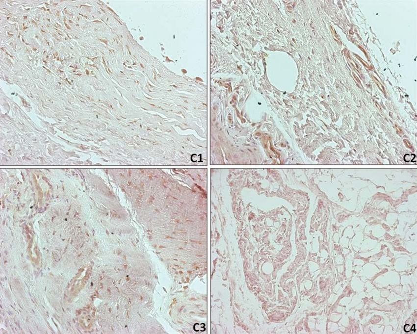
Figure C. Photomicrographs
of the immunohistochemical
reaction for the cytokine
interleukin-6 (IL-6); C1–C3.
* Pathological tissue in which
IL-6 is expressed at the level of
proliferative nodules (asterisks:
* C1, C2, C3, 20×); C4. Control
tissue (10×).
IL-1β in pathological samples, demonstrating positive level of the capillary endothelium, near the synovial
reactions in the cytoplasm of myo-fibroblastic cells cysts. IL-6 is appreciable in extracellular matrix, both
and in the extracellular matrix. IL-1β is complete- in proliferating myo-fibroblasts and in fibroblasts at
ly absent in the extracellular matrix of the palmar the level of proliferative nodules in patients affected
control fascia specimens (Fig. B4). Moreover, this by arthrogenic synovial cysts (Fig. C1). In the normal
pro-inflammatory cytokine is highly expressed at the tissue of palmar fascia IL-6 appears to be completely
136S. Taurone et al., Arthrogenic human synovial cysts
*
*
*
*
* *
* *
Figure D. Microphotographs
of the immunohistochemical
reaction for the proinflamma-
tory cytokine tumour necrosis
factor-alpha (TNF-α) expressed
in pathological myofibroblasts
* and in capillary endothelial cells
(D1, D2, D3, 20×). Myofibro-
blasts nodules (asterisks);
D4. Control tissue (20×).
absent at the level of the loose connective tissue, DISCUSSION
but moderately present in the endothelial cells of the Arthrogenic synovial cysts are benignly progress-
blood vessels and in the fibroblasts scattered in the ing fibro-proliferative disorders which often lead to
connective tissue (Fig. C2). Unlike the other cytokines, severe functional damage. The lack of knowledge
IL-6 shows an appreciable localisation in normal fi- related to the aetiopathogenesis of the disease has
broblasts (Fig. C1) and a more evident cytoplasmic meant that a specific therapy is not currently avail-
localisation in the pathological ones (Fig. C2). This able until now. Therefore, it is difficult to prevent
cytokine appears to be involved in the inflammatory its onset or to avoid its recurrence after surgical ex-
process that leads to the activation of the fibrotic cision. The absence of valid therapeutic targets has
process. Therefore, IL-6 appears to be synthesized led to the development of empirical therapies, such
in response to transforming growth factor-beta1 as local injection of steroids [9]. Inflammation plays
(TGF-β1) and acts by enhancing the proliferation and a fundamental role in the onset of fibrosis and this
differentiation of fibroblasts into myo-fibroblasts with finding is confirmed by the presence of proinflam-
deposition of amorphous substance in association matory cytokines [17]. In physiological conditions
with TGF-β1. TNF-α is expressed in the extracellular of wound healing or tissue repair activation of the
matrix of the myo-fibroblastic tissue at the prolifer- fibrotic process occurs. During this process fibro-
ation site observed in patients with synovial cysts blastic cells may differentiate into myo-fibroblasts,
(Fig. D1). TNF-α is present in pathological myo-fi- because their contractile activity is essential for tissue
broblasts and in capillary endothelial cells (Fig. D1). remodelling. The formation of myo-fibroblasts, con-
In the loose connective tissue of the normal palmar trolled by a variety of growth factors and numerous
fascia this inflammatory factor is however moder- mechanical stimuli, leads to an excessive deposition
ately positive in the cytoplasm of fibroblastic cells of extracellular matrix. Their action ends when the
and in the extracellular matrix (Fig. D2). This growth tissue is completely repaired or reabsorbed. In some
factor is also strongly expressed in the cytoplasm of pathological conditions the contractile activity of
secreting sweat glands (Fig. D2) in the dermis near myo-fibroblasts persists and leads to tissue defor-
the pathological proliferative nodules. mation [16]. IL-1β, an important pro-inflammatory
137Folia Morphol., 2021, Vol. 80, No. 1
cytokine, is involved in “in vitro” fibroblastic prolifer- the fibrotic reaction and its action depends exclusively
ation through the induction of the expression of tran- on its TNFR2 receptor, which is strongly expressed in
scriptional factors such as c-fos, c-jun and c-myc [6]. pathological conditions. Based upon our preliminary
The excessive expression of IL-1β alone could be re- results, local injections of anti-TNF-α drugs could be
sponsible for the local fibroblastic proliferation seen useful in preventing the progression of the disease
in the active phase of the disease. IL-1β seems to or avoiding its recurrence after surgical treatment.
have an important function in the activation and During the involutive phase a high ratio of collagen III
feeding of the inflammatory process, inducing the on collagen I was detected, differently from the nor-
synthesis of other cytokines such as IL-6 and IL-2, mal physiological condition [12].
interferons or chemokines, which are able to attract
macrophages and granulocytes towards the site of Conclusions
inflammation. It seems probable that the mitogen- These experimental results suggest, therefore,
ic effect of IL-1β is enhanced by the co-expression a possible application of these pro-inflammatory fac-
of other factors such as TGF-β and platelet-derived tors in identifying the degree of disease progression
growth factor alpha and beta. The combined expres- and in the use of some of these markers as prognostic
sion of these growth factors are probably responsible factors in the follow-up of patients undergoing sur-
for fibroblastic proliferation and excessive deposition gical resection of synovial cysts. Innovative therapies
of an amorphous substance and accumulation of could be characterised by the combined use of specific
synovial fluid, a condition typical of the disease. In inhibitors of the factors TNF-α, IL-1β and IL-6 and their
our experiments we also evaluated the level of IL-6 receptors in order to inhibit the progression of the
expression. This cytokine is directly involved in the disease through inactivation of the fibrotic process.
activation of the initial inflammatory process which
subsequently leads to fibrosis. The pro-inflammatory Acknowledgments
cytokine IL-6 plays an important role in the regulation This work was supported by a grant of the “Enrico
of inflammation and acts in association with TGF-β1, ed Enrica Sovena” Foundation, Italy.
thus leading to an increased pro-fibrotic response
[3, 14]. IL-6 acts by enhancing the TGF-β1 signal by REFERENCES
increasing endocytosis mediated by non-lipid endo- 1. Artico M, Cervoni L, Carloia S, et al. Synovial cysts: clinical
somes. This consequence is due to internalisation of and neuroradiological aspects. Acta Neurochir (Wien).
1997; 139(3): 176–181, doi: 10.1007/BF01844747, in-
the TGF-β1 receptors as a result of binding of their
dexed in Pubmed: 9143581.
ligand through endocytosis mediated by caveolin 2. Berenbaum F, Jacques C, Thomas G, et al. Synergistic
lipid vesicles and by non-lipid vesicles, although the effect of IL-1[beta] and TNF[alpha] on prostaglandin E2
TGF-β1 signal increases when the receptor endocyto- production by articular chondrocytes. Involvement of
cyclooxygenase without PLA2 stimulation. Exp Cell Res.
sis is mediated by non-lipidic vesicles. Therefore, IL-6
1996; 222(2): 379–384.
and TGF-β1 act synergistically, causing an increase 3. Bianchi E, Artico M, Di Cristofano C, et al. Growth factors,
in the expression of proinflammatory cytokines that their receptor expression and markers for proliferation of
appear to be the primary cause of the onset of the endothelial and neoplastic cells in human osteosarcoma.
disease. Verjee et al. [15] reported that TNF-α at low Int J Immunopathol Pharmacol. 2013; 26(3): 621–632,
doi: 10.1177/039463201302600306, indexed in Pubmed:
concentrations induce myo-fibroblastic contraction,
24067459.
while at high levels it induces reduction or complete 4. Calderazzi A, Eligi C, Guidetti F, et al. [Synovial chondroma-
inhibition of myo-fibroblastic contraction. It seems tosis of the temporomandibular joint: an occasional finding
that the action of TNF-α depends strictly on the TNFR in association with an arthrogenic cyst. A case report]. Radiol
Med. 1995; 89(4): 522–525, indexed in Pubmed: 7597236.
receptor type: TNFR2 causes fibroblastic proliferation,
5. De Haas WH, Van Heerde P. Synovial nature of pathologic
while TNFR1 activates programmed cell death. TNF-α periarticular structures, including subcutaneous nodules
could be considered a possible therapeutic target for descent from embryonic arthrogenic fibroblasts: a hypoth-
the treatment of the disease in the primary stages or esis. Z Rheumatol. 1979; 38(9-10): 318–329.
in preventing relapses following surgical removal. In 6. DiGiovine FS, Duff G. Interleukin 1: the first interleukin.
Immunology Today. 1990; 11: 13–20, doi: 10.1016/0167-
our experimental study we found that TNF-α showed
5699(90)90005-t.
a greater localisation in pathological fibroblasts. 7. El-Ghazaly MA, Nada AS, El-Hazek RM, et al. Effect of selec-
Therefore, this factor appears to be directly involved in tive COX-2 inhibitor, celecoxib on adjuvant-induced arthri-
138S. Taurone et al., Arthrogenic human synovial cysts
tis model in irradiated rats. Int J Radiat Biol. 2010; 86(12): 2010; 6(12): 715–726, doi: 10.1038/nrrheum.2010.180,
1079–1087, doi: 10.3109/09553002.2010.501839, indexed in Pubmed: 21060335.
indexed in Pubmed: 20698743. 13. Tatter SB, Cosgrove GR. Hemorrhage into a lumbar syno-
8. Gebhard HH, Zysk SP, Schmitt-Sody M, et al. The effects of vial cyst causing an acute cauda equina syndrome. Case
Celecoxib on inflammation and synovial microcirculation report. J Neurosurg. 1994; 81(3): 449–452, doi: 10.3171/
in murine antigen-induced arthritis. Clin Exp Rheumatol. jns.1994.81.3.0449, indexed in Pubmed: 8057153.
2005; 23(1): 63–70, indexed in Pubmed: 15789889. 14. Taurone S, Bianchi E, Attanasio G, et al. Immunohisto-
9. Huang AJ, Bos SA, Torriani M, et al. Long-term outcomes of chemical profile of cytokines and growth factors expressed
percutaneous lumbar facet synovial cyst rupture. Skeletal in vestibular schwannoma and in normal vestibular nerve
Radiol. 2017; 46(1): 75–80, doi: 10.1007/s00256-016- tissue. Mol Med Rep. 2015; 12(1): 737–745, doi: 10.3892/
2513-5, indexed in Pubmed: 27771754. mmr.2015.3415, indexed in Pubmed: 25738867.
10. Kusakabe T, Kasama F, Aizawa T, et al. Facet cyst in the 15. Verjee LS, Midwood K, Davidson D, et al. Myofibroblast
lumbar spine: radiological and histopathological findings distribution in Dupuytren’s cords: correlation with digital
and possible pathogenesis. J Neurosurg Spine. 2006; contracture. J Hand Surg Am. 2009; 34(10): 1785–1794, doi:
5(5): 398–403, doi: 10.3171/spi.2006.5.5.398, indexed 10.1016/j.jhsa.2009.08.005, indexed in Pubmed: 19910144.
in Pubmed: 17120888. 16. Wu M, Ben Amar M. Growth and remodelling for profound
11. Nucci F, Artico M, Santoro A, et al. Intraneural synovial circular wounds in skin. Biomech Model Mechanobiol.
cyst of the peroneal nerve: report of two cases and review 2015; 14(2): 357–370, doi: 10.1007/s10237-014-0609-1,
of the literature. Neurosurgery. 1990; 26(2): 339–344, indexed in Pubmed: 25183422.
doi: 10.1097/00006123-199002000-00028, indexed in 17. Wynn TA, Ramalingam TR. Mechanisms of fibrosis: ther-
Pubmed: 2155391. apeutic translation for fibrotic disease. Nat Med. 2012;
12. Shih B, Bayat A. Scientific understanding and clinical 18(7): 1028–1040, doi: 10.1038/nm.2807, indexed in
management of Dupuytren disease. Nat Rev Rheumatol. Pubmed: 22772564.
139You can also read