Publications de l'équipe - UMR3244 - Dynamique de l'information génétique - Centre de Recherche Institut Curie
←
→
Page content transcription
If your browser does not render page correctly, please read the page content below
Publications de l’équipe
UMR3244 – Dynamique de l’information génétique
Année de publication : 2021
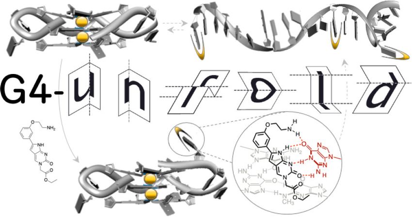
Jérémie Mitteaux, Pauline Lejault, Filip Wojciechowski, Alexandra Joubert, Julien Boudon, Nicolas
Desbois, Claude P. Gros, Robert H. E. Hudson, Jean-Baptiste Boulé, Anton Granzhan, David
Monchaud (2021 Aug 4)
Identifying G-Quadruplex-DNA-Disrupting Small Molecules
Journal of the American Chemical Society : 143 : 12567–12577 : DOI : 10.1021/jacs.1c04426
Résumé
The quest for small molecules that strongly bind to G-quadruplex-DNA (G4), so-called G4
ligands, has invigorated the G4 research field from its very inception. Massive efforts have
been invested to discover or rationally design G4 ligands, evaluate their G4-interacting
properties in vitro through a series of now widely accepted and routinely implemented
assays, and use them as innovative chemical biology tools to interrogate cellular networks
that might involve G4s. In sharp contrast, only uncoordinated efforts aimed at developing
small molecules that destabilize G4s have been invested to date, even though it is now
recognized that such molecular tools would have tremendous application in neurobiology as
many genetic and age-related diseases are caused by an overrepresentation of G4s. Herein,
we report on our efforts to develop in vitro assays to reliably identify molecules able to
destabilize G4s. This workflow comprises the newly designed G4-unfold assay, adapted from
the G4-helicase assay implemented with Pif1, as well as a series of biophysical and
biochemical techniques classically used to study G4/ligand interactions (CD, UV–vis, PAGE,
and FRET-melting), and a qPCR stop assay, adapted from a Taq-based protocol recently used
to identify G4s in the genomic DNA of Schizosaccharomyces pombe. This unique,
multipronged approach leads to the characterization of a phenylpyrrolocytosine (PhpC)-
based G-clamp analog as a prototype of G4-disrupting small molecule whose properties are
validated through many different and complementary in vitro evaluations.
INSTITUT CURIE, 20 rue d’Ulm, 75248 Paris Cedex 05, France | 1Publications de l’équipe
UMR3244 – Dynamique de l’information génétique
Marc Lavigne, Olivier Helynck, Pascal Rigolet, Rofia Boudria-Souilah, Mireille Nowakowski, Bruno
Baron, Sébastien Brülé, Sylviane Hoos, Bertrand Raynal, Lionel Guittat, Claire Beauvineau,
Stéphane Petres, Anton Granzhan, Jean Guillon, Geneviève Pratviel, Marie-Paule Teulade-Fichou,
Patrick England, Jean-Louis Mergny, Hélène Munier-Lehmann (2021 Jul 7)
SARS-CoV-2 Nsp3 unique domain SUD interacts with guanine quadruplexes and
G4-ligands inhibit this interaction.
Nucleic Acids Research : 49 : 7695–7712 : DOI : 10.1093/nar/gkab571
Résumé
The multidomain non-structural protein 3 (Nsp3) is the largest protein encoded by
coronavirus (CoV) genomes and several regions of this protein are essential for viral
replication. Of note, SARS-CoV Nsp3 contains a SARS-Unique Domain (SUD), which can bind
Guanine-rich non-canonical nucleic acid structures called G-quadruplexes (G4) and is
essential for SARS-CoV replication. We show herein that the SARS-CoV-2 Nsp3 protein also
contains a SUD domain that interacts with G4s. Indeed, interactions between SUD proteins
and both DNA and RNA G4s were evidenced by G4 pull-down, Surface Plasmon Resonance
and Homogenous Time Resolved Fluorescence. These interactions can be disrupted by
mutations that prevent oligonucleotides from folding into G4 structures and, interestingly, by
molecules known as specific ligands of these G4s. Structural models for these interactions
are proposed and reveal significant differences with the crystallographic and modeled 3D
structures of the SARS-CoV SUD-NM/G4 interaction. Altogether, our results pave the way for
further studies on the role of SUD/G4 interactions during SARS-CoV-2 replication and the use
of inhibitors of these interactions as potential antiviral compounds.
INSTITUT CURIE, 20 rue d’Ulm, 75248 Paris Cedex 05, France | 2Publications de l’équipe
UMR3244 – Dynamique de l’information génétique
V. Kapoor, C. Carabaña (2021 Jul 6)
Cell Tracking in 3D using deep learning segmentations
scipy
Résumé
Live-cell imaging is a highly used technique to study cell migration and dynamics over time.
Although many computational tools have been developed during the past years to
automatically detect and track cells, they are optimized to detect cell nuclei with similar
shapes and/or cells not clustering together. These existing tools are challenged when
tracking fluorescently labelled membranes of cells due to cell’s irregular shape, variability in
size and dynamic movement across Z planes making it difficult to detect and track them.
Here we introduce a detailed analysis pipeline to perform segmentation with accurate shape
information, combined with BTrackmate, a customized codebase of popular ImageJ/Fiji
software Trackmate, to perform cell tracking inside the tissue of interest. We developed
VollSeg, a new segmentation method able to detect membrane-labelled cells with low signal-
to-noise ratio and dense packing. Finally, we also created an interface in Napari, an Euler
angle based viewer, to visualize the tracks along a chosen view making it possible to follow a
cell along the plane of motion. Importantly, we provide a detailed protocol to implement this
pipeline in a new dataset, together with the required Jupyter notebooks.
Valentin Laplaud, Nicolas Levernier, Judith Pineau, Mabel San Roman, Lucie Barbier, Pablo J Sáez,
Ana-Maria Lennon-Duménil, Pablo Vargas, Karsten Kruse, Olivia du Roure, Matthieu Piel, Julien
Heuvingh (2021 Jul 3)
Pinching the cortex of live cells reveals thickness instabilities caused by myosin
II motors.
Science advances : DOI : eabe3640
Résumé
The cell cortex is a contractile actin meshwork, which determines cell shape and is essential
for cell mechanics, migration, and division. Because its thickness is below optical resolution,
there is a tendency to consider the cortex as a thin uniform two-dimensional layer. Using two
mutually attracted magnetic beads, one inside the cell and the other in the extracellular
medium, we pinch the cortex of dendritic cells and provide an accurate and time-resolved
measure of its thickness. Our observations draw a new picture of the cell cortex as a highly
dynamic layer, harboring large fluctuations in its third dimension because of actomyosin
contractility. We propose that the cortex dynamics might be responsible for the fast shape-
changing capacity of highly contractile cells that use amoeboid-like migration.
Emmanuelle Jeannot, Aurélien Latouche, Claire Bonneau, Marie-Ange Calméjane, Corine M
Beaufort, Kirsten Ruigrok-Ritstier, Guillaume Bataillon, Linda Larbi Chérif, Celia Dupain, Charlotte
Lecerf, Marina Popovic, Anne de la Rochefordière, Fabrice Lecuru, Virginie Fourchotte, Ekaterina
INSTITUT CURIE, 20 rue d’Ulm, 75248 Paris Cedex 05, France | 3Publications de l’équipe
UMR3244 – Dynamique de l’information génétique
S Jordanova, Heiko von der Leyen, Carine Tran-Perennou, Marie-Emmanuelle Legrier, Sylvain
Dureau, Laurence Raizonville, Diana Bello Roufai, Christophe Le Tourneau, Ivan Bieche, Roman
Rouzier, Els M J J Berns, Maud Kamal, Suzy Scholl (2021 Jul 2)
Circulating HPV DNA as a marker for early detection of relapse in patients with
cervical cancer.
Clinical cancer research : an official journal of the American Association for Cancer Research :
DOI : clincanres.0625.2021
Résumé
Almost all cervical cancers (CC) are caused by human papillomavirus (HPV) and patients with
advanced stage are at high risk for relapse. Circulating HPV DNA (HPV ctDNA) may serve as a
residual tumor marker at the end of chemo-radiation or to predict relapse during the follow-
up period.
M. Plays, S. Müller, R. Rodriguez (2021 Jul 1)
Chemistry and Biology of Ferritin
Metallomics : DOI : 10.1093/mtomcs/mfab021
Résumé
Piguel S., Le Bescont J., Mouawad L., Boddaert T., Bombard S. (2021 Jun 29)
Photoactivatable small-molecule inhibitors for light-controlled TAM kinase
activity
ChemPhotoChem : Accepted Author Manuscript : DOI : 10.1002/cptc.202100131
Résumé
The TAM kinase family arises as a promising therapeutical target for cancer therapy, auto-
immune, and viral diseases. In this study, we report the first photoactivatable caged
inhibitors of Tyro3 and Mer. This strategy enables spatial and temporal control of the
biological activity of the inhibitor upon irradiation with UV light. We describe the design, the
synthesis, the photocleavage properties, and the inhibitory activity of four Tyro3 and Mer
photoactivatable small molecules. The proof of concept on the TAM kinase family was
achieved in vitro , since irradiation by UV light restored the full inhibitory activity of two
prodrugs.
Jakub Muraszko, Karol Kramarz, Bilge Argunhan, Kentaro Ito, Gabriela Baranowska, Yumiko
Kurokawa, Yasuto Murayama, Hideo Tsubouchi, Sarah Lambert, Hiroshi Iwasaki, Dorota
Dziadkowiec (2021 Jun 22)
INSTITUT CURIE, 20 rue d’Ulm, 75248 Paris Cedex 05, France | 4Publications de l’équipe
UMR3244 – Dynamique de l’information génétique
Rrp1 translocase and ubiquitin ligase activities restrict the genome
destabilising effects of Rad51 in fission yeast.
Nucleic acids research : DOI : gkab511
Résumé
Rad51 is the key protein in homologous recombination that plays important roles during DNA
replication and repair. Auxiliary factors regulate Rad51 activity to facilitate productive
recombination, and prevent inappropriate, untimely or excessive events, which could lead to
genome instability. Previous genetic analyses identified a function for Rrp1 (a member of the
Rad5/16-like group of SWI2/SNF2 translocases) in modulating Rad51 function, shared with
the Rad51 mediator Swi5-Sfr1 and the Srs2 anti-recombinase. Here, we show that Rrp1
overproduction alleviates the toxicity associated with excessive Rad51 levels in a manner
dependent on Rrp1 ATPase domain. Purified Rrp1 binds to DNA and has a DNA-dependent
ATPase activity. Importantly, Rrp1 directly interacts with Rad51 and removes it from double-
stranded DNA, confirming that Rrp1 is a translocase capable of modulating Rad51 function.
Rrp1 affects Rad51 binding at centromeres. Additionally, we demonstrate in vivo and in vitro
that Rrp1 possesses E3 ubiquitin ligase activity with Rad51 as a substrate, suggesting that
Rrp1 regulates Rad51 in a multi-tiered fashion.
Katrina Cristall, Francois-Clement Bidard, Jean-Yves Pierga, Michael J Rauh, Tatiana Popova, Clara
Sebbag, Olivier Lantz, Marc-Henri Stern, Christopher R Mueller (2021 Jun 17)
A DNA methylation-based liquid biopsy for triple-negative breast cancer.
NPJ precision oncology : 53 : DOI : 10.1038/s41698-021-00198-9
Résumé
Here, we present a next-generation sequencing (NGS) methylation-based blood test called
methylation DETEction of Circulating Tumour DNA (mDETECT) designed for the optimal
detection and monitoring of metastatic triple-negative breast cancer (TNBC). Based on a
highly multiplexed targeted sequencing approach, this assay incorporates features that offer
superior performance and included 53 amplicons from 47 regions. Analysis of a previously
characterised cohort of women with metastatic TNBC with limited quantities of plasma
(Publications de l’équipe
UMR3244 – Dynamique de l’information génétique
Guillaume Jacquemin, Maria Benavente-Diaz, Samir Djaber, Aurélien Bore, Virginie Dangles-
Marie, Didier Surdez, Shahragim Tajbakhsh, Silvia Fre, Bethan Lloyd-Lewis (2021 Jun 17)
Longitudinal high-resolution imaging through a flexible intravital imaging
window.
Science advances : DOI : eabg7663
Résumé
Intravital microscopy (IVM) is a powerful technique that enables imaging of internal tissues
at (sub)cellular resolutions in living animals. Here, we present a silicone-based imaging
window consisting of a fully flexible, sutureless design that is ideally suited for long-term,
longitudinal IVM of growing tissues and tumors. Crucially, we show that this window, without
any customization, is suitable for numerous anatomical locations in mice using a rapid and
standardized implantation procedure. This low-cost device represents a substantial
technological and performance advance that facilitates intravital imaging in diverse contexts
in higher organisms, opening previously unattainable avenues for in vivo imaging of soft and
fragile tissues.
Graça Raposo, Guillaume van Niel, Philip D Stahl (2021 Jun 14)
Extracellular vesicles and homeostasis-An emerging field in bioscience research.
FASEB bioAdvances : 456-458 : DOI : 10.1096/fba.2021-00009
Résumé
To keep abreast of developments in the biological sciences and in parallel fields such as
medical education, () has created a special collections category, special collections ( SC),
that target, among other topics, emerging disciplines in the biomedical sciences. This SC is
focused on the emerging field of extracellular vesicles (EVs) and homeostasis. Leading
investigators in the biology of EVs around the globe have contributed to this collection of
articles that cover the gamut of research activities from biogenesis and secretion to
physiological function.
Angela Bellini, Ulrike Pötschger, Virginie Bernard, Eve Lapouble, Sylvain Baulande, Peter F
Ambros, Nathalie Auger, Klaus Beiske, Marie Bernkopf, David R Betts, Jaydutt Bhalshankar, Nick
Bown, Katleen de Preter, Nathalie Clément, Valérie Combaret, Jaime Font de Mora, Sally L
George, Irene Jiménez, Marta Jeison, Barbara Marques, Tommy Martinsson, Katia Mazzocco,
Martina Morini, Annick Mühlethaler-Mottet, Rosa Noguera, Gaelle Pierron, Maria Rossing, Sabine
Taschner-Mandl, Nadine Van Roy, Ales Vicha, Louis Chesler, Walentyna Balwierz, Victoria Castel,
Martin Elliott, Per Kogner, Geneviève Laureys, Roberto Luksch, Josef Malis, Maja Popovic-Beck,
Shifra Ash, Olivier Delattre, Dominique Valteau-Couanet, Deborah A Tweddle, Ruth Ladenstein,
Gudrun Schleiermacher (2021 Jun 11)
INSTITUT CURIE, 20 rue d’Ulm, 75248 Paris Cedex 05, France | 6Publications de l’équipe
UMR3244 – Dynamique de l’information génétique
Frequency and Prognostic Impact of Amplifications and Mutations in the
European Neuroblastoma Study Group (SIOPEN) High-Risk Neuroblastoma Trial
(HR-NBL1).
Journal of clinical oncology : official journal of the American Society of Clinical Oncology :
JCO2100086 : DOI : 10.1200/JCO.21.00086
Résumé
In neuroblastoma (NB), the ALK receptor tyrosine kinase can be constitutively activated
through activating point mutations or genomic amplification. We studied genetic alterations
in high-risk (HR) patients on the HR-NBL1/SIOPEN trial to determine their frequency,
correlation with clinical parameters, and prognostic impact.
GAUTIER Margot, THIRIANTCécile, DELATTRE Olivier, JANOUEIX-LEROSEY Isabelle (2021 Jun 10)
Plasticity in Neuroblastoma Cell Identity Defines a Noradrenergic-to-
Mesenchymal Transition (NMT)
Cancers (Basel) : 13(12):2904. : DOI : 10.3390/cancers13122904
Résumé
Neuroblastoma, a pediatric cancer of the peripheral sympathetic nervous system, is
characterized by an important clinical heterogeneity, and high-risk tumors are associated
with a poor overall survival. Neuroblastoma cells may present with diverse morphological
and biochemical properties in vitro, and seminal observations suggested that interconversion
between two phenotypes called N-type and S-type may occur. In 2017, two main studies
provided novel insights into these subtypes through the characterization of the
transcriptomic and epigenetic landscapes of a panel of neuroblastoma cell lines. In this
review, we focus on the available data that define neuroblastoma cell identity and propose to
use the term noradrenergic (NOR) and mesenchymal (MES) to refer to these identities. We
also address the question of transdifferentiation between both states and suggest that the
plasticity between the NOR identity and the MES identity defines a noradrenergic-to-
mesenchymal transition, reminiscent of but different from the well-established epithelial-to-
mesenchymal transition.
Daniel Lévy, Aurélie Di Cicco, Aurélie Bertin, Manuela Dezi (2021 Jun 7)
[Cryo-electron microcopy for a new vision of the cell and its components]
Medecine/Sciences : 379-385 : DOI : 10.1051/medsci/2021034
Résumé
Cryo-electron microscopy (cryo-EM) is a technique for imaging biological samples that plays
a central role in structural biology, with high impact on research fields such as cell and
developmental biology, bioinformatics, cell physics and applied mathematics. It allows the
determination of structures of purified proteins within cells. This review describes the main
INSTITUT CURIE, 20 rue d’Ulm, 75248 Paris Cedex 05, France | 7Publications de l’équipe
UMR3244 – Dynamique de l’information génétique
recent advances in cryo-EM, illustrated by examples of proteins of biomedical interest, and
the avenues for future development.
Eugenio de la Mora, Manuela Dezi, Aurélie Di Cicco, Joëlle Bigay, Romain Gautier, John Manzi,
Joël Polidori, Daniel Castaño Díez, Bruno Mesmin, Bruno Antonny, Daniel Lévy. (2021 Jun 7)
Nanoscale architecture of a VAP-A-OSBP tethering complex at membrane
contact sites
Nature Communications : DOI : 10.1038/s41467-021-23799-1
Résumé
Membrane contact sites (MCS) are subcellular regions where two organelles appose their
membranes to exchange small molecules, including lipids. Structural information on how
proteins form MCS is scarce. We designed an in vitro MCS with two membranes and a pair of
tethering proteins suitable for cryo-tomography analysis. It includes VAP-A, an ER
transmembrane protein interacting with a myriad of cytosolic proteins, and oxysterol-binding
protein (OSBP), a lipid transfer protein that transports cholesterol from the ER to the trans
Golgi network. We show that VAP-A is a highly flexible protein, allowing formation of MCS of
variable intermembrane distance. The tethering part of OSBP contains a central, dimeric, and
helical T-shape region. We propose that the molecular flexibility of VAP-A enables the
recruitment of partners of different sizes within MCS of adjustable thickness, whereas the T
geometry of the OSBP dimer facilitates the movement of the two lipid-transfer domains
between membranes.
INSTITUT CURIE, 20 rue d’Ulm, 75248 Paris Cedex 05, France | 8You can also read