COVID-19 and Kounis Syndrome: Deciphering Their Relationship - Balkan Medical Journal
←
→
Page content transcription
If your browser does not render page correctly, please read the page content below
Editorial 145 Balkan Med J 2021;38:145-149
Content of this journal is licensed under a Creative Commons
Attribution-NonCommercial-NoDerivatives 4.0 International License.
COVID-19 and Kounis Syndrome: Deciphering Their Relationship
Nicholas G. Kounis1 , Ioanna Koniari2 , Cesare de Gregorio3
1
Department of Cardiology, Patras University School of Medicine, Patras, Greece
2
Department of Cardiology, University Hospital of South Manchester NHS Foundation Trust, Manchester, UK
3
Department of Clinical and Experimental Medicine, University of Messina Medical School, Messina, Italy
Coronavirus infection is not a newly discovered condition. Cur- Anaphylaxis and Cytokines A Common Pathway Toward
rently, 7 coronaviruses that can cause human disease have been Kounis Syndrome
identified. Coronaviruses hCoV-HKU1, hCoV-OC43, hCoV- In severe anaphylaxis accompanied by hypotension or hypoxemia,
NL63, and hCoV-229E can principally cause asymptomatic or a range of mediators––similar to those of the cytokine storm in
mild respiratory and gastrointestinal infections accounting for coronavirus disease 2019 (COVID-19) patients––are reported to
approximately 5-30% of common colds.1 In the last 2 decades, 3 increase in both animal models and in vitro cell stimulation studies.6
human coronaviruses resulted in outbreaks that raised considerable In immune-mediated anaphylaxis and in the COVID-19 cytokine
global health concerns. These include the severe acute respiratory storm, the implicated molecules are released from inflammatory
syndrome coronavirus (SARS-CoV), the Middle East respiratory cells and include IL-1α, IL-1β, IL-6, IL-10, IL-17A, IL-12 p70,
syndrome coronavirus (MERS-CoV), and the current new human IL-18, IFNα, TNF, and an additional inflammatory cluster defined
coronavirus, SARS-CoV-2 that has rapidly spread from Wuhan in by thrombopoietin, IL-33, IL-16, IL-21, IL-23, IFNλ, eotaxin,
China to over 223 countries and regions in the world, causing a and eotaxin-3, demonstrating an increased correlation with severe
global pandemic affecting 114 million people with over 2.5 million disease.7 Furthermore, a marked increase in multiple type 2 effec-
deaths by March 1, 2021, with a case fatality ratio ranging from 1 tors, namely interleukin-5 (IL-5), IL-13, immunoglobulin E (IgE),
to 3.5% in most countries.2 Interestingly, these highly pathogenic eosinophils, and type 2 antibody isotype IgE, was found in severe
human coronaviruses have evolutionarily acquired the ability to COVID-19 disease; the increase continued during the course of
encode numerous proteins, allowing them to escape recognition the disease.7,8 Eosinophils and basophils are activated by monocyte
and response by the immune system, while they can simultane- chemotactic protein 3, which is the most effective activating che-
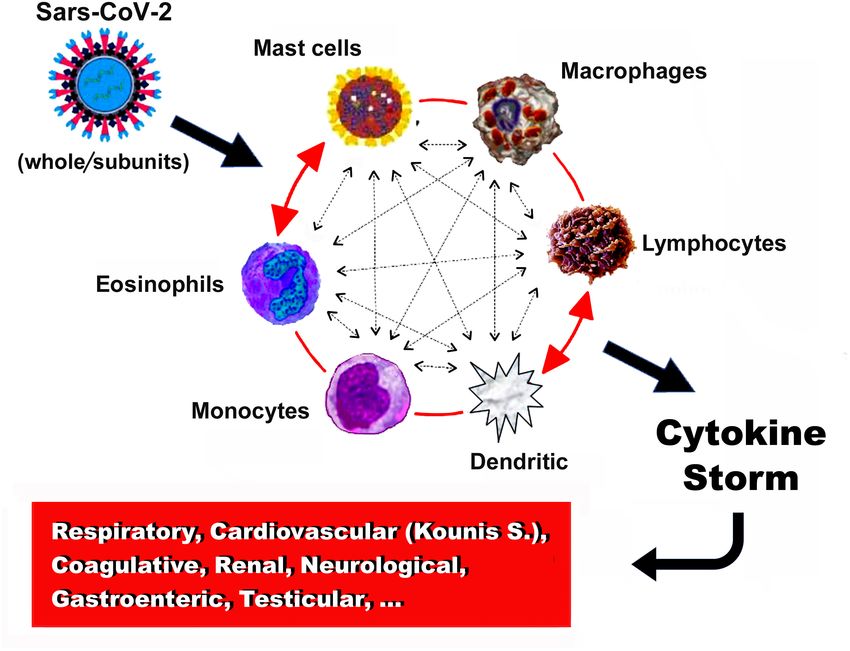
ously enable inflammatory and immune cell overactivation induc- mokine.9 Dendritic cells may also initiate autoimmune responses
ing a cytokine storm.3 Severe systemic complications such as and stimulate T cells, resulting in macrophage activation.10 Inter-
coagulopathy with thromboses, acute cardiac/coronary injury such actions between lymphocytes, monocytes, macrophages, and mast
as myocardial infarction and stent thrombosis, brain or liver injury, cells can increase vascular permeability, via release of TNF, IL-1β,
and multiple organ dysfunction have been associated with the cyto- IL-6, CXCL8 (IL-8), macrophage migration inhibitory factor,
kine storm and cytokine release syndrome, increasing the risk of CCL2 (also known as monocyte chemoattractant protein-1 (MCP-
mortality (Table 1). Various types of cytokines have been associ- 1)), high-mobility group box-1 protein, and matrix metalloprotein-
ated with the cytokine storm, including interleukins, chemokines, ases.11 The latter can promote plaque disruption or rupture, leading
interferons, tumor necrosis factors, and colony-stimulating factors. to myocardial infarction via activation of their zymogen forms
The cytokine storm can occur as a result of inappropriate recog- such as interstitial collagenase, gelatinase, and stromelysin.12 All
nition, for instance, in case of hypersensitivity or an ineffective types of inflammatory cells participate in an inflammatory vicious
response due to immune evasion.4 Indeed, a storm of proinflamma- cycle in which they can activate each other, like a “ball of thread,”
tory cytokines that can lead to catastrophic events and to Kounis via multidirectional signals (Figure 1). For example, mast cells
acute hypersensitivity-associated syndrome has been encountered can activate βmacrophages and may enhance T-cell activation.
in human anaphylaxis with profuse hypotension or hypoxemia.5,6 Inducible macrophage protein 1a may activate mast cells, while
Address for correspondence: Nicholas G. Kounis, Department of Cardiology, Patras University School of Medicine, Patras, Greece
e-mail: ngkounis@otenet.gr
Received: April 13, 2021 Accepted: April 23, 2021 • DOI: 10.5152/balkanmedj.2021.21097
Available at www.balkanmedicaljournal.org
ORCID iDs of the authors: N.G.K. 0000-0002-9751-6710; I.K. 0000 -0002-1033-5299; C.d.G. 0000-0003-3022-266X.
Cite this article as:
Kounis NG, Koniari I, de Gregorio C. COVID-19 and Kounis syndrome: Deciphering their relationship. Balkan Med J. 2021; 38(3):145-149.
Copyright@Author(s) - Available online at http://balkanmedicaljournal.org/146 Kounis et al. COVID-19 and Kounis Syndrome
TABLE 1. Symptoms and signs of Cytokine storm such as IL-6 and IL-1β, thus potentially contributing to COVID-19
Symptoms pathology.13 These cells enter the circulation from bone marrow as
Anosmia mononuclear cell precursors and circulate as mast cell precursors,
Ageusia
disposing in their surface KIT receptors (cytokine receptors) for
stem cell factor (SCF). SCF is a major cytokine which is essen-
Anorexia,
tial for mast cell growth, survival, differentiation, proliferation,
Arthralgia adhesion, and homing. Mast cells can adhere to all human tissues,
Diarrhea, even to the brain tissue that does not suffer from allergic reac-
Difficulty in Breathing tions because IgE antibodies cannot cross the blood–brain barrier
Dyspnea (BBB). Therefore, they differentiate and mature in these tissues, a
Fatigue process that might take several days to occur or even weeks. On
the contrary, basophils mature in bone marrow from granulocyte
Fever
precursors and enter the circulation as mature cells; they do not go
Headache
into the tissues except during the late stage of an allergic reaction.
Myalgia Mast cells are located perivascularly in the coronary arteries where
Neuropsychiatric Symptoms they can mature under the influence of local micro-environmental
Skin Itching and Rash factors, resulting in different phenotypes.
Tachypnea
Signs Mast cells can be typically activated via the following pathways:
Acute Liver Injury a. By allergens cross-linking allergen-specific IgE bound to
Acute-Phase Hypoalbuminemia high affinity Fc epsilon receptor 1.6
Acute Respiratory Distress Syndrome b. By non-IgE-mediated mast cell degranulation via activation
Anasarca
of the complement C1q, C3a C4, C5a, and Factor B, which
are called anaphylatoxins. This complement pathway activa-
Capillary Leak Syndrome
tion involves IL-5 and tryptase and is much more common
Catastrophic Hemorrhages than recognized, in patients who develop renal failure or fatal
Cholestasis cerebral events.14
Coagulopathy c. By the low affinity mas-related G protein-coupled receptor
Disseminated Intravascular Coagulation X2 (MRGPRX2) that may activate mast cells via non-Fcε
Encephalopathy receptors.14
d. By neuropeptides, including corticotropin-releasing hor-
Endothelial-Cell Death
mone, neurotensin (NT), and substance P (SP) via high-affin-
Hemostatic Imbalance
ity receptors.15
Hyperinflammation
Hypotension In complement and MRGPRX2 direct mast cell activation, the spe-
Hypoxemia cific IgEs may remain undetected, and tryptase levels may be nor-
mal, even in a condition as serious as the Kounis syndrome.
Low Platelet Counts
Neurotoxicity Syndrome Following activation, mast cells rapidly secrete the preformed,
Renal Failure granule-stored, heparin, histamine, tryptase, and TNF, as well
Spontaneous Hemorrhage
as newly synthesized leukotrienes, PAF, prostaglandin D2, cyto-
kines, and chemokines that are released 6-24 hours later. Mast
Takotsubo Cardiomyopathy
cell-derived vasoactive mediators, especially cytokines, can also
Vascular Occlusion increase the permeability of the BBB that explains the SARS-
Vasodilatory Shock and Death CoV-2 “COVID-19 brain fog” either directly via activation of mast
cells or by permitting cytokines to enter through a disrupted BBB.16
CD169- macrophages activate CD8 T cells. T cells may mediate It has been proposed that co-stimulation of human and rodent
mast cell activation and proliferation and further regulate macro- mast cell populations in vitro with FcεRI and certain TLRs can
phage activity.6 induce release of the above described proinflammatory mediators,
suggesting another mechanism through which bacterial or viral
Mast Cells and COVID-19
infections might exacerbate atopic asthma and other IgE- and mast
Mast cells constitute a key source of proinflammatory cytokines in
cell-associated disorders such as Kounis syndrome in vivo.17
COVID-19.12 These cells are typically activated by allergic triggers,
but they can also be triggered by virus-associated molecular pat- Kounis syndrome and COVID-19
terns, via activation of toll-like receptors (TLRs), including SARS- The allergic angina syndrome, representing a manifestation of
CoV-2, in order to release a variety of proinflammatory mediators endothelial dysfunction or microvascular angina that belongs to
Balkan Med J, Vol. 38, No.3, 2021Kounis et al. COVID-19 and Kounis Syndrome 147
FIG. 1. The vicious cycle of inflammatory cells that activate each other like a “ball of thread”.
the group of myocardial infarctions with nonobstructive coro- As noted above, serum cytokine levels are elevated in both
nary arteries (MINOCA), was described some 30 years ago and COVID-19 and in Kounis acute hypersensitivity-associated syn-
was named by American researchers as the Kounis syndrome, in drome. The following indicate and support the view of etiopatho-
2005.6,18 The pathophysiology of this syndrome includes inflam- genetic similarities between COVID-19 associated with cytokine
matory mediators released during an anaphylactic event from mast storm and Kounis /MINOCA, and thrombotic variants:
cells that are activated according to the above described 4 types of
1. COVID-19 affects the coronary arterial tree as it can induce
mast cell activation. In such an activation cascade, other interacting
coronary spasm, direct endothelial or vascular injury, plaque
cells including T-lymphocytes, macrophages, eosinophils, mono-
rupture and microthrombi, hypoxic injury, cytokine storm,
cytes, and dendritic cells can also participate. Despite the fact that
and a higher than expected incidence of stent thrombosis,
mast cells are a numerical minority in this inflammatory cascade,
that is attributed to the underlying hypercoagulable state that
they do decisively influence the inflammatory process. Addition-
clinically coincides with the main 3 Kounis syndrome vari-
ally, platelets also participate via activation of their correspond-
ants namely coronary spasm, acute myocardial infarction,
ing receptors by thromboxane, histamine, and platelet-activating
and stent thrombosis.21
factor. Moreover, platelets also express high-affinity IgE fragment
crystallizable (FcεRI), low-affinity IgE FcεRII/CD23, and low- 2. COVID-19 is associated with an increase in effector cells
affinity IgG FcγRIIA/CD32 receptors19 that constitute potential including IgEs and eosinophils.7
targets for many antigens.
3. Antihistamines (famotidine, rupatadine, ebastine) that block
The SARS-CoV-2 invades the endothelial cells that contain histamine receptors, and corticosteroids (dexamethasone)
ACE-2 receptors. The ACE-2 receptors constitute the main which are potent anti-inflammatory and immunomodulating
pathways through which the virus enters the endothelial cells. agents, are the drugs of choice for treating both COVID-19
ACE-2 metabolizes angiotensin II to the vasodilatory and anti- and Kounis syndrome.20
inflammatory peptide angiotensin. Entry of the SARS-CoV-2 into 4. COVID-19-induced activation of the immune system in
the endothelial cells, especially in the early phases of the infection, asymptomatic subjects could increase the risk of conver-
interrupts the metabolism of angiotensin II, which further results in sion from asymptomatic, subclinical, or atherosclerotic dis-
increased angiotensin II levels that induce a significant proinflam- ease into an unstable state with vulnerable plaques prone to
matory-cytokine release, leading to a cytokine storm.20 Angiotensin thrombosis, as in Kounis syndrome.23
II can exert several prothrombotic effects such as vasoconstriction,
endothelial and platelet activation, and proinflammatory cytokine 5. COVID-19-induced cytokine storm and hypercoagulopa-
release. thy can present with large cerebral vessel occlusion and
Balkan Med J, Vol. 38, No.3, 2021148 Kounis et al. COVID-19 and Kounis Syndrome
Kounis syndrome as a result of increased risk of fatal arterial especially in women who mainly use products that contain poly-
thrombosis.24 sorbate might solve this very serious problem.
6. COVID-19 cardiac arrest due to Prinzmetal’s angina resem- Author Contributions: Concept - N.G.K.; Literature Review - I.K.; Writing - N.G.K.;
Critical Review - C.d.G.
bling Kounis syndrome in a previously normal heart has been
described recently.25 Conflict of Interest: The authors have no conflicts of interest to declare.
7. Cerebral venous sinus and other body organ thromboses with Funding: The authors declared that this study had received no financial support.
thrombocytopenia have been reported, especially in women,
REFERENCES
following adenoviral Ad26.COV2.S and ChAdOx1 nCov-19
vaccination. Heparin platelet factor 4 (PF4) antibody was 1. Zhu Z, Lian X, Su X et al. From SARS and MERS to COVID-19: A brief summary
and comparison of severe acute respiratory infections caused by three highly patho-
positive, nearly in all patients, unrelated to heparin use.26
genic human coronaviruses. Respir Res. 2020;21(1):224. ([CrossRef]
Although the presence of antibodies that may represent a 2. Hopkins J Coronavirus Resource Center (available at: https://coronavirus.jhu.edu/
link between the immune response to the vaccines and the data/mortality March 1, 2021)
clotting syndromes has been already speculated,27 the mecha- 3. Ye Q, Wang B, Mao J. The pathogenesis and treatment of the ‘cytokine Storm’ in
nisms of PF4 complex formation leading to thrombus cre- COVID-19. J Infect. 2020;80(6):607-613. [CrossRef]
4. Fajgenbaum DC, June CH. Cytokine Storm. N Engl J Med. 2020 ;383(23):2255-2273.
ation have not been elaborated. [CrossRef]
PF4 is a highly positive protein present in the a-granules of plate- 5. Stone SF, Cotterell C, Isbister GK, Emergency Department Anaphylaxis Investiga-
tors et al. Elevated serum cytokines during human anaphylaxis: identification of
lets that can quickly bind and neutralize heparin. The pair PF4/ potential mediators of acute allergic reactions. J Allergy Clin Immunol.
heparin has antigenic properties and induces anti-PF4/heparin 2009;124(4):786-92.e4. [CrossRef]
antibodies of IgG class but the reason of its action as autoantigen 6. Kounis NG, Koniari I, Velissaris D, Tzanis G, Hahalis G. Kounis syndrome—not a
remains largely unclear. PF4-heparin-IgG complex antibodies have single-organ arterial disorder but a multisystem and multidisciplinary disease. Balk
Med J. 2019;36(4):212-221. [CrossRef]
been detected in 3.1-4.4% of healthy subjects.
7. Lucas C,Wong P, Klein J et al. Longitudinal analyses reveal immunological misfiring
The heparin-PF4-IgG immune complex can cause thrombosis, via in severe COVID-19. Nature. 2020;584(7821):463-469. [CrossRef]
8. Chen N, Zhou M, Dong X et al. Epidemiological and clinical characteristics of 99
activation of the specific low affinity FcγRIIa receptors on the plate- cases of 2019 novel coronavirus pneumonia in Wuhan, China: a descriptive study.
let surface whereas platelet surface brings additional high affinity Lancet. 2020;395(10223):507-513. [CrossRef]30211-7)
for IgE FcεRI and low affinity for IgE FcεRII/CD32 receptors.28 9. Dahinden CA, Geiser T, Brunner T et al. Monocyte chemotactic protein 3 is a most
effective basophil- and eosinophil-activating chemokine. J Exp Med. 1994;179(2):751-
Heparin is naturally occurring glycosaminoglycan (GAG), thus is 756. [CrossRef]
not required to be given externally to cause thrombosis. PF4 has 10. Ferenbach D, Hughes J. Macrophages and dendritic cells: What is the difference?
ability to bind to cell-surface GAGs and other negatively charged Kidney Int. 2008;74(1):5-7. [CrossRef]
11. Dollery CM, Libby P. Atherosclerosis and proteinase activation. Cardiovasc Res.
molecules and forms complexes with heparin and also with dextran
2006;69(3):625-635. [CrossRef]
sulfate and fucoidan which contain sulfate groups per monosaccha- 12. Newby AC. Metalloproteinases promote plaque rupture and myocardial infarction: A
ride residue.GAG heparin is found exclusively in mast cells whilst persuasive concept waiting for clinical translation. Matrix Biol. 2015;44-46:157-166.
heparan sulphate is expressed on the surface of mast cells and play [CrossRef]
important part in regulating of allergic inflammation.29 13. Theoharides TC. Potential association of mast cells with coronavirus disease 2019.
Ann Allergy Asthma Immunol. 2021;126(3):217-218. [CrossRef]
The raised question is why thromboses occur only with these 2 vac- 14. Khan S. Mast cell tryptase level should be checked in all patients with suspected
Kounis syndrome. Eur Heart J. 2020;41(31):3018. [CrossRef]
cines and also only in women. Is any common path that connects
15. Theoharides TC. Neuroendocrinology of mast cells: challenges and controversies.
these 2 conditions? Exp Dermatol. 2017;26(9):751-759. [CrossRef]
16. Theoharides TC, Conti P. COVID-19 and multisystem inflammatory syndrome, or is
Only these vaccines contain polysorbate excipient-a mixture of it mast cell activation syndrome? J Biol Regul Homeost Agents. 2020;34(5) :1633-
esters and etherates synthesized by oleic acid, ethylene oxide, sor- 1636. [CrossRef]
bitan and isosorbide that abound in creams, lotions, and cosmetics 17. Galli SJ, Tsai M. Mast cells in allergy and infection: versatile effector and regulatory
and mainly used by women. These polysorbate components can cells in innate and adaptive immunity. Eur J Immunol. 2010;40(7):1843-1851.
[CrossRef]
induce hypersensitivity reactions and hemolysis. Anaphylaxis and
18. Rich MW. Is vasospastic angina an inflammatory disease? Am J Cardiol.
acute heparin-induced thrombocytopenia (HIT) have occurred in 2005;96(11):1612. [CrossRef]
a patient with heparin-induced antibodies.30 Indeed, in this patient 19. Hasegawa S, Tashiro N, Matsubara T, Furukawa S, Ra C. A comparison of Fcepsil-
the antibodies were exclusively IgGs! onRI-mediated RANTES release from human platelets between allergic patients and
healthy individuals. Int Arch Allergy Immunol. 2001;125(suppl 1):42-47. [CrossRef]
Therefore, the cascade of GAG heparins, polysorbate and sulfates, 20. Kounis NG, Koniari I, de Gregorio C et al. Allergic reactions to current available
could explain the “non heparin” HIT-like thrombus formation.31 We COVID-19 vaccinations: pathophysiology, causality, and therapeutic considerations.
believe that confirmation of this assumption and application of Vaccines (Basel). 2021;9(3):221. [CrossRef]
21. Prieto-Lobato A, Ramos-Martínez R, Vallejo-Calcerrada N, Corbí-Pascual M, Cór-
diagnostic measures e.g. skin prick tests to polysorbate and pre- doba-Soriano JG. A case series of stent thrombosis During the COVID-19 pandemic.
ventive measures e.g. avoidance vaccines that contain polysorbate JACC Case Rep. 2020;2(9):1291-1296. [CrossRef]
Balkan Med J, Vol. 38, No.3, 2021Kounis et al. COVID-19 and Kounis Syndrome 149
2 2. Saba L, Gerosa C, Wintermark M et al. COVID19 trigger the plaque vulnerability-a 27. Karron RA, Key NS, Sharfstein JM. Assessing a rare and serious adverse event fol-
Kounis syndrome warning for “asymptomatic subjects”. Cardiovasc Diagn Ther. lowing administration of the Ad26.COV2.S vaccine. JAMA. 2021. [CrossRef] [Epub
2020;10:1352-1355. ahead of print]
2 3. Ertekin A. Triggered by Covid-19? Large vascular occlusion resulting in cytokine 28. Hasegawa S, Tashiro N, Matsubara T, Furukawa S, Ra C. A comparison of Fcepsil-
Storm syndrome and Kounis syndrome: A case report. J Biomed Res Environ Sci. onRI-mediated RANTES release from human platelets between allergic patients and
2021;2:030-033. healthy individuals. Int Arch Allergy Immunol. 2001;125(suppl 1):42-47. [CrossRef]
2 4. Wang M, Talon A, Saririan M. Covid-19 cardiac arrest due to Prinzmetal's angina in 29. Rose MJ, Page C. Glycosaminoglycans and the regulation of allergic inflammation.
a previously normal heart. Authorea. March 28, 2021. [CrossRef] Curr Drug Targets Inflamm Allergy. 2004 ;3(3):221-225. [CrossRef]
2 5. Greinacher A, Thiele T, Warkentin TE et al. Thrombotic thrombocytopenia after 30. Hewitt RL, Akers DL, Leissinger CA, Gill JI, Aster RH. Concurrence of anaphylaxis
ChAdOx1 nCov-19 Vaccination. N Engl J Med. 2021 April 9. [CrossRef] and acute heparin-induced thrombocytopenia in a patient with heparin-induced anti-
26. See I, Su JR, Lale A et al. US Case Reports of Cerebral Venous Sinus Thrombosis bodies. J Vasc Surg. 1998 ;28(3):561-565. [CrossRef]
With Thrombocytopenia After Ad26.COV2.S Vaccination, March 2 to April 21, 2021. 31. Kounis NG, Soufras GD, Almpanis G, Tsigkas G, Mazarakis A. Acute stent throm-
JAMA.COV2. 2021;26:e217517. [CrossRef] [Epub ahead of print]. bosis and heparin induced thrombocytopenia: another manifestation of kounis syn-
drome? Korean Circ J. 2013 ;43(4):221-222. [CrossRef]
Balkan Med J, Vol. 38, No.3, 2021You can also read