Circular RNA hsa_circ_0007121 regulates proliferation, migration, invasion, and epithelial-mesenchymal transition of trophoblast cells by ...
←
→
Page content transcription
If your browser does not render page correctly, please read the page content below
Open Medicine 2020; 15: 1061–1071
Research Article
Shukun Gai#, Li Sun#, Huiying Wang, Ping Yang*
Circular RNA hsa_circ_0007121 regulates proliferation,
migration, invasion, and epithelial–mesenchymal transition of
trophoblast cells by miR-182-5p/PGF axis in preeclampsia
https://doi.org/10.1515/med-2020-0230 HTR-8/SVneo cell proliferation, migration, invasion, and
received June 09, 2020; accepted August 26, 2020 EMT, while upregulation of hsa_circ_0007121 promoted
Abstract this process. Besides, miR-182-5p was a target gene of
Background ‒ Mounting evidence has revealed that hsa_circ_0007121 and could target PGF. Further analysis
abnormal expression of circular RNAs play pivotal roles indicated that hsa_circ_0007121 regulated the proliferation,
in many human diseases including preeclampsia (PE). migration, invasion, and EMT of HTR-8/SVneo cells via
While human sapiens circular RNA 0007121 (hsa_ altering PGF expression by interacting with miR-182-5p.
circ_0007121) has been verified to be downregulated in Conclusion ‒ Hsa_circ_0007121 mediated the progres-
human placental tissues, the underlying mechanisms sion of PE via miR-182-5p/PGF axis.
were still unclear. This research aims to investigate the Keywords: PE, hsa_circ_0007121, miR-182-5p, PGF
effect and underlying mechanisms of hsa_circ_0007121
in preeclampsia.
Methods ‒ The expression of hsa_circ_0007121,
microRNA (miR)-182-5p, and placental growth factor 1 Introduction
(PGF) was assessed by quantitative reverse transcription
polymerase chain reaction in PE placentas relative to the Preeclampsia (PE) affected 2–8% of pregnancies world-
expression in normal pregnancy placentas. After trans- wide and led to 46,900 deaths in 2015 [1]. Hypertension,
fection, cell counting kit-8 assay was employed to detect diabetes mellitus, proteinuria, obesity, family history,
cell proliferation. Cell migration and invasion were multiple pregnancies, and thrombotic vascular disease
tested by the transwell assay. The relative level of are the risk factors for PE [2]. Previous studies showed
epithelial–mesenchymal transition (EMT)-related pro- that the inadequate trophoblast invasion was correlated
teins in HTR-8/SVneo cells and PGF in placentas with PE [3–5]. Also, growing evidence indicated that
samples were measured by western blot. The relation- epithelial–mesenchymal transition (EMT) was related to
ship between miR-182-5p and hsa_circ_0007121 or PGF the development of PE [6,7]. HTR-8/SVneo cell line is
was predicated by circular RNA interactome or ENCORI human being chorial trophocyte cell that was always
and verified by dual-luciferase reporter assay and RNA used for the study of trophoblast biology and placental
immunoprecipitation assay. function, which may improve our understanding of
Results ‒ The levels of hsa_circ_0007121 and PGF were diseases related to tumor progression, abnormal placen-
significantly declined in PE placental tissues and HTR-8/ tation hypoinvasiveness in preeclampsia, and hyperin-
SVneo cells, whereas miR-182-5p had an opposite result. vasiveness in trophoblastic neoplasms [8,9]. Although
Downregulation of hsa_circ_0007121 obviously inhibited the potential pathogenesis of PE is barely elucidated, we
chose HTR-8/Svneo cell as a study subject in vitro.
Circular RNAs (CircRNAs) could accumulate in specific
# Contributed equally. cell types in a temporally regulated manner owing its high
stability, which was presumably the result of their
covalently closed ring structure protecting these molecules
* Corresponding author: Ping Yang, Department of Obstetrics, from exonuclease-mediated degradation [10]. Increasing
Yantai Yuhuangding Hospital, 20 Yudong Road, Zhifu District, evidence has suggested that CircRNAs play a vital role in
Shandong Province, 264000, Yantai, Shandong, China,
many diseases including PE. Garikipati et al. reports
e-mail: a4g8y0@163.com, tel: +86-0535-669-1999
Shukun Gai, Li Sun, Huiying Wang: Department of Obstetrics,
indicate that CircFndc3b modulated cardiac repair after
Yantai Yuhuangding Hospital, 20 Yudong Road, Zhifu District, myocardial infarction via FUS/VEGF-A axis [11]. Holdt et al.
Shandong Province, 264000, Yantai, Shandong, China found that circRNA antisense noncoding RNA in the INK4
Open Access. © 2020 Shukun Gai et al., published by De Gruyter. This work is licensed under the Creative Commons Attribution 4.0
International License.
1062 Shukun Gai et al.
locus (ANRIL) modulated ribosomal RNA maturation and 2 Material and methods
atherosclerosis [12]. Furthermore, recent reports indicated
that circRNAs functioned in regulating PE progression
2.1 Samples and cell culture
[13–15]. Researchers attempted to investigate the profile of
circRNAs in placental tissues of preeclampsic women and
also examined the potential effects of circRNAs dysregu- Thirty-five patients with PE and 35 gestational and
lation on the progression of PE. From a total of 22,796 maternal age-match healthy women were included in
circRNAs, Bai et al. identified 300 differentially expressed this study. PE diagnosis was according to American
circRNAs and found that the potential noninvasive College of Obstetricians and Gynecologists 2013 diag-
biomarker hsa_circ_0007121, which could help to predict nostic criteria [34], with systolic blood pressure more
PE [16]. Thus, hsa_circ_0007121 is a noninvasive bio- than 140 mm Hg or diastolic blood pressure more than
marker for the prediction of PE, which still needs further 90 mm Hg, either accompanied by proteinuria or edema.
investigation due to its uncharted mechanisms. The subjects were limited to nulliparous women with a
MicroRNAs (miRNAs) are a type of small RNAs singleton pregnancy at 20 + 0 to 24 + 6 weeks gestation.
(about 22 nucleotides), and they combine with mes- Exclusion criteria were as follows: underlying medical
senger RNAs (mRNAs) in the 3′-untranslated region (3′ disease, previous cervical surgery, history of pregnancy
UTR) to modulate its expression [17]. Emerging reports losses, known fetal abnormality or abnormal karyotype,
manifested that miRNAs played a pivotal role in a variety or accepted obstetric intervention at recruitment. The PE
of pregnancy-related complications such as pree- placental tissues (n = 35) and normal placental tissues
clampsia and fetal growth restriction [18]. Lv et al. (n = 35) were collected from participants at Yantai
uncover that miR-145-5p facilitated the trophoblast cell Yuhuangding Hospital (Yantai, China) between March
growth and invasion via targeting FLT1 [19]. Yuan et al. 2017 and October 2019. Each participant signed the
found that miR-16 regulated the pathogenesis of PE via informed consent, and this research was authorized by
targeting Notch2 [20]. More recently, Fang et al. the Ethics Committee of Yantai Yuhuangding Hospital.
confirmed that the upregulated miR-182-5p promotes Placental samples were taken from a representative
PE progression [21]. MiR-182, a precursor to miR-182-5p, block of the central portion of tissue below one-third of
also linked to altered angiogenesis in PE [22]. Yet, it is the placenta near maternal side and preserved in a
very significant to clarify the role of different miRNAs in freezer at −80°C for later use. The human trophoblast
orchestrating the placental vascular development. cells (HTR-8/SVneo) were purchased from American
The placental growth factor (PGF) belongs to the Type Culture Collection (Manassas, VA, USA) and then
vascular endothelial growth factor (VEGF) family. The was cultivated in the McCoy’s 5A medium (Sigma, St
overexpression of VEGF is linked to trophoblastic failed Louis, MO, USA) with 10% fetal bovine serum (FBS;
invasion, which was widely accepted as one of the PE Sigma) and 5% CO2. Transcription inhibition experiment
key factors [23,24]. PGF can regulate angiogenesis, was performed by adding 2 μg/mL actinomycin D
which is important for the development of the embryo (Sigma) to the medium, and dimethylsulphoxide
[25]. PGF levels were found significantly lower during (DMSO; Sigma) was used as the control.
PE, and its levels correlated with the severity of the
disease, which was possible to predict the development
of PE [26–30]. Wu et al. found that decreased PGF might
lead to trophoblast dysfunction in fetal growth restric- 2.2 Cell transfection
tion [31]. Kurtoglu et al. reported that serum PGF might
be a significant marker to predict the severity of PE [32]. Small interfering RNA for hsa_circ_0007121 (si-hsa_
Besides, Gao et al. reported that PGF was clearly circ_0007121), miR-182-5p mimic (miR-182-5p), miR-182-
downregulated in PE placental tissues [33]. Therefore, 5p inhibitor (anti-miR-182-5p), small interfering RNA for
PGF could be a potential target, and corresponding PGF (si-PGF), and the controls (si-NC, NC, anti-NC, and
regulators should be explored. scramble) were sourced from GenePharma (Shanghai,
In our study, we checked the levels of hsa_ China). Hsa_circ_0007121 overexpression plasmid
circ_0007121, miR-182-5p, and PGF in PE placental (named as hsa_circ_0007121), PGF overexpression
tissues and HTR-8/SVneo cells. In addition, the role plasmid (PGF), and corresponding matched controls
and the possible regulatory mechanism of hsa_ (circ-NC and vector) were acquired from RiboBio
circ_0007121 in PE were also studied. (Guangzhou, China). Lipofectamine 3000 (Solarbio,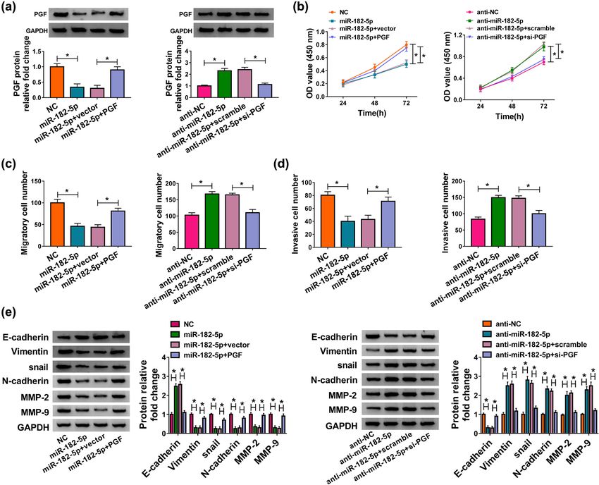
Hsa_circ_0007121/miR-182-5p/PGF axis regulates preeclampsia progression 1063
Beijing, China) was purchased from Sigma and used to (Solarbio) and were analyzed using the microscope (MTX
transfect cells following the provided procedures. Lab Systems, Bradenton, FL, USA).
2.6 Western blot
2.3 Quantitative reverse transcription
polymerase chain response (qRT-PCR) Western blot was executed according to the previous report
and RNase treatment [12]. Briefly, after the extraction and separation, proteins
were incubated with the primary antibodies and the
The TRIzol reagent (Sigma) was applied for RNA extrac- secondary antibody. The protein band was observed using
tion, and PrimeScript RT Master Mix kit (Takara, Dalian, ECL kit (Solarbio). Antibodies used in this research were
China) was used for reverse transcription. Then, the as follows: anti-E-cadherin (1:1,000, ab15148, Abcam,
QuantiTect SYBR Green RT-PCR Kit (Qiagen, Shanghai, Cambridge, United Kingdom), anti-Vimentin (1:3,000,
China) was used to perform the qRT-PCR for hsa_ ab137321, Abcam), anti-snail (1:1,000, ab82846, Abcam),
circ_0007121 and PGF. The miScript SYBR Green PCR kit anti-N-cadherin (1:2,500, ab18203, Abcam), anti-matrix
(Qiagen) was used for the qRT-PCR of miR-182-5p. Beta- metalloprotein (MMP)-2 (1:3,000, ab97779, Abcam), anti-
actin (β-actin) was used to normalize hsa_circ_0007121 MMP-9 (1:1,000, ab38898, Abcam), anti-PGF (1:2,500,
and PGF expression, and U6 was used to normalize miR- ab196666, Abcam), anti-glyceraldehyde 3-phosphate de-
182-5p expression. The data were computed using the hydrogenase (1:2,500, ab9485, Abcam), and Goat Anti-
2−ΔΔCt method. The following primers were used (5′ to 3′): Rabbit IgG H&L (HRP) (1:3,000, ab205718, Abcam).
hsa_circ_0007121 (F, GGGGGTTTTATTTCAGGTGGA; R, AGG
GGAAAAATAGTCCTCACAGA); linear mRNA primer (F,
AGTTTTAGGCGTGGCTGTGA; R, CACGATTGCTCACAATGG
AGG); miR-182-5p (F, ATCACTTTTGGCAATGGTAGAACT; R, 2.7 Dual-luciferase reporter assay
TATGGTTTTGACGACTGTGTGAT); PGF (F, CCCACCTGGATG
CTGTT; R, ATAGAGGGTAGGTACCAG); β-actin (F, GCACCA The potential target sequences in hsa_circ_0007121 or
CACCTTCTACAATG; R, TGCTTGCTGATCCACATCTG); U6 (F, PGF of miR-182-5p were predicated by CircRNA inter-
TCCGGGTGATGCTTTTCCTAG; R, CGCTTCACGAATTTGCGT actome or ENCORI, respectively. The sequence of
GTCAT). RNase R (Sigma) was utilized to treat purified hsa_circ_0007121 or PGF 3′UTR was inserted into pGL3
RNAs to check the circular form of hsa_circ_0007121. vector (Promega, Madison, WI, USA) for the establish-
ment of hsa_circ_0007121-wt or PGF-wt reporter vector.
Also, the hsa_circ_0007121-mut or PGF-mut reporter
vector was constructed by mutating the possible binding
2.4 Cell counting kit-8 (CCK-8) assay sites. Then, HTR-8/SVneo cells were cotransfected with
reporter vector and miR-182-5p or miR-NC. The luciferase
HTR-8/SVneo cells were seeded into a 96-well plate and activity was checked by using the Dual-Glo® Luciferase
added with 10 µL CCK-8 solution (MedChemExpress, Assay System kit (Promega).
Shanghai, China). After 2 h, the optical density at 450 nm
wavelength was checked with a microplate reader (Bio-
Rad, Richmond, Virginia, USA).
2.8 RNA immunoprecipitation (RIP) assay
Magna RIP RNA-Binding Protein Immunoprecipitation
2.5 Transwell assay Kit (Millipore, Billerica, MA, USA) was introduced for RIP
in line with the given protocols. In brief, cells were lysed
Transwell chamber precoated with or without Matrigel and incubated with anti-Argonaute 2 antibody (Anti-
(Solarbio) was utilized to evaluate cell invasion or Ago2; Millipore) with conjugated magnetic beads for
migration, respectively. Cells with serum-free medium 24 h, and then, the beads were treated with proteinase K
were added into the upper chamber, and medium to remove protein. The immunoglobulin G (IgG) was
containing fetal bovine serum was added into the lower used as a control. The immune precipitated RNA was
chamber. Then, the cells were treated with crystal violet purified and analyzed by qRT-PCR.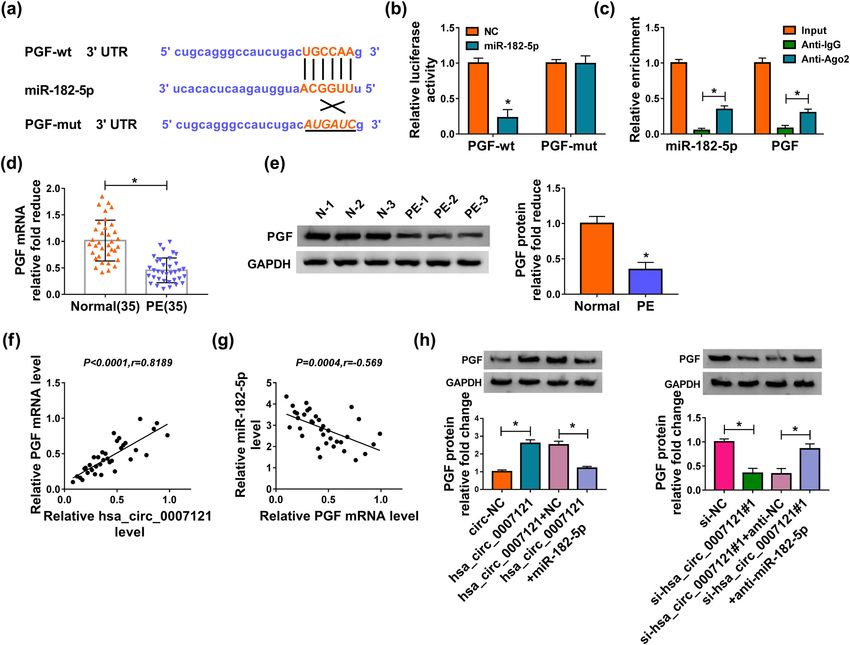
1064 Shukun Gai et al.
2.9 Statistical analysis 3.2 Hsa_circ_0007121 regulated HTR-8/
SVneo cell proliferation, migration,
Experimental data were presented by mean ± standard invasion, and EMT
deviation and analyzed by GraphPad Prism (GraphPad, La
Jolla, CA, USA). Two independent groups were compared The effect of hsa_circ_0007121 on PE was further investi-
via using Student’s t-test. The one-way analysis of gated, and we detected its level in HTR-8/SVneo cells after
variance was utilized to assess the difference for more transfection with circ-NC, hsa_circ_0007121, si-NC, or
than two groups. The correlation among miR-182-5p, si-hsa_circ_0007121 (si-hsa_circ_0007121#1, si-hsa_
hsa_circ_0007121, and PGF in PE placental tissues was circ_0007121#2, and si-hsa_circ_0007121#3). The result
analyzed by Pearson’s correlation coefficient. Each showed that hsa_circ_0007121 was conspicuously upregu-
experiment was carried out with at least three replica- lated in hsa_circ_0007121 group relative to circ-NC
tions. P < 0.05 indicated the statistical significance. group, and it was significantly downregulated in the
si-hsa_circ_0007121 group compared with the si-NC group
(Figure 2a). Overexpression of hsa_circ_0007121 promoted
cell proliferation, while an opposite result was obtained
3 Results when hsa_circ_0007121 was knocked down (Figure 2b).
Meanwhile, the transwell assay indicated that cell migra-
tion and invasion were boosted by upregulated hsa_
3.1 Hsa_circ_0007121 is downregulated in circ_0007121, while repressed by downregulation of
PE placental tissues hsa_circ_0007121 (Figure 2c and d). Moreover, EMT-related
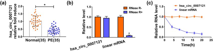
First, we measured hsa_circ_0007121 level in PE pla- protein levels were checked, and the results revealed that
cental tissues and compared them with those in normal hsa_circ_0007121 overexpression reduced the level of
placentas. The results showed that relative to the normal E-cadherin and elevated the levels of Vimentin, snail,
placental tissues, hsa_circ_0007121 was significantly N-cadherin, MMP2, and MMP9, while hsa_circ_0007121
downregulated in PE placental tissues (Figure 1a). silencing exhibited opposite results (Figure 2e). On the
Then, the levels of hsa_circ_0007121 and the linear whole, these results illustrated that hsa_circ_0007121 was
mRNA were checked, and the data indicated that involved in the modulation of PE progression.
hsa_circ_0007121 level was not clearly changed under
treatment with RNase R, while the level of linear mRNA
was apparently declined under RNase R treatment
(Figure 1b). Besides, the transcript half-life of hsa_ 3.3 Hsa_circ_0007121 directly targeted
circ_0007121 (nearly 20 h) was longer than the half-life miR-182-5p to regulate its expression
of linear mRNA (less than 5 h) after the treatment with
actinomycin D (Figure 1c). These data suggested that To explore how hsa_circ_0007121 participates in the
hsa_circ_0007121 was downregulated with high stability modulation of PE progression, CircRNA interactome was
in HTR-8/SVneo cells than linear mRNA. used to explore its potential target, and we found that
Figure 1: Hsa_circ_0007121 was downregulated in PE placental tissues. (a) The level of hsa_circ_0007121 in PE placental tissues (n = 35)
and normal placental tissues (n = 35) was measured by qRT-PCR. (b) Hsa_circ_0007121 and the linear mRNA levels in HTR-8/SVneo cells
treated with or without RNase R were detected by qRT-PCR. (c) hsa_circ_0007121 and the linear mRNA levels in HTR-8/SVneo cells treated
with actinomycin D at the pointed time were checked by qRT-PCR. *P < 0.05.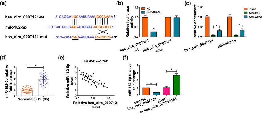
Hsa_circ_0007121/miR-182-5p/PGF axis regulates preeclampsia progression 1065 Figure 2: Hsa_circ_0007121 was involved in the regulation of PE. (a) The overexpression efficiency of hsa_circ_0007121 and the knockdown efficiency of si-hsa_circ_0007121 (si-hsa_circ_0007121#1, si-hsa_circ_0007121#2, and si-hsa_circ_0007121#3) were evaluated by qRT- PCR. (b) The proliferation of HTR-8/SVneo cells transfected with circ-NC, hsa_circ_0007121, si-NC, or si-hsa_circ_0007121#1 was checked by the CCK8 assay. (c and d) Cell migration and invasion were checked by the transwell assay. (e) The protein levels of EMT-related proteins (E-cadherin, Vimentin, snail, N-cadherin, MMP-2, and MMP-9) in transfected HTR-8/SVneo cells were measured by western blot assay. *P < 0.05. hsa_circ_0007121 contained the complementary se- comparison to Anti-IgG (Figure 3c). Next, miR-182-5p quences of miR-182-5p, which suggested that miR-182- level was checked, and we found that it was strikingly 5p might be bound to hsa_circ_0007121 (Figure 3a). higher in PE placental tissues than that in normal Then, the luciferase activity of hsa_circ_0007121-wt in placental tissues (Figure 3d). Moreover, miR-182-5p was HTR-8/SVneo cells was notably diminished by miR-182- negatively associated with hsa_circ_0007121 in PE 5p, while there was no change in the hsa_circ_0007121- placental tissues (Figure 3e). In addition, hsa_ mut group (Figure 3b). Besides, RIP assay exhibited circ_0007121 overexpression significantly decreased that both hsa_circ_0007121 and miR-182-5p were the level of miR-182-5p in HTR-8/SVneo cells, whereas enriched when incubation with Anti-Ago2 in hsa_circ_0007121 knockdown evidently increased the
1066 Shukun Gai et al.
Figure 3: Hsa_circ_0007121 directly interacted with miR-182-5p. (a) The potential target sites between miR-182-5p and hsa_circ_0007121
were predicated by CircRNA interactome. (b and c) The dual-luciferase reporter assay and RIP assay were performed to investigate the
interaction between miR-182-5p and hsa_circ_0007121. (d) The level of miR-182-5p in PE placental tissues (n = 35) and normal placental
tissues (n = 35) was detected by qRT-PCR. (e) The correlation between miR-182-5p and hsa_circ_0007121 in PE placental tissues was
analyzed by Pearson’s correlation coefficient. (f) The expression level of miR-182-5p was measured by qRT-PCR in HTR-8/SVneo cells
transfected with circ-NC, hsa_circ_0007121, si-NC, or si-hsa_circ_0007121#1. *P < 0.05.
levels miR-182-5p (Figure 3f). Collectively, these results inhibitory effect on cell proliferation (Figure 4b).
illustrated that hsa_circ_0007121 negatively regulated Besides, the transwell assay indicated that miR-182-5p
miR-182-5p via directly targeting. mimic rescued hsa_circ_0007121 overexpression in-
duced migration and invasion, and its inhibitor inverted
the inhibited migration and invasion caused by hsa_
3.4 Hsa_circ_0007121 regulated HTR-8/ circ_0007121 knockdown (Figure 4c and d). Moreover,
SVneo cell proliferation, migration, the levels of EMT-related proteins in the hsa_
circ_0007121 group or the si-hsa_circ_0007121 group
invasion, and EMT through miR-182-5p
were reversely changed after miR-182-5p was over-
expressed or knockdown, respectively (Figure 4e).
To investigate the functional mechanism between
In general, these findings disclosed that hsa_
hsa_circ_0007121 and miR-182-5p, HTR-8/SVneo cells
circ_0007121 regulated PE development by targeting
were transfected with hsa_circ_0007121, hsa_circ_
miR-182-5p.
0007121 + miR-182-5p, si-hsa_circ_0007121#1, or si-hsa_
circ_0007121#1 + anti-miR-182-5p, as well as matched
controls. QRT-PCR result shows that the expression of
miR-182-5p was inhibited in the cell transfected 3.5 Hsa_circ_0007121 regulated PGF
hsa_circ_0007121, while this inhibition effect was expression via targeting miR-182-5p
reversed when miR-182-5p was upregulated; meanwhile
anti-miR-182-5p reversed the promotion effect on miR- ENCORI was used to find the possible targets of miR-182-
182-5p expression induced by circ_0007121 knockdown 5p. It was displayed that the existence of binding sites
(Figure 4a). Subsequently, CCK-8 results exhibited between miR-182-5p and PGF 3’UTR (Figure 5a), and the
that upregulation of miR-182-5p reversed the promo- dual-luciferase reporter assay and RIP assay further
tion effect on cell proliferation induced by hsa_ verified this interaction (Figure 5b and c). We then
circ_0007121 overexpression, and miR-182-5p knock- discovered that the PGF level was clearly decreased in PE
down overturned hsa_circ_0007121 silencing-mediated placental tissues (Figure 5d and e). Moreover, PGF mRNAHsa_circ_0007121/miR-182-5p/PGF axis regulates preeclampsia progression 1067 Figure 4: Hsa_circ_0007121 regulated PE progression by interacting with miR-182-5p. (a) The level of miR-182-5p in HTR-8/SVneo cells transfected with hsa_circ_0007121, hsa_circ_0007121 + miR-182-5p, si-hsa_circ_0007121#1, or si-hsa_circ_0007121#1 + anti-miR-182-5p, as well as matched controls was checked by qRT-PCR. (b) The proliferation of transfected HTR-8/SVneo cells was checked by the CCK8 assay. (c and d) The abilities of migration and invasion of transfected HTR-8/SVneo cells were estimated by the transwell assay. (e) The protein levels of EMT-related proteins in samples were detected by Western blot. *P < 0.05. level was positively associated with hsa_circ_0007121 3.6 MiR-182-5p-/PGF axis-modulated HTR- (Figure 5f) and had an opposite correlation with miR- 8/SVneo cell proliferation, migration, 182-5p in PE placental tissues (Figure 5g). Further invasion, and EMT analysis demonstrated that the elevated protein level of PGF in the hsa_circ_0007121 group was reversed when To dissect the mechanism of miR-182-5p and PGF in PE miR-182-5p overexpressed, and the decreased protein progression, we first measured the protein level of PGF in level of PGF in the si-hsa_circ_0007121 group was also transfected HTR-8/SVneo cells. The declined protein level inverted by miR-182-5p inhibitor (Figure 5h). Our of PGF was observed in the miR-182-5p group, and this findings indicated that PGF was a target of miR-182-5p- trend was reversed by PGF overexpression, and the and hsa_circ_0007121-modulated PGF expression via increased protein level of PGF due to anti-miR-182-5p was miR-182-5p. reversed following the transfection with si-PGF (Figure 6a).
1068 Shukun Gai et al. Figure 5: MiR-182-5p bound to the 3’UTR of PGF and negatively regulated PGF expression. (a) The putative binding sites between miR-182- 5p and PGF were predicated by ENCORI. (b and c) The interaction between miR-182-5p and PGF was explored by the dual-luciferase reporter assay and RIP assay. (d and e) The mRNA and the protein levels of PGF in PE placental tissues (n = 35) and normal placental tissues (n = 35) were checked by qRT-PCR and western blot, respectively. (f and g) The correlation between PGF and hsa_circ_0007121 or miR-182-5p in PE placental tissues was analyzed using Pearson’s correlation coefficient. (h) The protein level of PGF in HTR-8/SVneo cells transfected with hsa_circ_0007121, hsa_circ_0007121 + miR-182-5p, si-hsa_circ_0007121#1, or si-hsa_circ_0007121#1 + anti-miR-182-5p, as well as corresponding controls was checked by qRT-PCR. *P < 0.05. CCK8 assay showed that PGF overexpression inverted 4 Discussion miR-182-5p upregulation-mediated suppressive cell pro- liferation, while the promoted cell proliferation due to PE is a growing threat to the pregnant woman, and miR-182-5p downregulation was recovered by PGF nearly, 76,000 pregnant women died from PE and knockdown (Figure 6b). Meanwhile, miR-182-5p over- related hypertensive disorders every year [35]. Therefore, expression weakened migration and invasion was it is a crying need for exploring the underlying receded by PGF overexpression, and PGF silencing mechanism and discovering new therapeutic strategies revoked miR-182-5p depletion-mediated boosted effects for PE. The previous research showed that circRNAs were on cell migration and invasion (Figure 6c and d). closely related to the regulation of PE. Deng et al. Analogously, upregulation of PGF rescued the effect of confirmed that hsa_circ_0011460 might serve as a miR-182-5p on EMT, whereas downregulation of PGF biomarker for the treatment of severe PE [13]. Zhou rescued miR-182-5p depletion-mediated promoted effect et al. reported that knockdown of circPAPPA facilitated on EMT (Figure 6e). These data demonstrated that miR- the onset and development of PE via inhibiting 182-5p targeted PGE to regulate PE development. trophoblast cells [14]. hsa_circ_0007021, which was
Hsa_circ_0007121/miR-182-5p/PGF axis regulates preeclampsia progression 1069
Figure 6: MiR-182-5p regulated PE progression via targeting PGF. (a) The protein level of PGF in HTR-8/SVneo cells transfected with miR-
182-5p, miR-182-5p + PGF, anti-miR-182-5p, and anti-miR-182-5p + si-PGF was measured by western blot. (b) The CCK8 assay was
conducted to check the proliferation of transfected HTR-8/SVneo cells. (c and d) The transwell assay was executed to evaluate the abilities
of cell migration and invasion. (e) The protein levels of EMT-related proteins were determined by western blot. *P < 0.05.
found to be decreased in PE plasma before the disease reported that circRNA MFACR modulated cardiomyocyte
phenotype presents, might be a novel biomarker of death by sponging miR-652-3p [37]. In this research,
preeclampsia [16]. In our study, hsa_circ_0007121 level miR-182-5p was confirmed to be bound and negatively
was reduced in PE placental tissues compared with the regulated by hsa_circ_0007121. Besides, miR-182-5p
normal placental tissues, which was in line with the overexpression or knockdown reversed hsa_circ_0007121
previous report [16]. Here, we first proposed the upregulation- or silencing-mediated effect on HTR-8/
regulatory network of hsa_circ_0007121/miR-182-5p/ SVneo cell proliferation, migration, invasion, and EMT,
PGF and revealed the effect and underlying mechanisms indicating that hsa_circ_0007121 plays roles in PE
of hsa_circ_0007121 in PE. development by regulating miR-182-5p.
Growing evidence have elucidated that circRNAs act To further understand the mechanism of miR-182-5p
as a competing endogenous RNA (ceRNA) and could also in regulating PE, we predicated and verified its target
sponging miRNAs to regulate the expression of the gene, PGF, which was tightly associated with PE
downstream genes. Wu et al. reported that circTADA2A [25,31,32]. In this study, we found a decreased expres-
promoted cell proliferation and metastasis in osteo- sion of PEG in PE placental tissues, which was in
sarcoma by binding to miR-203a-3p [36]. Wang et al. accordance with a recent report [33]. In addition, PGF1070 Shukun Gai et al.
was positively correlated with hsa_circ_0007121 and [7] Zou Y, Li S, Wu D, Xu Y, Wang S, Jiang Y, et al. Resveratrol
negatively associated with miR-182-5p in PE placental promotes trophoblast invasion in pre-eclampsia by inducing
tissues. Moreover, hsa_circ_0007121 altered PGF expres- epithelial-mesenchymal transition. J Cell Mol Med.
2019;23(4):2702–10. doi: 10.1111/jcmm.14175.
sion via sponging miR-182-5p. Also, PGF overexpression
[8] Graham CH, Hawley TS, Hawley RG, MacDougall JR, Kerbel RS,
or downregulation rescued miR-182-5p mimic- or in- Khoo N, et al. Establishment and characterization of
hibitor-mediated impact on proliferation, migration, first trimester human trophoblast cells with extended life-
invasion, and EMT in HTR-8/SVneo cells. Therefore, span. Exp Cell Res. 1993;206(2):204–11. doi: 10.1006/
these results suggested that hsa_circ_0007121 could excr.1993.1139.
[9] Chakraborty C, Gleeson LM, McKinnon T, Lala PK. Regulation
regulate the expression of PGF by sponging miR-182-
of human trophoblast migration and invasiveness. Can J
5p, eventually influencing the progression of PE. Physiol Pharmacol. 2002;80(2):116–24. doi: 10.1139/y02-016.
Although our research provides the theoretical support [10] Kristensen LS, Andersen MS, Stagsted LVW, Ebbesen KK,
for the application of hsa_circ_0007121 in PE therapy, Hansen TB, Kjems J. The biogenesis, biology and character-
other function of has_circ_007121 in PE still needs ization of circular RNAs. Nat Rev Genet. 2019;20(11):675–91.
doi: 10.1038/s41576-019-0158-7.
further exploration, and animal model of PE is required
[11] Garikipati VNS, Verma SK, Cheng Z, Liang D, Truongcao MM,
for further study to better elucidate the mechanism of
Cimini M, et al. Circular RNA CircFndc3b modulates cardiac
hsa_circ_0007121 in PE. repair after myocardial infarction via FUS/VEGF-A axis. Nat
In conclusion, our studies suggested that hsa_ Commun. 2019;10(1):4317. doi: 10.1038/s41467-019-11777-7
circ_0007121 was notably downregulated in PE placental [12] Holdt LM, Stahringer A, Sass K, Pichler G, Kulak NA, Wilfert W,
tissues and HTR-8/SVneo cells, and hsa_circ_0007121 et al. Circular non-coding RNA ANRIL modulates ribosomal
RNA maturation and atherosclerosis in humans. Nat Commun.
mediated the EMT of trophoblast cells proliferation,
2016;7:12429. doi: 10.1038/ncomms12429.
migration, invasion, and EMT via miR-182-5p/PGF axis. [13] Deng N, Lei D, Huang J, Yang Z, Fan C, Wang S. Circular RNA
This novel mechanism might provide a new light for the expression profiling identifies hsa_circ_0011460 as a novel
therapy of PE. molecule in severe preeclampsia. Pregnancy Hypertens.
2019;17:216–25. doi: 10.1016/j.preghy.2019.06.009.
Conflict of interest: The authors declare that they have [14] Zhou W, Wang H, Yang J, Long W, Zhang B, Liu J, et al. Down-
regulated circPAPPA suppresses the proliferation and invasion
no financial conflicts of interest.
of trophoblast cells via the miR-384/STAT3 pathway. Biosci
Rep. 2019;39(9):bsr20191965. doi: 10.1042/bsr20191965.
[15] Shen XY, Zheng LL, Huang J, Kong HF, Chang YJ, Wang F, et al.
CircTRNC18 inhibits trophoblast cell migration and epithelial-
References mesenchymal transition by regulating miR-762/Grhl2 pathway
in pre-eclampsia. RNA Biol. 2019;16(11):1565–73. doi:
[1] GBD 2015 Mortality and Causes of Death Collaborators. 10.1080/15476286.2019.1644591.
Global, regional, and national life expectancy, all-cause [16] Bai Y, Rao H, Chen W, Luo X, Tong C, Qi H. Profiles of circular
mortality, and cause-specific mortality for 249 causes of death, RNAs in human placenta and their potential roles related to
1980–2015: a systematic analysis for the Global Burden of preeclampsia. Biol Reprod. 2018;98(5):705–12. doi: 10.1093/
Disease Study 2015. Lancet. 2016;388(10053):1459–544. biolre/ioy034.
doi: 10.1016/s0140-6736(16)31012-1. [17] Bartel DP. MicroRNAs: genomics, biogenesis, mechanism, and
[2] Al-Jameil N, Aziz Khan F, Fareed Khan M, Tabassum H. A brief function. Cell. 2004;116(2):281–97. doi: 10.1016/s0092-
overview of preeclampsia. J Clin Med Res. 2014;6(1):1–7. 8674(04)00045-5.
doi: 10.4021/jocmr1682w. [18] Chiofalo B, Laganà AS, Vaiarelli A, La Rosa VL, Rossetti D,
[3] Goldman-Wohl D, Yagel S. Regulation of trophoblast invasion: Palmara V, et al. Do miRNAs play a role in fetal growth
from normal implantation to pre-eclampsia. Mol Cell restriction? A fresh look to a busy corner. Biomed Res Int.
Endocrinol. 2002;187(1–2):233–8. doi: 10.1016/s0303- 2017;2017:6073167. doi: 10.1155/2017/6073167.
7207(01)00687-6. [19] Lv Y, Lu X, Li C, Fan Y, Ji X, Long W, et al. miR-145-5p promotes
[4] Mustafa R, Ahmed S, Gupta A, Venuto RC. A comprehensive trophoblast cell growth and invasion by targeting FLT1. Life
review of hypertension in pregnancy. J Pregnancy. Sci. 2019;239:117008. doi: 10.1016/j.lfs.2019.117008.
2012;2012:105918. doi: 10.1155/2012/105918. [20] Yuan Y, Wang X, Sun Q, Dai X, Cai Y. MicroRNA-16 is involved
[5] Romero R, Chaiworapongsa T. Preeclampsia: a link between in the pathogenesis of pre-eclampsia via regulation of
trophoblast dysregulation and an antiangiogenic state. J Clin Notch2. J Cell Physiol. 2020;235(5):4530–44. doi: 10.1002/
Invest. 2013;123(7):2775–7. doi: 10.1172/jci70431. jcp.29330.
[6] Sun YY, Lu M, Xi XW, Qiao QQ, Chen LL, Xu XM, et al. [21] Fang YN, Huang ZL, Li H, Tan WB, Zhang QG, Wang L, et al.
Regulation of epithelial-mesenchymal transition by homeobox Highly expressed miR-182-5p can promote preeclampsia
gene DLX4 in JEG-3 trophoblast cells: a role in preeclampsia. progression by degrading RND3 and inhibiting HTR-8/SVneo
Reprod Sci. 2011;18(11):1138–45. doi: 10.1177/ cell invasion. Eur Rev Med Pharmacol Sci.
1933719111408112. 2018;22(20):6583–90. doi: 10.26355/eurrev_201810_16132.Hsa_circ_0007121/miR-182-5p/PGF axis regulates preeclampsia progression 1071
[22] Noack F, Ribbat-Idel J, Thorns C, Chiriac A, Axt-Fliedner R, [30] Polsani S, Phipps E, Jim B. Emerging new biomarkers of
Diedrich K, et al. miRNA expression profiling in formalin-fixed preeclampsia. Adv Chronic Kidney Dis. 2013;20(3):271–9.
and paraffin-embedded placental tissue samples from preg- doi: 10.1053/j.ackd.2013.01.001.
nancies with severe preeclampsia. J Perinat Med. [31] Wu WB, Xu YY, Cheng WW, Yuan B, Zhao JR, Wang YL, et al.
2011;39(3):267–71. doi: 10.1515/jpm.2011.012. Decreased PGF may contribute to trophoblast dysfunction in
[23] Laganà AS, Vitale SG, Sapia F, Valenti G, Corrado F, Padula F, fetal growth restriction. Reproduction. 2017;154(3):319–29.
et al. miRNA expression for early diagnosis of preeclampsia doi: 10.1530/rep-17-0253.
onset: hope or hype? J Matern Fetal Neonatal Med. [32] Kurtoglu E, Avci B, Kokcu A, Celik H, Cengiz Dura M,
2018;31(6):817–21. doi: 10.1080/14767058.2017.1296426. Malatyalioglu E, et al. Serum VEGF and PGF may be significant
[24] Hu Y, Li P, Hao S, Liu L, Zhao J, Hou Y. Differential expression markers in prediction of severity of preeclampsia. J Matern
of microRNAs in the placentae of Chinese patients with severe Fetal Neonatal Med. 2016;29(12):1987–92. doi: 10.3109/
pre-eclampsia. Clin Chem Lab Med. 2009;47(8):923–9. 14767058.2015.1072157.
doi: 10.1515/cclm.2009.228. [33] Gao Y, Guo X, Li Y, Sha W, She R. The decreased lncRNA ZEB2-
[25] Chau K, Hennessy A, Makris A. Placental growth factor and AS1 in pre-eclampsia controls the trophoblastic cell line HTR-
pre-eclampsia. J Hum Hypertens. 2017;31(12):782–6. 8/SVneo’s invasive and migratory abilities via the miR-149/
doi: 10.1038/jhh.2017.61. PGF axis. J Cell Biochem. 2019;120(10):17677–86. doi:
[26] Laganà AS, Favilli A, Triolo O, Granese R, Gerli S. Early serum 10.1002/jcb.29034.
markers of pre-eclampsia: are we stepping forward? J Matern [34] Hypertension in pregnancy. Report of the American college of
Fetal Neonatal Med. 2016;29(18):3019–23. doi: 10.3109/ obstetricians and gynecologists’ task force on hypertension in
14767058.2015.1113522. pregnancy. Obstet Gynecol. 2013;122(5):1122–31. doi:
[27] Levine RJ, Maynard SE, Qian C, Lim KH, England LJ, Yu KF, et al. 10.1097/01.aog.0000437382.03963.88.
Circulating angiogenic factors and the risk of preeclampsia. [35] Kuklina EV, Ayala C, Callaghan WM. Hypertensive disorders
N Engl J Med. 2004;350(7):672–83. doi: 10.1056/ and severe obstetric morbidity in the United States. Obstet
NEJMoa031884. Gynecol. 2009;113(6):1299–306. doi: 10.1097/
[28] Widmer M, Villar J, Benigni A, Conde-Agudelo A, AOG.0b013e3181a45b25.
Karumanchi SA, Lindheimer M. Mapping the theories of [36] Wu Y, Xie Z, Chen J, Chen J, Ni W, Ma Y, et al. Circular RNA
preeclampsia and the role of angiogenic factors: a systematic circTADA2A promotes osteosarcoma progression and metastasis
review. Obstet Gynecol. 2007;109(1):168–80. doi: 10.1097/ by sponging miR-203a-3p and regulating CREB3 expression.
01.AOG.0000249609.04831.7c. Mol Cancer. 2019;18(1):73. doi: 10.1186/s12943-019-1007-1.
[29] Moore Simas TA, Crawford SL, Solitro MJ, Frost SC, Meyer BA, [37] Wang K, Gan TY, Li N, Liu CY, Zhou LY, Gao JN, et al. Circular
Maynard SE. Angiogenic factors for the prediction of RNA mediates cardiomyocyte death via miRNA-dependent
preeclampsia in high-risk women. Am J Obstet Gynecol. upregulation of MTP18 expression. Cell Death Differ.
2007;197(3):244.e1–8. doi: 10.1016/j.ajog.2007.06.030. 2017;24(6):1111–20. doi: 10.1038/cdd.2017.61.You can also read