Adipose derived mesenchymal stem cells inhibit cell proliferation and migration and suppress extracellular matrix synthesis in hypertrophic scar ...
←
→
Page content transcription
If your browser does not render page correctly, please read the page content below
EXPERIMENTAL AND THERAPEUTIC MEDICINE 21: 139, 2021

   Adipose‑derived mesenchymal stem cells inhibit cell proliferation
     and migration and suppress extracellular matrix synthesis
             in hypertrophic‑scar and keloid fibroblasts
                               FANG XIE, LI TENG, JIAJIE XU, JIANJIAN LU, CHAO ZHANG,
                                   LIYA YANG, XIAOYANG MA and MINGHAO ZHAO

                           Cranio‑Maxillo‑Facial Surgery Department 2, Plastic Surgery Hospital,
             Chinese Academy of Medical Sciences and Peking Union Medical College, Beijing 100144, P.R. China

                                       Received September 10, 2019; Accepted March 26, 2020

                                                   DOI: 10.3892/etm.2020.9571

Abstract. Pathological scars occur during skin wound               Introduction
healing, and the use of adipose‑derived stem cells (ADSCs)
is one of the various treatments. The present study aimed to       A pathological scar is a fibroproliferative disorder that is char‑
investigate the in vitro effects of ADSCs on the biological        acterized by the excessive repair by tissue repair cells, mainly
properties of hypertrophic scar fibroblasts (HSFs) and keloid      fibroblasts, through the excessive synthesis and secretion of
fibroblasts (KFs), such as proliferation, migration, and the       extracellular matrix during skin wound healing (1). Not only
synthesis of extracellular matrix proteins. Transwell cham‑        do pathological scars seriously affect the physical appear‑
bers were used to establish a co‑culture system of ADSCs           ance, but they are also usually accompanied with infection,
with normal skin fibroblasts (NFs), HSFs or KFs. The effect        itching, pain and ulceration (2,3). In addition, they can cause
of ADSCs on the proliferation of fibroblasts was evaluated         serious dysfunction or disfigurement, which obviously affects
by CCK8 measurement, while the migration ability of fibro‑         the quality of life of the patient (2,3). Despite the existence
blasts was assessed using cell scratch assay. The expression       of different clinical treatments for pathological scars, such as
of extracellular matrix proteins was measured by immunob‑          surgical resection, laser treatment, cortisol injection therapy,
lotting. Co‑culture of NFs with ADSCs did not affect cell          and compression therapy, no treatment method is known to
proliferation and migration, nor the expression of extracellular   achieve a satisfactory therapeutic effect (4,5).
matrix proteins [collagen‑I, collagen‑III, fibronectin (FN) and        Mesenchymal stem cells, derived from the mesoderm at
α‑smooth muscle actin (α‑SMA)] in NFs. However, as with            the embryonic stage, are adult stem cells with self‑renewal
the inhibitor SB431542, ADSCs significantly inhibited cell         and multi‑directional differentiation potential. During wound
proliferation and migration and the expression of extracellular    healing, mesenchymal stem cells have been shown to regulate
matrix proteins (collagen‑I, collagen‑III, FN and α‑SMA), but      macrophages and T‑cell function (6,7), neutralize oxidizing
also suppressed the protein expression of transforming growth      substances (8), secrete anti‑fibrotic factors (9), strengthen the
factor β1 (TGF‑ β1), phosphorylated (p‑) mothers against           function of dermal fibroblasts (10), promote vascularization
decapentaplegic homolog (Smad) 2, p‑Smad3 and Smad7 in             and stability of blood vessels, and induce the differentiation of
HSFs and KFs. The results show that ADSCs inhibited cell           dermal layer cells, which can help in healing of the tissue (11).
proliferation and migration and the expression of extracellular    In addition, previous studies have shown that mesenchymal
matrix proteins in HSCs and KFs in vitro, possibly through         stem cells, bone marrow mesenchymal stem cells (12,13),
inhibition of the TGF‑β1/Smad pathway.                             umbilical cord mesenchymal stem cells (14) and chorionic
                                                                   mesenchymal stem cells (15) can promote wound healing and
                                                                   treat various types of fibrotic diseases.
                                                                       Adipose‑derived stem cells (ADSCs), which have been
                                                                   isolated from human adipose tissue suspensions, have
                                                                   multipotential differentiation capacity (16,17). In addition to
Correspondence to: Professor Li Teng, Cranio‑Maxillo‑Facial
Surgery Department 2, Plastic Surgery Hospital, Chinese            possessing the characteristics of general stem cells, ADSCs
Academy of Medical Sciences and Peking Union Medical College,      have the ability of self‑renewal and multiplication, and can also
33 Ba‑Da‑Chu Road, Shi Jing Shan, Beijing 100144, P.R. China       differentiate into many specific functional cell lines (16,17).
E‑mail: tenglpshcams@163.com                                       Compared with other mesenchymal stem cells, ADSCs have
                                                                   a wide range of sources, only lead to minor damage in the
Key words: adipose‑derived mesenchymal stem cells, fibroblasts,    donor site, have a good tissue compatibility, are easy to culture
migration, proliferation, extracellular matrix                     in vitro, have weak immunogenicity and relatively uncontro‑
                                                                   versial ethically (16,17). It has been shown that ADSCs can
                                                                   help repair tissue and organ damage (18,19), as well as promote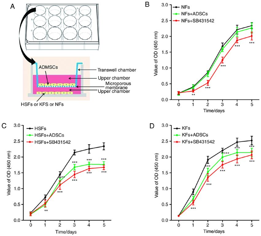
2           XIE et al: ADIPOSE-DERIVED MESENCHYMAL STEM CELLS IN HYPERTROPHIC SCAR AND KELOID FIBROBLASTS

wound healing through their paracrine effects in diabetic and          were incubated in a cell culture dish containing low‑glucose
nude mice (20,21). However, the molecular mechanisms by                DMEM medium (cat. no. 21885108; Gibco; Thermo Fisher
which ADSCs promote wound healing remain to be elucidated.             Scientific, Inc.) supplemented with 1% penicillin‑streptomycin
   The present study demonstrated that co‑culture with                 (cat. no. 15140163; Thermo Fisher Scientific, Inc.), 10% fetal
ADSCs inhibited the proliferation, migration, and protein              bovine serum (cat. no. 10437028; Thermo Fisher Scientific,
expression of extracellular matrix, and also inhibited the             Inc.), and 640 µg/ml glutamine (cat. no. G3126; Sigma‑Aldrich;
transforming growth factor β1 (TGF‑ β1)/mothers against                Merck KGaA). The incubation was continued until a dense
decapentaplegic homolog (Smad) pathway in hypertrophic                 monolayer (80% confluency) of cells formed around the tissue
scar fibroblasts and keloid fibroblasts.                               pieces. Cells from passages 3‑6 were used in the experiments.

Materials and methods                                                  Transwell chamber co‑culture system. A Transwell chamber
                                                                       (cat. no. 140652; Thermo Fisher Scientific, Inc.) was used to
Tissue specimens and patients. Adipose tissue, used to extract         culture ADSCs with fibroblasts. The co‑culture system was
adipose‑derived mesenchymal stem cells, was derived from               performed as follows: 1.5x10³ ADSCs were added per well in
5 healthy subjects (2 males and 3 females; 25‑42 years old)            the upper chamber of a 12‑well plate Transwell chamber, with
undergoing local liposuction from October 2018 to May 2019             0.5 ml of culture medium, and 3x10³ ADSCs per well were
at Plastic Surgery Hospital, Chinese Academy of Medical                inoculated into the lower chamber with 1.5 ml culture medium.
Sciences and Peking Union Medical College (Beijing, China).            For the single culture system only 3x10³ ADSCs were inocu‑
Hypertrophic scar tissues were obtained from 9 patients                lated per well into the lower chamber, with or without adding
with hypertrophic scars, keloid tissues were obtained from             SB431542 (cat. no. s4317; Sigma‑Aldrich; Merck KGaA) into
14 keloid patients, and 5 normal skin tissues were obtained            the culture medium.
from post‑reconstruction cat ear malformation and cosmetic
outpatient surgeries. Patients with the following criteria were        Cell proliferation assay. The viability of fibroblasts was evalu‑
excluded from the study: i) Less than 6 months with the condi‑         ated using the MTT Cell Proliferation and Cytotoxicity Assay
tion; ii) infection in the lesion; iii) radiation therapy or steroid   kit and the BrdU Cell Proliferation Assay kit (cat. no. C0075S;
injection; iv) pathological scar disease combined with other           Beyotime Institute of Biotechnology). Briefly, after 4 h of
hereditary diseases, body fluid transmission diseases (such            incubation with MTT (10 µl, 10 mg/ml), the supernatant
as HIV and HBV), malignant tumors and skin diseases; and,              was removed and 100 µl DMSO was added. After 30 min,
v) age >55 years or
EXPERIMENTAL AND THERAPEUTIC MEDICINE 21: 139, 2021 3 Figure 1. Effects of ADSCs on the proliferation of NFs, HSFs and KFs. (A) Co‑culture system of ADSCs with NFs, HSFs and KFs. (B) The proliferation of NFs in different culture environments. **P
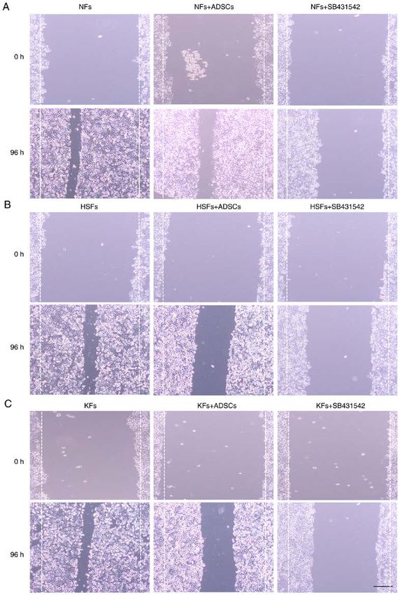
4            XIE et al: ADIPOSE-DERIVED MESENCHYMAL STEM CELLS IN HYPERTROPHIC SCAR AND KELOID FIBROBLASTS

Figure 2. Effects of ADSCs on the proliferation of NFs, HSFs and KFs. (A) Cell scratch assays were used to measure the migration of NFs in different culture
environments. (B) Cell scratch assays were used to measure the migration of HSFs in different culture environments. (C) Cell scratch assays were used to
measure the migration of KFs in different culture environments. Scale bar=100 µm. ADSCs, adipose‑derived mesenchymal stem cells; NFs, normal skin
fibroblasts; HSFs, hypertrophic scar fibroblasts; KFs, keloid fibroblasts.

and KFs, were measured in the single culture systems and in the                 collagen‑I, collagen‑III, FN and α‑SMA proteins in HSFs
co‑cultured cells (Fig. 1A). There was no significant difference                and KFs from the single culture system were significantly
in the expression of collagen‑I, collagen‑III, FN and α‑SMA                     lower than those from the co‑culture system with ADSCs
protein in NFs cultured alone and in those co‑cultured with                     (Fig. 3A‑C). On the other hand, SB431542 reduced the protein
ADSCs. However, the expression of collagen‑I, collagen‑III,                     expression of collagen‑I, collagen‑III, FN and α‑SMA in HSFs
FN and α‑SMA proteins was significantly decreased in NFs                        and KFs from the single culture system (Fig. 3A‑C). In addi‑
treated with SB431542. Of importance, the expression of                         tion, the concentration of hydroxyproline (HYP) in HSFs and
EXPERIMENTAL AND THERAPEUTIC MEDICINE 21: 139, 2021                                                            5

Figure 3. Effects of ADSCs on the synthesis of extracellular matrix in NFs, HSFs and KFs. Western blotting was used to detect the expression of collagen‑I,
collagen‑III, FN and α‑SMA protein in (A) NFs (***P
6            XIE et al: ADIPOSE-DERIVED MESENCHYMAL STEM CELLS IN HYPERTROPHIC SCAR AND KELOID FIBROBLASTS

Figure 4. Effects of ADSCs on TGF‑β1/Smad pathway in NFs, HSFs and KFs. A‑C, Western blot was used to detect the expression of TGF‑β1, Smad2, Smad3,
p‑Smad 2, p‑Smad 3 and Smad 7 protein in (A) NFs (***P
EXPERIMENTAL AND THERAPEUTIC MEDICINE 21: 139, 2021                                            7

Discussion                                                            fibroblasts, using a Transwell chamber wherein ADSCs were
                                                                      not in direct contact with fibroblasts. However, in animal
The complex causes and mechanisms have led to a number                experiments, ADSCs are in direct contact with scar fibroblasts.
of hypotheses to explain pathological scars formation, such           While a study has indicated that local injection of adipose stem
as the immunoinflammatory over‑the‑sun holiday hypothesis             cells can promote healing and reduce the risk of scar forma‑
(i.e. excessive inflammation results in extracellular matrix          tion during healing of the injury site (43), ADSCs‑conditioned
deposition and tissue fibrosis), the cytokine regulatory disorder     medium was alone able to alter the biological behavior of target
hypothesis, the cell matrix line disorder hypothesis and the          cells (44,45). Therefore, the interaction between the two cell
epigenetic hypothesis (26,27). However, no single hypothesis          types could be achieved through the influence of receptors, in
can fully explain the mechanism of pathological scars forma‑          addition to their direct interaction.
tion. Despite that, various hypotheses can hold several views             The present study observed that co‑culture with ADSCs
on the causes of pathological scar formation; excessive fibro‑        inhibited the protein expression of TGF‑β1, p‑Smad2/Smad2,
blast proliferation and deposition of extracellular matrix are        p‑Smad3/Smad3 and Smad7 in HSFs and KFs. The TGF‑β
considered the most significant pathological changes during           family is highly conserved and its members are widely
the development of pathological scars (26,27). Therefore,             expressed during embryonic and tissue development, where
inhibition of fibroblasts proliferation and the suppression of        they have been shown to exhibit different biological functions
extracellular matrix synthesis by fibroblasts could be poten‑         in a cell‑dependent and condition‑dependent manner (46).
tial targets for the prevention and treatment of pathological         TGF‑ β1 is a representative cytokine of the TGF‑ β family
scars (28).                                                           that plays an important role in the regulation of the biological
    Previous studies have shown that the transplantation of           behavior of different cell types at different stages of develop‑
mesenchymal stem cells into the large area of wounds can              ment (46). TGF‑β1 exists in complex regulatory networks with
accelerate wound healing, improve healing quality and reduce          different cell signaling pathway molecules that can regulate
scar formation (29,30). This suggests that mesenchymal stem           the expression of each other (46). In the process of wound
cells can inhibit scar formation, which provides an approach          healing, moderate secretion of TGF‑β1 can promote the prolif‑
for the treatment of wounds and pathological scars (29,30).           eration and migration of fibroblasts, and can also accelerate
Previous studies have shown that mesenchymal stem cells               the healing of wounds (47,48). Jung et al (49) demonstrated
can inhibit scar hyperplasia through myofibroblasts regula‑           that ADSCs can downregulate the expression of type‑1
tion (31,32), immune response regulation (33), ROS/RNS                collagen and hyaluronic acid at the mRNA level via paracrine
homeostasis (34) and angiogenesis induction (35). The present         TGF‑β1 activity. Overexpression of TGF‑β1 has been reported
study demonstrated that ADSCs inhibited cell proliferation            to promote the secretion of extracellular matrix, which leads to
and migration, as well as the protein expression of collagen‑I,       scars formation (50,51).
collagen‑III, FN and α‑SMA in hypertrophic scar fibroblasts               In summary, the present study demonstrated that ADSCs
and keloid fibroblasts. Evidently, the present study only inves‑      can affect the biological behavior of HSFs and KFs in vitro,
tigated the effect of ADSCs on proliferation, migration and the       specifically proliferation, migration and extracellular matrix
synthesis of extracellular matrix in HSFs and KFs in vitro. The       synthesis, by regulating the TGF‑β1/Smad pathway.
current study was limited to outside the body to circumvent the
complex environment inside the body, and its conclusion needs         Acknowledgements
to be confirmed in vivo. With the advancements in cell therapy
and stem cells understanding, ADSCs are regarded as model             Not applicable.
seed cells for cell therapy due to their ability to secrete a large
number of active factors (36,37) that can act through paracrine       Funding
mechanisms to exert multiple effects, such as the induction of
wound healing (19), angiogenesis (22), the inhibition of scar         No funding was received.
formation following myocardial infarction (38) and multi‑direc‑
tional differentiation (39). Yoshihiko et al (40) demonstrated        Availability of data and materials
that adipose‑derived stem/stromal cells can inhibit the forma‑
tion of vocal cord scars through the regulation of the biological     The datasets used and/or analyzed during the current study are
behavior of vocal fold fibroblasts and through the regulation         available from the corresponding author on reasonable request.
of vocal folds inflammation. Yun et al (41) demonstrated that
human ADSCs can stimulate scar remodeling in a pig wound              Authors' contributions
model by decreasing the activity of mast cells, inhibiting the
effects of TGF‑β on fibroblasts and decreasing the expression of      LT conceived and designed the current study and contributed
MMP molecules. In vitro, human ADSCs were shown to inhibit            to writing the manuscript. FX and JX performed the experi‑
TGF‑β1‑induced differentiation of human dermal fibroblasts            ments. JL, CZ, LY, XM and MZ analyzed and interpreted the
and keloid scar‑derived fibroblasts in a paracrine manner (42).       data. All authors read and approved the final manuscript.
    The mode of action of ADSCs in the regulation of scar
fibroblasts can occur either through direct contact, or through       Ethics approval and consent to participate
indirect non‑contact mechanisms (16,17). The present study
established an indirect co‑culture system of ADSCs and                All participants in the present study signed informed consents,
fibroblasts, including hypertrophic scar fibroblasts and keloid       and the study was approved by the Ethics Committee of Plastic
8 XIE et al: ADIPOSE-DERIVED MESENCHYMAL STEM CELLS IN HYPERTROPHIC SCAR AND KELOID FIBROBLASTS
Surgery Hospital, Chinese Academy of Medical Sciences and 16. Bunnell BA: Adipose‑derived stem cells. Methods Mol Biol 5:
59‑67, 2008.
Peking Union Medical College (Beijing, China). 17. Ma T, Sun J, Zhao Z, Lei W, Chen Y, Wang X, Yang J and Shen Z:
A brief review: Adipose‑derived stem cells and their therapeutic
Patient consent for publication potential in cardiovascular diseases. Stem Cell Res Ther 8: 124,
2017.
18. Yang D, Wang W, Li L, Peng Y, Chen P, Huang H, Guo Y, Xia X,
All the participants in the present study signed informed Wang Y and Wang H: The relative contribution of paracine effect
consents. versus direct differentiation on adipose‑derived stem cell trans‑
plantation mediated cardiac repair. PLoS One 8: e59020, 2013.
19. Kim WS, Park BS, Sung JH, Yang JM, Park SB, Kwak SJ and
Competing interests Park JS: Wound healing effect of adipose‑derived stem cells:
A critical role of secretory factors on human dermal fibroblasts.
The authors declare that they have no competing interests. J Dermatol Sci 48: 15‑24, 2007.
20. Lee SH, Lee JH and Cho KH: Effects of human adipose‑derived
stem cells on cutaneous wound healing in nude mice.
References Ann Dermatol 23: 150‑155, 2011.
21. Maharlooei MK, Bagheri M, Solhjou Z, Jahromi BM, Akrami M,
Rohani L, Monabati A, Noorafshan A and Omrani GR: Adipose
1. Fearmonti RM, Bond JE, Erdmann D, Levin LS, Pizzo SV and tissue derived mesenchymal stem cell (AD‑MSC) promotes
Levinson H: The modified patient and observer scar assessment skin wound healing in diabetic rats. Diabetes Res Clin Pract 93:
scale: A novel approach to defining pathologic and nonpathologic 228‑234, 2011.
scarring. Plast Reconstr Surg 129: 242‑247, 2012. 22. Gao W, Qiao X, Ma S and Cui L: Adipose‑derived stem cells
2. Bock O, Schmid‑Ott G, Malewski P and Mrowietz U: Quality accelerate neovascularization in ischaemic diabetic skin flap via
of life of patients with keloid and hypertrophic scarring. expression of hypoxia‑inducible factor‑1α. J Cell Mol Med 15:
Arch Dermatol Res 297: 433‑438, 2006. 2575‑2585, 2011.
3. Rohrer TE and Gold M: Introduction to special issue on hyper‑ 23. Bock O, Yu H, Zitron S, Bayat A, Ferguson MWJ and
trophic scars and keloids. Dermatol Surg 43 (Suppl 1): S1‑S2, Mrowietz U: Studies of transforming growth factors beta 1‑3 and
2017.
4. Seifert O and Mrowietz U: Keloid scarring: Bench and bedside. their receptors I and II in fibroblast of keloids and hypertrophic
Arch Dermatol Res 301: 259‑272, 2009. scars. Acta Derm Venereol 85: 216‑220, 2005.
5. Gauglitz GG, Korting HC, Pavicic T, Ruzicka T and Jeschke MG: 24. Wong VW, You F, Januszyk M, Gurtner GC and Kuang AA:
Hypertrophic Scarring and Keloids: Pathomechanisms and Transcriptional profiling of rapamycin‑treated fibroblasts from
current and emerging treatment strategies. Mol Med 17: 113‑125, hypertrophic and keloid scars. Ann Plast Surg 72: 711‑719,
2011. 2014.
6. Nakajima H, Uchida K, Guerrero AR, Watanabe S, Sugita D, 25. Tao J, Zhang J, Ling Y, Mccall CE and Liu TF: Mitochondrial
Takeura N, Yoshida A, Long G, Wright KT and Johnson WE: sirtuin 4 resolves immune tolerance in monocytes by rebalancing
Transplantation of mesenchymal stem cells promotes an alterna‑ glycolysis and glucose oxidation homeostasis. Front Immunol 9:
tive pathway of macrophage activation and functional recovery 419, 2018.
after spinal cord injury. J Neurotrauma 29: 1614‑1625, 2012. 26. O'Leary R, Wood EJ and Guillou PJ: Pathological scarring:
7. Schurgers E, Kelchtermans H, Mitera T, Geboes L and Strategic interventions. Eur J Surg 168: 523‑534, 2002.
Matthys P: Discrepancy between the in vitro and in vivo effects 27. Sarrazy V, Billet F, Micallef L, Coulomb B and Desmoulière A:
of murine mesenchymal stem cells on T‑cell proliferation and Mechanisms of pathological scarring: Role of myofibroblasts
collagen‑induced arthritis. Arthritis Res Ther 12: R31, 2010. and current developments. Wound Repair Regen 19 (Suppl 1):
8. Kim WS, Park BS, Kim HK, Park JS, Kim KJ, Choi JS, Chung SJ, S10‑S15, 2011.
Kim DD and Sung JH: Evidence supporting antioxidant action 28. Wagner JA: Therapy of pathological scars. J Dtsch Dermatol
of adipose‑derived stem cells: Protection of human dermal Ges 11: 1139‑1157, 2013.
fibroblasts from oxidative stress. J Dermatol Sci 49: 133‑142, 29. Liu YL, Liu WH, Sun J, Hou TJ, Liu YM, Liu HR, Luo YH,
2008. Zhao NN, Tang Y and Deng FM: Mesenchymal stem
9. Hiwatashi N, Bing R, Kraja I and Branski RC: Mesenchymal cell‑mediated suppression of hypertrophic scarring is p53 depen‑
stem cells have antifibrotic effects on transforming growth dent in a rabbit ear model. Stem Cell Res Ther 5: 136, 2014.
factor‑β1‑stimulated vocal fold fibroblasts. Laryngoscope 127: 30. Zhang Q, Liu LN, Yong Q, Deng JC and Cao WG: Intralesional
E35‑E41, 2017. injection of adipose‑derived stem cells reduces hypertrophic
10. Smith AN, Willis E, Chan VT, Muffley LA, Isik FF, Gibran NS scarring in a rabbit ear model. Stem Cell Res Ther 6: 145, 2015.
and Hocking AM: Mesenchymal stem cells induce dermal fibro‑ 31. Jia SS, Li WY, Liu X and Li LY: Transforming growth factor‑β1
blast responses to injury. Exp Cell Res 316: 48‑54, 2010. induces differentiation of bone marrow‑derived mesenchymal
11. Harvestine JN, Orbay H, Chen JY, Sahar DE and Leach JK: stem cells into myofibroblasts via production of reactive oxygen
Cell‑secreted extracellular matrix, independent of cell source, species. Beijing Da Xue Xue Bao Yi Xue Ban 47: 737‑742,
promotes the osteogenic differentiation of human stromal 2015 (In Chinese).
vascular fraction. J Mater Chem B 6: 4104‑4115, 2018. 32. Lichtman MK, Otero‑Vinas M and Falanga V: Transforming
12. Antoniou KM, Papadaki HA, Soufla G, Kastrinaki MC, growth factors β (TGF‑β) isoforms in wound healing and fibrosis.
Damianaki A, Koutala H, Spandidos DA and Siafakas NM: Wound Repair Regen 24: 215‑222, 2016.
Investigation of bone marrow mesenchymal stem cells (BM 33. Stagg J: Immune regulation by mesenchymal stem cells: Two
MSCs) involvement in idiopathic pulmonary fibrosis (IPF). sides to the coin. Tissue Antigens 69: 1‑9, 2010.
Respir Med 104: 1535‑1542, 2010. 34. Park JE, Seo YK, Yoon HH, Kim CW, Park JK and Jeon S:
13. Yang D, Sun S, Wang Z, Zhu P, Yang Z and Zhang B: Stromal Electromagnetic fields induce neural differentiation of human
cell‑derived factor‑1 receptor CXCR4‑overexpressing bone bone marrow derived mesenchymal stem cells via ROS mediated
marrow mesenchymal stem cells accelerate wound healing by EGFR activation. Neurochem Int 62: 418‑424, 2013.
migrating into skin injury areas. Cell Reprogram 15: 206‑215, 35. Manieri NA, Mack MR, Himmelrich MD, Worthley DL,
2013. Hanson EM, Lars E, Wang TC and Stappenbeck TS: Mucosally
14. Yuben M, Daniel A, Ursula M, Samuel CS, Jorge T, Sivakami I, transplanted mesenchymal stem cells stimulate intestinal healing
Richard B and Alan T: Human umbilical cord mesenchymal by promoting angiogenesis. J Clin Invest 125: 3606‑3618, 2015.
stem cells reduce fibrosis of bleomycin‑induced lung injury. Am 36. Zuk P, Zhu M, Mizuno H, Huang J, Futrell J, Katz A, Benhaim P,
J Pathol 175: 303‑313, 2009. Lorenz H and Hedrick M: Multilineage cells from human
15. Lee MJ, Jung J, Na KH, Moon JS, Lee HJ, Kim JH, Kim GI, adipose tissue: Implications for cell‑based therapies. Tissue
Kwon SW, Hwang SG and Kim GJ: Anti‑fibrotic effect of chori‑ Eng 7: 211‑228, 2001.
onic plate‑derived mesenchymal stem cells isolated from human 37. Zuk PA, Zhu M, Ashjian P, De Ugarte DA, Huang JI, Mizuno H,
placenta in a rat model of CCl(4)‑injured liver: Potential applica‑ Alfonso ZC, Fraser JK, Benhaim P and Hedrick MH: Human
tion to the treatment of hepatic diseases. J Cell Biochem 111: adipose tissue is a source of multipotent stem cells. Mol Biol
1453‑1463, 2010. Cell 13: 4279‑4295, 2002.EXPERIMENTAL AND THERAPEUTIC MEDICINE 21: 139, 2021 9
38. Bayesgenis A, Soler‑Botija C, Farré J, Sepúlveda P, Raya A, 46. Pardali E, Sanchez‑Duff hues G, Gomez‑Puerto MC and
Roura S, Prat‑Vidal C, Gálvez‑Montón C, Montero JA and Dijke PT: TGF‑β‑induced endothelial‑mesenchymal transition in
Büscher D: Human progenitor cells derived from cardiac adipose fibrotic diseases. Int J Mol Sci 18: 2157, 2017.
tissue ameliorate myocardial infarction in rodents. J Mol Cell 47. Yichiang H, Chen MJ, Yu YM, Shunyao K and Chang CC:
Cardiol 49: 771‑780, 2010. Suppression of TGF‑β1/SMAD pathway and extracellular matrix
39. Cao Y, Sun Z, Liao L, Meng Y, Han Q and Zhao RC: Human production in primary keloid fibroblasts by curcuminoids: Its
adipose tissue‑derived stem cells differentiate into endothelial potential therapeutic use in the chemoprevention of keloid.
cells in vitro and improve postnatal neovascularization in vivo. Arch Dermatol Res 302: 717‑724, 2010.
Biochem Biophys Res Commun 332: 370‑379, 2005. 48. Emami A, Halim AS, Salahshifar I, Yussof SJ, Khoo TL and
40. Yoshihiko K, Kobler JB, Herrera VLM and Zeitels SM: Kannan TP: Association of TGFβ1 and SMAD4 variants in the
Perspectives on adipose‑derived stem/stromal cells as potential etiology of keloid scar in the Malay population. Arch Dermatol
treatment for scarred vocal folds: Opportunity and challenges. Res 304: 541‑547, 2012.
Curr Stem Cell Res Ther 5: 175‑181, 2010. 49. Jung H, Kim HH, Lee DH, Hwang YS, Yang HC and Park JC:
41. Yun IS, Jeon YR, Lee WJ, Lee JW, Rah DK, Tark KC and Transforming growth factor‑beta 1 in adipose derived stem
Lew DH: Effect of human adipose derived stem cells on scar cells conditioned medium is a dominant paracrine mediator
formation and remodeling in a pig model: A pilot study. Dermatol determines hyaluronic acid and collagen expression profile.
Surg 38: 1678‑1688, 2012. Cytotechnology 63: 57‑66, 2011.
42. Spiekman M, Przybyt E, Plantinga JA, Gibbs S, van der Lei B 50. Shah M, Foreman DM and Ferguson MW: Neutralisation of
and Harmsen MC: Adipose tissue‑derived stromal cells inhibit TGF‑beta 1 and TGF‑beta 2 or exogenous addition of TGF‑beta 3
TGF‑ β1‑induced differentiation of human dermal fibroblasts to cutaneous rat wounds reduces scarring. J Cell Sci 108:
and keloid scar‑derived fibroblasts in a paracrine fashion. 985‑1002, 1995.
Plast Reconstr Surg 134: 699‑712, 2014. 51. Pohlers D, Brenmoehl J, Löffler I, Müller CK, Leipner C,
43. Zonari A, Martins TM, Paula AC, Boeloni JN, Novikoff S, Schultze‑Mosgau S, Stallmach A, Kinne RW and Wolf G: TGF‑β
Ma rques A P, Cor relo VM, Reis R L and Goes A M: and fibrosis in different organs‑molecular pathway imprints.
Polyhydroxybutyrate‑co‑hydroxyvalerate structures loaded with Biochim Biophys Acta 1792: 746‑756, 2009.
adipose stem cells promote skin healing with reduced scarring.
Acta Biomater 17: 170‑181, 2015.
44. Zhang Y, Dong W, Wang J, Cai J and Wang Z: Human omental This work is licensed under a Creative Commons
adipose‑derived mesenchymal stem cell‑conditioned medium Attribution-NonCommercial-NoDerivatives 4.0
alters the proteomic profile of epithelial ovarian cancer cell lines International (CC BY-NC-ND 4.0) License.
in vitro. Onco Targets Ther 10: 1655‑1663, 2017.
45. Ivanova‑Todorova E, Bochev I, Dimitrov R, Belemezova K,
Mourdjeva M, Kyurkchiev S, Kinov P, Altankova I and
Kyurkchiev D: Conditioned medium from adipose tissue‑derived
mesenchymal stem cells induces CD4 +FOXP3 + cells and
increases IL‑10 secretion. J Biomed Biotechnol 2012: 295167,
2012.You can also read