A two-microRNA signature predicts the progression of male thyroid cancer
←
→
Page content transcription
If your browser does not render page correctly, please read the page content below
Open Life Sciences 2021; 16: 981–991
Research Article
Bingyang Liu, Haihong Shi, Weigang Qiu, Xinquan Wu*, Liqiong Li, Wenyi Wu
A two-microRNA signature predicts the
progression of male thyroid cancer
https://doi.org/10.1515/biol-2021-0099 miR-16-1-3p, nor a prediction model based on both miRNAs
received May 31, 2021; accepted July 27, 2021 effectively predicted survival in female TC patients. In con-
Abstract: In various cancers, microRNAs (miRNAs) are clusion, both miR-451a and miR-16-1-3p may play impor-
abnormally expressed, including thyroid cancer (TC). In tant roles in the processes of male TC. The two-miRNA
recent years, the incidence of TC has increased annually signature involving miR-1258 and miR-193a may serve as
around the world. Compared with female patients, male a novel prognostic biomarker for male TC patients.
TC patients are more likely to have a postoperative Keywords: male thyroid cancer, microRNA signature,
recurrence and lymph node metastasis, and hence need tumour progression, miR-451a, miR-16-1-3p
second treatments. However, the molecular biological
processes underlying this phenomenon are not under-
stood. Therefore, we collected data on miRNA expression
and clinical information of male TC patients from The 1 Introduction
Cancer Genome Atlas (TCGA) database. Differentially
expressed miRNAs were identified between male TC tissues Over the past few decades, the incidence of thyroid
and matched normal tissues. The Kaplan–Meier method, cancer (TC) has increased substantially in many coun-
univariate and multivariate Cox regressions, and receiver tries [1]. TC is three times more frequent in women than
operating characteristic curve analyses were performed to in men [2], but in multiple studies, the male sex has been
assess the association between miRNAs and the disease-free shown to be a risk factor for mortality in patients with TC
survival of male TC patients. Gene Ontology (GO) and the [3–5]. Thus far, the studies present conflicting evidence
Kyoto Encyclopaedia of Gene and Genome (KEGG) enrich- concerning the impact of female hormonal and reproduc-
ment analyses were then used to explore the function of tive processes [6–9]. One study suggested that the BRAF
miRNA target genes. Furthermore, we evaluated the ability V600E mutation could be a vital independent risk factor
of the miRNA biomarker to predict survival in female TC for male TC patients [10]. To date, the role that male sex
patients. As a result, a total of 118 differentially expressed plays in TC remains unclear and has not been extensively
miRNAs were identified, including 25 upregulated and studied. The microRNAs (miRNAs) are a class of endo-
93 downregulated miRNAs. Among them, miR-451a and genous, non-coding small RNAs. These RNAs play crucial
miR-16-1-3p were confirmed to be independent prognostic roles in many types of cancer by regulating gene expres-
factors for the disease-free survival rate. The target genes sion [11,12]. Although miRNAs themselves do not encode
of miR-451a and miR-16-1-3p were identified, and functional any substances, their abnormal expression may cause
analysis showed that these genes were enriched in 25 Go tumours through a number of ways [13–15]. Many studies
and KEGG accessions, including cell signal transduction, have shown that there is a large number of abnormal
motor adhesion, phagocytosis, regulation of transcription, expressions of miRNAs in the pathogenesis of TC, such
cell proliferation, angiogenesis, etc. Neither miR-451a and as miR-220, miR-22, let-7, and miR-345 [16]. Other
miRNAs (e.g., miR-21 and miR-192) are involved in dif-
ferent cell death pathways [17]. However, there is no clear
* Corresponding author: Xinquan Wu, Department of Thyroid and evidence that explains the role of miRNA in the patho-
Breast Surgery, The Second Affiliated Hospital of Fujian Medical genesis of male TC, particularly with regard to miRNA
University, Quanzhou, Fujian 362000, People’s Republic of China, and patient prognosis. We identified prognostic miRNAs
e-mail: xinquan.wu@fjmu.edu.cn
associated with disease-free survival (DFS) time in male
Bingyang Liu, Haihong Shi, Weigang Qiu, Liqiong Li, Wenyi Wu:
Department of Thyroid and Breast Surgery, The Second Affiliated
TC patients using comprehensive bioinformatic ana-
Hospital of Fujian Medical University, Quanzhou, Fujian 362000, lysis. The miRNA-seq data and clinical information origi-
People’s Republic of China nated from the TCGA database. The functions of miRNA
Open Access. © 2021 Bingyang Liu et al., published by De Gruyter. This work is licensed under the Creative Commons Attribution 4.0
International License.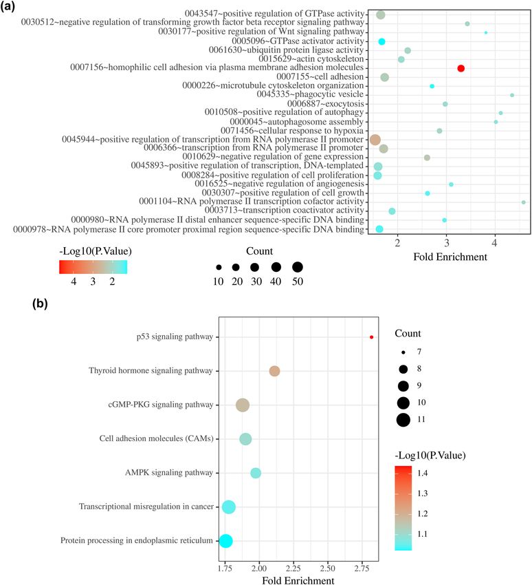
982 Bingyang Liu et al.
target genes were explored using GO and KEGG enrichment (version 3.2-7) in R. In the first step, a total of 129 patients
analyses. Finally, we developed a two-miRNA expression were divided into high- and low-risk groups based on the
signature to predict the DFS rate in male TC patients. median expression level of a differentially expressed
Furthermore, we evaluated the ability of the miRNA bio- miRNA. Then, the Kaplan–Meier method with the log-
marker to predict survival in female TC cancer patients, rank test and univariate Cox regression analysis were
and the results showed that neither miR-451a and miR-16- employed to evaluate the relationship between the expres-
1-3p nor the two-miRNA expression signature were effective sion level of this miRNA and the DFS time of patients.
in predicting survival in female TC patients. A P-value 2.0 and a P-value risk score and other clinical features as the explanatory
60 vs ≤60), histological type (non-
papillary thyroid carcinoma [nonTPC] vs TPC), tumour
2.3 Identifying survival-related miRNAs size (T3–4 vs T1–2), lymph node status (N1 vs N0 and
using survival analysis Nx), metastasis (M1 vs M0 and Mx), and pathological
stage (S3–4 vs S1–2). A P-value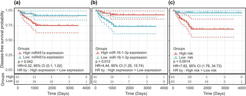
A two-microRNA signature predicts progression of male thyroid cancer 983 expression levels occurred in the two clinical groups. 3 Results Data are reported as the mean value and standard devia- tion. A P-value
984 Bingyang Liu et al.
Figure 1: (a) Volcano plot of differentially expressed miRNAs. Yellow dots represent upregulated miRNAs, and blue dots represent down-
regulated miRNAs. (b) The heat map shows the top 10 upregulated and top 10 downregulated differentially expressed miRNAs. miR-451a and
miR-16-1-3p are included in this heat map. A red box represents upregulated expression, whereas a blue box represents downregulated
expression. For log fold change (lfc), red represents upregulated expression, whereas green represents downregulated expression; the
colour scale for lfc ranges from −4 (green) to 6 (red), and the x-axis above the plot indicates grouping of samples in which red is the tumour
tissue group and blue is the normal tissue group.
differentially expressed between any groups of clinical vari- low-risk group, patients in the high-risk group had a
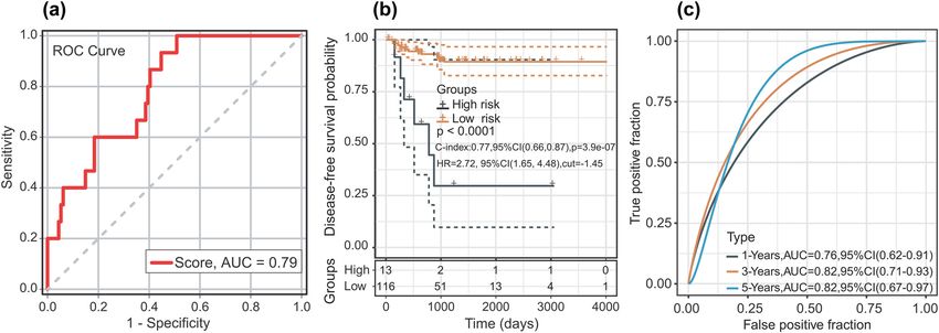
ables (Table 1), which ruled out an association between the shorter DFS time (HR = 2.72; 95% CI, 1.65–4.48; P < 0.01)
expression level of the two miRNAs and clinical groups and (Figure 3b). We evaluated the ROC curve of 1-, 3-, and
allowed for co-linearity between variables to be excluded. 5-year DFS, and the ROC curves demonstrated that the
According to the ROC analysis, the optimal cut-off two-miRNA signature harboured a promising ability to
risk score for DFS was −3.13 (AUC = 0.789; P < 0.01; predict DFS (1-year AUC = 0.76, 3-year AUC = 0.82, and
and 95% CI 0.69 and 0.89). The sensitivity and specificity 5-year AUC = 0.82) (Figure 3c). In the univariate analysis,
were 100% (95% CI: 0.78 and 0.99) and 49.12% (95% pathological stage (HR = 4.579; P = 0.009; and 95% CI,
CI: 0.40 and 0.59), respectively, at the best cut-off point 1.458–14.386) and the two-miRNA signature (HR = 7.823;
(Figure 3a). The patients were divided into low-risk P = 0.007; and 95% CI, 1.762–34.728) were associated with
groups (n = 116) and high-risk groups (n = 13) based on DFS time in patients with TC. In the multivariate analysis,
the best cut-off point. Compared with the patients in the the two-miRNA signature (HR = 6.937; P = 0.015; and 95%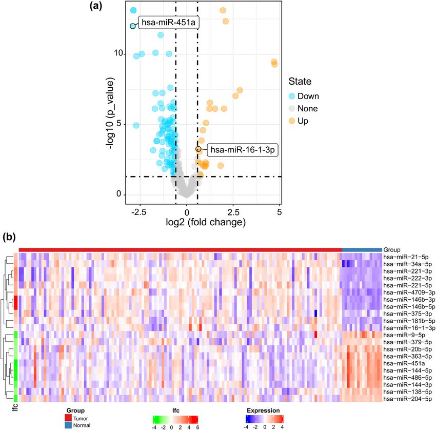
A two-microRNA signature predicts progression of male thyroid cancer 985
Figure 2: Kaplan–Meier curve with the log-rank test for miRNAs and the miRNA signature. (a) miR-16-1-3p, (b) miR-451a, and (c) two-miRNA
signature.
Table 1: Associations between the two miRNAs and clinical features
Variables Numbers miR-451a P-value miR-16-1-3p P-value
Patient age at diagnosis
≤60 89 9.22 ± 1.23 0.98 2.54 ± 0.59 0.33
>60 40 9.23 ± 1.49 2.66 ± 0.64
Clinical stage(AJCC)
SI–II 79 9.39 ± 1.28 0.07 2.53 ± 0.57 0.30
SIII–IV 50 8.96 ± 1.32 2.65 ± 0.66
T stage (AJCC)
T1–2 69 9.36 ± 1.42 0.20 2.52 ± 0.50 0.22
T3–4 60 9.06 ± 1.16 2.65 ± 0.70
N stage (AJCC)
N0 & Nx 60 9.20 ± 1.38 0.87 2.54 ± 0.58 0.47
N1 69 9.24 ± 1.26 2.61 ± 0.63
M stage (AJCC)
M0 & Mx 126 9.25 ± 1.31 0.19 2.57 ± 0.61 0.38
M1 3 8.25 ± 1.22 2.88 ± 0.56
Histologic type
TPC 125 9.21 ± 1.32 0.48 2.57 ± 0.60 0.12
Non TPC 4 9.68 ± 0.82 2.81 ± 0.80
Tumour location
One lobe 97 9.15 ± 1.26 0.32 2.63 ± 0.55 0.12
More than 32 9.42 ± 1.46 2.43 ± 0.73
one lobe
CI, 1.466–32.819) was the only independent risk factor for (Figure 4a and b). GO enrichment analysis revealed that
the DFS time of male TC patients (Table 2). these overlapping genes were mainly enriched in cell
signal transduction, motor adhesion, phagocytosis, reg-
ulation of transcription, cell proliferation, and angio-
3.3 KEGG and GO analyses of target genes of genesis (Figure 5a). KEGG enrichment analysis revealed
the miRNAs in the two-miRNA signature significant enrichment for seven KEGG signalling path-
ways: p53 signalling pathway, thyroid hormone signalling
A total of 12 overlapping target genes of miR-451a and 673 pathway, cGMP-PKG signalling pathway, cell adhesion
overlapping target genes of miR-16-1-3p were identified molecules, AMPK signalling pathway, transcriptional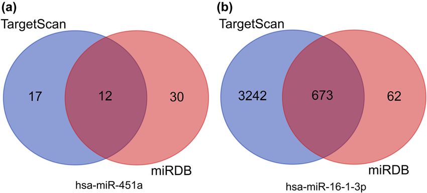
986 Bingyang Liu et al.
Figure 3: (a) ROC curve of sample for male TC patients. (b) Kaplan–Meier curve after optimal cut-off value; 13 patients were placed into the
high-risk group and 116 patients were placed in the low-risk group (optimal cut-off value = −1.45). (c) The AUC curve of the research subject
for 1, 3, and 5 years.
Table 2: Univariate and multivariate Cox regression analyses in male TC patients
Univariate analysis Multivariate analysis
P value HR (95% CI) P value HR (95% CI)
Age (>60 vs ≤60) 0.071 2.551 (0.923–7.044) 0.928 0.943 (0.266–3.339)
Histological type (NON TPC vs TPC) 0.617 0.047 (0.001–7444.315) 0.988 0.001 (0.001–∞)
Tumour size (T3–4 vs T1–2) 0.056 3.054 (0.972–9.595) 0.552 1.481 (0.406–5.406)
Lymph node status (N1 vs N0 & Nx) 0.871 0.919 (0.332–2.538) 0.447 0.637 (0.201–2.034)
Metastasis (M1 vs M0 & Mx) 0.199 3.784 (0.497–28.819) 0.629 1.717 (0.192–15.361)
Pathological stage (S3–4 vs S1–2) 0.009 4.579 (1.458–14.386) 0.098 3.666 (0.787–17.072)
two-miRNA signature (high risk vs low risk) 0.007 7.823 (1.762–34.728) 0.015 6.937 (1.466–32.819)
Figure 4: Venn diagrams showing the overlap of target genes that were predicted using the TargetScan and miRDB online tools. (a) hsa-miR-
451a and (b) hsa-miR-16-1-3p.A two-microRNA signature predicts progression of male thyroid cancer 987
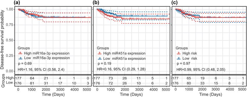
misregulation in cancer, and protein processing in endo- 0.56–2.4) (Figure 6a). Similarly, the difference in the
plasmic reticulum (Figure 5b). DFS time between the high and low hsa-miR-451a expres-
sion groups was not statistically significant (HR = 0.61;
P = 0.19; and 95% CI, 0.29–1.28) (Figure 6b). There was
3.4 Predictive value of miRNA biomarkers in also no statistically significant difference in DFS time
female TC patients between the high-risk group and the low-risk group
(HR = 0.99; P = 0.97; and 95% CI, 0.48–2.05) (Figure 6c).
In the Kaplan–Meier analysis, there were no significant Neither miR-451a and miR-16-1-3p nor a prediction model
difference in DFS time between the high and low miR-16-1-3p based on both miRNAs were effective in predicting the
expression groups (HR = 1.16; P = 0.69; and 95% CI, survival of female TC patients.
Figure 5: (a) Gene Ontology analysis of two-miRNA signature target genes. (b) KEGG pathway analysis of two-miRNA signature target genes.988 Bingyang Liu et al.
Figure 6: Predictive value of the miRNA biomarkers in female thyroid cancer patients using the Kaplan–Meier curve: (a) miR-16-1-3p,
(b) miR-451a, and (c) two-miRNA signature.
4 Discussion expression of miR-451a is downregulated in TC tissues,
and lower expression of miR-451a correlates with aggres-
In this study, we identified a pathological stage-related sive clinicopathological features of papillary thyroid carci-
two-miRNA signature as a promising predictor of DFS for noma (PTC). These effects may be related to the AKT/
male TC patients. In the multivariate Cox model, this mTOR pathway [30]. All these results indicate that miR-
signature was identified as an independent predictive 451a might play a protective role in combating cancer in
factor. We evaluated the ability of the miRNA biomarker the occurrence and development of TC, which is consistent
to predict the survival in female TC patients. The results with our conclusions. miR-451a might be used as a ther-
showed that neither miR-451a and miR-16-1-3p nor a two- apeutic target for certain drugs by upregulating the ex-
miRNA signature based on both miRNAs were effective in pression of miR-451a to inhibit tumour cell invasiveness.
predicting the survival in female TC patients. These two miR-16 has been extensively studied in other cancer
miRNAs might play a unique role in male TC develop- tissues. In ovarian cancer, studies have found that the
ment. KEGG and GO analyses revealed that the two- high expression of miR-16-1-3p is related to the recur-
miRNA signature plays crucial roles in cell adhesion, rence of ovarian cancer [31]. In a study on cholangiocar-
cell motility, cell signalling, transcription control and cinoma (CCA), miR-16 was one of the largest nodes in the
regulation of gene expression, cell proliferation, angio- ceRNA regulatory network, which indicates that miR-16
genesis, and cellular responses to hypoxia. The above might play an essential role in CCA development [32].
results suggest that the aforementioned two miRNAs Another study showed that the upregulation of miR-16 can
are closely related to the biological behaviour of male increase the invasiveness of cancer cells through the cell
TC, particularly invasion and metastasis. signalling factor pathway in triple-negative breast cancer
The molecule miR-451a has been studied in many patients with brain metastases. This process could be related
types of cancer such as stomach cancer, hepatocellular to epithelial-mesenchymal transition [33]. Bladen et al. [34]
carcinoma, bladder cancer, colorectal cancer, and basal also suggested that miR-16 is the central factor leading to
cell carcinoma. Several studies have suggested that a low high invasiveness in sebaceous gland carcinoma. All the
expression level of miR-451a positively correlates with the above results support the suggestion that miR-16 is a risk
metastatic ability and invasiveness of cancer cells [23–27]. factor, which is consistent with our results.
Some studies suggest that miR-451a can be used as a bio- The KEGG pathway analysis showed that the target
logical marker to predict the risk of recurrence [23,24]. genes of miR-451a and miR-16-1-3p were involved in the
In the field of TC research, experimental data from mul- p53 and AMPK signalling pathways. Mutations in p53
tiple cell types have confirmed that miR-451a could sup- have long been noticed in TC [35]. Maroof et al. [36]
press tumour cell proliferation and invasion by targeting reported that miRNAs could regulate apoptosis in TC cells
PSMB8 and ZEB1 [28,29]. One study revealed that the by targeting p53. The AMPK signal transduction pathwayA two-microRNA signature predicts progression of male thyroid cancer 989
has also been extensively investigated, and researchers may be related to VEGF-A overexpression in TC cells [55].
hold that the AMPK signalling pathway may contribute to In conclusion, the progression of male TC may be affected
the initiation, progression, and recurrence of cancer [37,38]. by miR-451a and miR-16-1-3p via angiogenesis.
When using AMPK inhibitors to inhibit the AMPK signalling
pathway, there is a strong anti-cancer effect in cell induc-
tion [39]. Several studies have suggested that activation of
the AMPK signalling pathway leads to nuclear translocation 5 Conclusion
of pyruvate kinase M2, which helps cancer cells survive
under metabolic stress and promotes cancer cell invasion Overall, this study identified two miRNAs that are related
and metastasis [40,41]. In TC research, Xu et al. [42] found to the DFS of male TC patients. These two miRNAs may
that TMP21 regulates TCP1 cell growth by inducing auto- play an important role in the development of male TC.
phagy, which may lead to activation of the AMPK/mTOR The two-miRNA signature involving miR-1258 and miR-
pathway. All these studies provide indirect evidence in sup- 193a may serve as a novel prognostic biomarker for male
port of our conclusion that miR-451a and miR-16-1-3p may TC patients. This is the first study to provide evidence for
influence male TC progression by regulating the activity of the relationship between miRNAs and male TC. Our study
the p53 and AMPK pathways. has some limitations. The results obtained from TGCA
Transcriptional regulation plays important roles in the need to be verified by in vitro cell experiments and
processes of eukaryotic gene expression. Transcriptional large-sample clinical trials, and the molecular mechan-
regulation modifies the gene expression level by altering isms involving miRNA signatures in male TC also need to
the efficiency of gene transcription [43]. Studies have be investigated further.
proven that the occurrence of cervical cancer is related
to transcription failure [44]. Cell experiments have also Acknowledgments: We thank TCGA for generating the
confirmed that miRNAs can upregulate or downregulate miRNA-seq data and providing data access.
the transcription of target genes in breast cancer by acti-
vating cell signalling pathways that promote breast cancer Funding information: The authors state no funding involved.
[45]. Many transcription factors can regulate transcription
products by regulating the activity of RNA polymerase. Conflict of interest: The authors state no conflict of
Runt-related transcription factor 2 (RUNX2) is an impor- interest.
tant regulator of osteogenic transcription. This factor was
found to be involved in the process of TC calcification and Data availability statement: The datasets generated during
invasiveness. In vitro cell experiments found that the and/or analysed during the current study are available
enhanced activity of the RUNX2 promoter can enhance from the corresponding author on reasonable request.
the activity of alkaline phosphatase, thereby promoting
calcification and the migration and invasion of cancer cells Disclosure: TCGA database belongs to public databases.
[46]. Recent studies have also shown that cell prolifera- The data resources had obtained informed consent from
tion, the cell cycle, apoptosis, and autophagy are key tech- all participants and had obtained approval from their
niques to control the development and progression of PTC research ethics committees or institutional review boards.
[47,48]. These findings suggest that miR-451a and miR-16- Users can download relevant data for free for research and
1-3p may regulate the progression of male TC through the publish relevant articles. This study is based on open-
regulation of transcription and cell proliferation. source data, so there are no ethical issues and other con-
Cell adhesion and the actin cytoskeleton are closely flicts of interest.
related to haematogenous metastatic spread of tumour
cells [49]. E-cadherin, which plays an important role in
normal cell-cell adhesion, was found to be expressed at
low levels in TC tumour tissue. This leads to the loss of
cell-cell adhesion, allowing TC cell migration [50]. A References
structural variant of the actin cytoskeleton is an impor-
tant process in cancer cell metastasis [51]. One study indi- [1] Kitahara CM, Sosa JA. The changing incidence of thyroid
cancer. Nat Rev Endocrinol. 2016;12(11):646–53.
cated that TC cells are three- to five-fold softer than normal
[2] Nilubol N, Zhang L, Kebebew E. Multivariate analysis of the
thyroid cells [52]. Angiogenesis also plays a pivotal role in relationship between male sex, disease-specific survival, and
tumour progression [53]. Abnormal angiogenesis has been features of tumor aggressiveness in thyroid cancer of follicular
found in several histopathologic subtypes of TC [54]. This cell origin. Thyroid. 2013;23(6):695–702.990 Bingyang Liu et al.
[3] Micheli A, Ciampichini R, Oberaigner W, Ciccolallo L, [21] Wang X. Improving microRNA target prediction by modeling
de Vries E, Izarzugaza I, et al. The advantage of women in with unambiguously identified microRNA-target pairs from
cancer survival: an analysis of EUROCARE-4 data. Eur J Cancer. CLIP-ligation studies. Bioinformatics. 2016;32(9):1316–22.
2009;45(6):1017–27. [22] Huang da W, Sherman BT, Lempicki RA. Bioinformatics
[4] Cunningham MP, Duda RB, Recant W, Chmiel JS, Sylvester JA, enrichment tools: paths toward the comprehensive functional
Fremgen A. Survival discriminants for differentiated thyroid analysis of large gene lists. Nucleic Acids Res.
cancer. Am J Surg. 1990;160(4):344–7. 2009;37(1):1–13.
[5] Bray F, Ferlay J, Soerjomataram I, Siegel RL, Torre LA, Jemal A. [23] Wang Y, Lin Z, Song J, Wei S, Ye Z, Chen S, et al. MicroRNA-451a
Global cancer statistics 2018: GLOBOCAN estimates of inci- targets caveolin-1 in stomach cancer cells. Int J Clin Exp Pathol.
dence and mortality worldwide for 36 cancers in 185 countries. 2020;13(10):2524–33.
CA Cancer J Clin. 2018;68(6):394–424. [24] Zhang Z, Zhang D, Cui Y, Qiu Y, Miao C, Lu X. Identification of
[6] Rajoria S, Suriano R, Shanmugam A, Wilson YL, Schantz SP, microRNA-451a as a novel circulating biomarker for colorectal
Geliebter J, et al. Metastatic phenotype is regulated by cancer diagnosis. Biomed Res Int. 2020;2020:5236236.
estrogen in thyroid cells. Thyroid. 2010;20(1):33–41. [25] Xu Y, Cao L, Chen G, Chen L, Li Y, Lai Y, et al. Human umbilical
[7] Rahbari R, Zhang L, Kebebew E. Thyroid cancer gender dis- cord mesenchymal stem cells-derived exosomal microRNA-
parity. Future Oncol. 2010;6(11):1771–9. 451a represses epithelial-mesenchymal transition of hepato-
[8] Peterson E, De P, Nuttall R. BMI, diet and female reproductive cellular carcinoma cells by inhibiting ADAM10. RNA Biol.
factors as risks for thyroid cancer: a systematic review. 2020;31(12):1–16.
PLoS One. 2012;7(1):e29177. [26] Xu K, Zhang YY, Han B, Bai Y, Xiong Y, Song Y, et al.
[9] Mack WJ, Preston-Martin S, Bernstein L, Qian D, Xiang M. Suppression subtractive hybridization identified differentially
Reproductive and hormonal risk factors for thyroid cancer in expressed genes in colorectal cancer: microRNA-451a as a
Los Angeles County females. Cancer Epidemiol Biomarkers novel colorectal cancer-related gene. Tumour Biol.
Prev. 1999;8(11):991–7. 2017;39(5):1010428317705504.
[10] Wang F, Zhao S, Shen X, Zhu G, Liu R, Viola D, et al. BRAF [27] Sun H, Jiang P. MicroRNA-451a acts as tumor suppressor in
V600E confers male sex disease-specific mortality risk in cutaneous basal cell carcinoma. Mol Genet Genomic Med.
patients with papillary thyroid cancer. J Clin Oncol. 2018;6(6):1001–9.
2018;36(27):2787–95. [28] Wang Q, Shang J, Zhang Y, Zhou Y, Tang L. miR-451a restrains
[11] Su Z, Ni L, Yu W, Yu Z, Chen D, Zhang E, et al. MicroRNA-451a the growth and metastatic phenotypes of papillary thyroid
is associated with cell proliferation, migration and carcinoma cells via inhibiting ZEB1. Biomed Pharmacother.
apoptosis in renal cell carcinoma. Mol Med Rep. 2020;127:109901.
2015;11(3):2248–54. [29] Fan X, Zhao Y. miR-451a inhibits cancer growth, epithelial-
[12] Mirzaei H, Hamblin MR. Regulation of glycolysis by non-coding mesenchymal transition and induces apoptosis in papillary
RNAs in cancer: switching on the warburg effect. Mol Ther thyroid cancer by targeting PSMB8. J Cell Mol Med.
Oncolytics. 2020;19:218–39. 2019;23(12):8067–75.
[13] Laengsri V, Kerdpin U, Plabplueng C, Treeratanapiboon L, [30] Minna E, Romeo P, Dugo M, De Cecco L, Todoerti K, Pilotti S,
Nuchnoi P. Cervical cancer markers: epigenetics and et al. miR-451a is underexpressed and targets AKT/mTOR
microRNAs. Lab Med. 2018;49(2):97–111. pathway in papillary thyroid carcinoma. Oncotarget.
[14] Rawat M, Kadian K, Gupta Y, Kumar A, Chain PSG, 2016;7(11):12731–47.
Kovbasnjuk O, et al. MicroRNA in pancreatic cancer: from [31] Delfino KR, Rodriguez-Zas SL. Transcription factor-microRNA-
biology to therapeutic potential. Genes (Basel). 2019;10:10. target gene networks associated with ovarian cancer survival
[15] Iqbal MA, Arora S, Prakasam G, Calin GA, Syed MA. MicroRNA and recurrence. PLoS One. 2013;8(3):e58608.
in lung cancer: role, mechanisms, pathways and therapeutic [32] Xu F, Zhao Y, Qin G, Huan Y, Li L, Gao W. Comprehensive
relevance. Mol Aspects Med. 2019;70:3–20. analysis of competing endogenous RNA networks associated
[16] Borran S, Ahmadi G, Rezaei S, Anari MM, Modabberi M, with cholangiocarcinoma. Exp Ther Med. 2019;18(5):4103–12.
Azarash Z, et al. Circular RNAs: new players in thyroid cancer. [33] Li Z, Peng Z, Gu S, Zheng J, Feng D, Qin Q, et al. Global analysis
Pathol Res Pract. 2020;216(10):153217. of miRNA-mRNA interaction network in breast cancer with
[17] Sadri Nahand J, Shojaie L, Akhlagh SA, Ebrahimi MS, brain metastasis. Anticancer Res. 2017;37(8):4455–68.
Mirzaei HR, Bannazadeh Baghi H, et al. Cell death pathways [34] Bladen JC, Wang J, Sangaralingam A, Moosajee M, Fitchett C,
and viruses: role of microRNAs. Mol Ther Nucleic Acids. Chelala C, et al. MicroRNA and transcriptome analysis in
2021;24:487–511. periocular sebaceous gland carcinoma. Sci Rep.
[18] Lossos IS, Czerwinski DK, Alizadeh AA, Wechser MA, 2018;8(1):7531.
Tibshirani R, Botstein D, et al. Prediction of survival in diffuse [35] Ito T, Seyama T, Mizuno T, Tsuyama N, Hayashi T, Hayashi Y,
large-B-cell lymphoma based on the expression of six genes. et al. Unique association of p53 mutations with undifferen-
N Engl J Med. 2004;350(18):1828–37. tiated but not with differentiated carcinomas of the thyroid
[19] Lewis BP, Shih IH, Jones-Rhoades MW, Bartel DP, Burge CB. gland. Cancer Res. 1992;52(5):1369–71.
Prediction of mammalian microRNA targets. Cell. [36] Maroof H, Irani S, Arianna A, Vider J, Gopalan V, Lam AK.
2003;115(7):787–98. Interactions of vascular endothelial growth factor and p53 with
[20] Wong N, Wang X. miRDB: an online resource for microRNA miR-195 in thyroid carcinoma: possible therapeutic targets in
target prediction and functional annotations. Nucleic Acids aggressive thyroid cancers. Curr Cancer Drug Targets.
Res. 2015;43(Database issue):D146–52. 2019;19(7):561–70.A two-microRNA signature predicts progression of male thyroid cancer 991
[37] Liu J, Ma L, Xu J, Liu C, Zhang J, Liu J, et al. Co-expression of CD44 [46] Jin Y, Kim HK, Lee J, Soh EY, Kim JH, Song I, et al. Transcription
and ABCG2 in spheroid body-forming cells of gastric cancer cell factor HOXA9 is linked to the calcification and invasion of
line MKN45. Hepatogastroenterology. 2013;60(125):975–80. papillary thyroid carcinoma. Sci Rep. 2019;9(1):6773.
[38] Feng JQ, Xu ZY, Shi LJ, Wu L, Liu W, Zhou ZT. Expression of cancer [47] Chou CK, Liu RT, Kang HY. MicroRNA-146b: a novel biomarker
stem cell markers ALDH1 and Bmi1 in oral erythroplakia and the and therapeutic target for human papillary thyroid cancer.
risk of oral cancer. J Oral Pathol Med. 2013;42(2):148–53. Int J Mol Sci. 2017;18:3.
[39] Wu Y, Yan B, Xu W, Guo L, Wang Z, Li G, et al. Compound C [48] Bi W, Huang J, Nie C, Liu B, He G, Han J, et al. CircRNA
enhances the anticancer effect of aspirin in HER-2-positive circRNA_102171 promotes papillary thyroid cancer progression
breast cancer by regulating lipid metabolism in an AMPK- through modulating CTNNBIP1-dependent activation of beta-
independent pathway. Int J Biol Sci. 2020;16(4):583–97. catenin pathway. J Exp Clin Cancer Res. 2018;37(1):275.
[40] Yang YC, Chien MH, Liu HY, Chang YC, Chen CK, Lee WJ, et al. [49] Xin Y, Chen X, Tang X, Li K, Yang M, Tai WC, et al. Mechanics
Nuclear translocation of PKM2/AMPK complex sustains cancer and actomyosin-dependent survival/chemoresistance of
stem cell populations under glucose restriction stress. Cancer suspended tumor cells in shear flow. Biophys J.
Lett. 2018;421:28–40. 2019;116(10):1803–14.
[41] Wang R, Cheng Y, Su D, Gong B, He X, Zhou X, et al. Cpt1c [50] Brabant G, Hoang-Vu C, Cetin Y, Dralle H, Scheumann G,
regulated by AMPK promotes papillary thyroid carcinomas Molne J, et al. E-cadherin: a differentiation marker in thyroid
cells survival under metabolic stress conditions. J Cancer. malignancies. Cancer Res. 1993;53(20):4987–93.
2017;8(18):3675–81. [51] Khakshour S, Labrecque MP, Esmaeilsabzali H, Lee FJS,
[42] Xu X, Gao H, Qin J, He L, Liu W. TMP21 modulates cell growth in Cox ME, Park EJ, et al. Retinoblastoma protein (Rb) links
papillary thyroid cancer cells by inducing autophagy through hypoxia to altered mechanical properties in cancer cells as
activation of the AMPK/mTOR pathway. Int J Clin Exp Pathol. measured by an optical tweezer. Sci Rep. 2017;7(1):7833.
2015;8(9):10824–31. [52] Prabhune M, Belge G, Dotzauer A, Bullerdiek J, Radmacher M.
[43] Casamassimi A, Ciccodicola A. Transcriptional regulation: Comparison of mechanical properties of normal and malignant
molecules, involved mechanisms, and misregulation. thyroid cells. Micron. 2012;43(12):1267–72.
Int J Mol Sci. 2019;20:6. [53] Hanahan D, Weinberg RA. Hallmarks of cancer: the next
[44] Meng Q, Wang S, Tang W, Wu S, Gao N, Zhang C, et al. XRCC1 generation. Cell. 2011;144(5):646–74.
mediated the development of cervival cancer through a novel [54] Sprindzuk MV. Angiogenesis in malignant thyroid tumors.
Sp1/Krox-20 swich. Oncotarget. 2017;8(49):86217–26. World J Oncol. 2010;1(6):221–31.
[45] Gorbatenko A, Sokilde R, Sorensen EE, Newie I, Persson H, [55] Song YS, Kim MJ, Sun HJ, Kim HH, Shin HS, Kim YA, et al.
Morancho B, et al. HER2 and p95HER2 differentially regulate Aberrant thyroid-stimulating hormone receptor signaling
miRNA expression in MCF-7 breast cancer cells and downre- increases VEGF-A and CXCL8 secretion of thyroid cancer cells,
gulate MYB proteins through miR-221/222 and miR-503. Sci contributing to angiogenesis and tumor growth. Clin Cancer
Rep. 2019;9(1):3352. Res. 2019;25(1):414–25.You can also read