The miR 136 5p/ROCK1 axis suppresses invasion and migration, and enhances cisplatin sensitivity in head and neck cancer cells
←
→
Page content transcription
If your browser does not render page correctly, please read the page content below

EXPERIMENTAL AND THERAPEUTIC MEDICINE 21: 317, 2021

 The miR‑136‑5p/ROCK1 axis suppresses invasion and migration,
  and enhances cisplatin sensitivity in head and neck cancer cells
                   BO YANG1, JIAN ZANG1, WEILI YUAN2, XUEJUN JIANG1 and FANG ZHANG3

        1
        Department of Otorhinolaryngology Head and Neck Surgery, The First Hospital of China Medical University,
      Shenyang, Liaoning 110001; Departments of 2Oral and Maxillofacial Surgery and 3Otorhinolaryngology Head and
      Neck Surgery, The Fourth Affiliated Hospital of China Medical University, Shenyang, Liaoning 110032, P.R. China

                                    Received March 4, 2020; Accepted December 15, 2020

                                                  DOI: 10.3892/etm.2021.9748

Abstract. Laryngeal squamous cell carcinoma (LSCC) and             ROCK1 and attenuated activity of the Akt/mTOR signaling
hypopharyngeal squamous cell carcinoma (HPSCC) are two             pathway in cisplatin‑treated LSCC and HPSCC cells.
types of head and neck cancers with high incidence rates and       Conversely, miR‑136‑5p knockdown increased ROCK1 levels
relatively poor prognoses. The aim of the present study was to     and decreased cisplatin sensitivity of the LSCC and HPSCC
determine the effects of microRNA (miR/miRNA)‑136‑5p and           cells by increasing cell viability and inhibiting cell apoptosis,
its downstream target, Rho‑associated coiled‑coil containing       which was reversed by ROCK1 inhibition using the ROCK1
protein kinase 1 (ROCK1), on LSCC and HPSCC progression            inhibitor, Y27632. Taken together, the results showed that the
and cisplatin sensitivity. The miRNA and protein expression        miR‑136‑5p/ROCK1 axis inhibits cell invasion and migration,
levels in head and neck cancer cell lines were evaluated using     and increases the sensitivity of LSCC and HPSCC cells to
reverse transcription‑quantitative PCR and western blotting,       cisplatin.
respectively. MTT, wound healing assays, transwell assays
and flow cytometry analysis were performed to measure cell         Introduction
properties. The binding between miR‑136‑5p and ROCK1 was
detected using a dual‑luciferase reporter assay. Autophagy         Head and neck cancer is one of the most common types
double‑labeled adenoviral infection assays were used to            of cancer worldwide which includes the mouth, pharynx
assess cell autophagy. The results showed that miR‑136‑5p          (nasopharynx, oropharynx and hypopharynx), larynx and
was expressed in LSCC and HPSCC cells. Functional                  paranasal sinus. According to the biological characteristics
experiments showed that the expression of miR‑136‑5p in            of head and neck cancer, it can be divided into several
LSCC and HPSCC cells was negatively correlated with cell           types. The most common type is head and neck squamous
viability, invasion and migration. Additionally, miR‑136‑5p        cell carcinoma (HNSCC), which accounts for >95% of all
overexpression inhibited epithelial‑mesenchymal transition,        cases of head and neck cancer (1). HNSCC is the sixth most
whereas miR‑136‑5p knockdown had the opposite effect.              common type of cancer worldwide, with 600,000 cases
Dual‑luciferase reporter assays confirmed the targeting            diagnosed each year and a mortality rate of 40‑50% (2,3). The
relationship between miR‑136‑5p and ROCK1. miR‑136‑5p              majority of patients with locally advanced HNSCC usually
overexpression increased the cisplatin sensitivity of LSCC and     develop a locoregional recurrence and/or distant metastases,
HPSCC cells by reducing cell viability, as well as promoting       and only a few patients with a locoregional recurrence may
cell apoptosis and autophagy. miR‑136‑5p overexpression            recover following treatment surgery and/or re‑irradiation (3).
decreased the expression levels of its downstream target           Overall, the 5‑year survival rate for patients with HNSCC has
                                                                   remained at 40‑60% and is accompanied by a relatively poor
                                                                   prognosis (1,4,5). Laryngeal squamous cell carcinoma (LSCC)
                                                                   and hypopharyngeal squamous cell carcinoma (HPSCC) are
                                                                   two types of HNSCC with high incidence rates and a relatively
Correspondence      to: Dr Fang Zhang, Department of
                                                                   poor prognosis following treatment (6‑8). Although multiple
Otorhinolaryngology Head and Neck Surgery, The Fourth Affiliated
Hospital of China Medical University, 4 East Chongshan Road,
                                                                   proteins and pathways have been shown to be associated
Shenyang, Liaoning 110032, P.R. China                              with the development of LSCC and HPSCC, the exact
E‑mail: zhangfang_cmu4h@163.com                                    molecular mechanisms of action and potential therapeutic
                                                                   targets affecting proliferation and migration remain to be
Key words: cell invasion, cell migration, cisplatin sensitivity,   elucidated (9).
laryngeal squamous cell carcinoma, hypopharyngeal squamous             MicroRNAs (miRNAs/miRs) are noncoding small
cell carcinoma, microRNA‑136‑5p, Rho‑associated coiled‑coil        RNA molecules that regulate the expression of target genes
containing protein kinase 1                                        by binding to the 3'‑untranslated region (UTR). miRNAs
                                                                   are involved in various biological processes including cell
                                                                   proliferation, apoptosis, invasion and migration (10). Various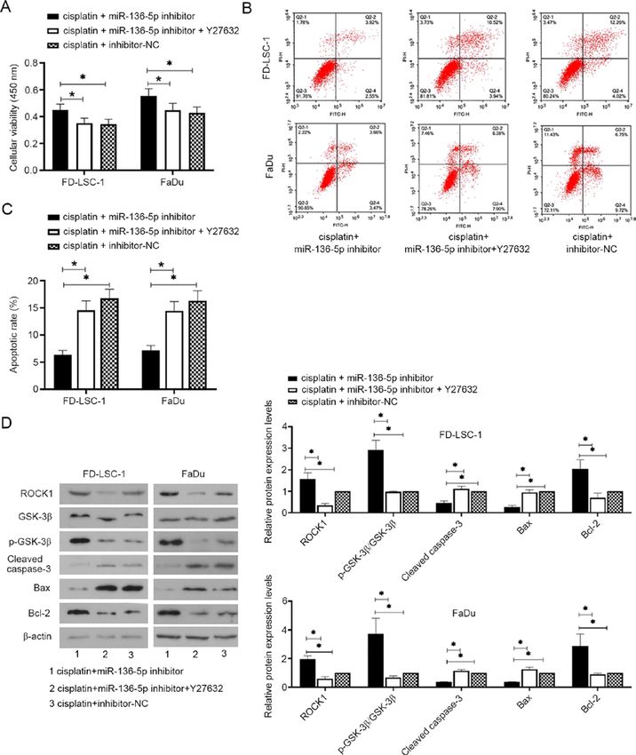
2           YANG et al: miR-136-5P/ROCK1 AXIS ENHANCES CISPLATIN SENSITIVITY IN HEAD AND NECK CANCER CELLS

miRNAs exhibit altered expression levels in cancers and serve       were immediately treated with cisplatin (2.6 µM,
crucial roles in the development of several types of cancer (11).   Sigma‑Aldrich; Merck KGaA) and/or Y2763 (25 µM,
Previous studies have found that the expression levels of           MedChem Express, Ltd.) for 24 h. After transfection and treat‑
miR‑136 are significantly decreased at the tumor site and that      ment, the cells were taken immediately for further experiments.
miR‑136 acts as a tumor suppressor in various tumor types,          The sequences for the miRNA mimics and inhibitors were as
participating in the development of tumors by regulating            follows: hsa‑miR‑136‑5p mimics, 5'‑ACUCCAUUUGUUU
the expression levels of downstream apoptosis‑related               UGAUGAUGGA‑3'; 5'‑CAUCAUCAAAACAAAUGGAGU
genes (12,13). Additionally, miR‑136 has also been found to         UU‑3'; NC mimics, 5'‑UUCUCCGAACGUGUCACGUTT‑3';
be associated with cisplatin resistance in human epithelial         5'‑ACGUGACACGUUCGGAGAATT‑3'; hsa‑miR‑136‑5p
ovarian and gastric cancers (14,15). Therefore, miR‑136 may         inhibitor, 5'‑UCCAUCAUCAAAACAAAUGGAGU‑3'; NC
be a potential target for cancer therapy.                           inhibitor, 5'‑UUGUACUACACAAAAGUACUG‑3'. The
    A previous study reported the relationship between              sequences were purchased from Jintuosi Biological Technology
miR‑136 and Rho‑associated coiled‑coil containing protein           Co., Ltd.
kinase (ROCK) 1 (16,17). ROCK1, one of the isoforms of the
ROCK, is a downstream effector of Rho A and is activated            Reverse transcription‑quantitative PCR (RT‑qPCR). Total
when it selectively binds to GTP (18,19). Activated ROCK            RNA was extracted from cells using a Total RNA extraction kit
interacts with the actin cytoskeleton to promote the formation      (Tiangen Biotech, Co., Ltd.). cDNA was synthesized using an
of stress fibers and focal adhesions, which in turn promote the     RNase inhibitor (Tiangen Biotech, Co., Ltd.), Super M‑MLV
metastatic ability of tumor cells (20‑22). ROCK overexpres‑         reverse transcriptase (BioTeke Corporation), 5x PCR buffer
sion has been reported to be associated with the progression        and dNTP (2.5 mM each). The miR‑136‑5p primer used for
of various malignancies, including bladder cancer, liver cancer     reverse transcription was: 5'‑GTTGGCTCTGGTGCAGGGTC
and breast cancer (23,24). ROCK downregulation inhibits             CGAGGTATTCGCACCAGAGCCAACTCCATC‑3'. The
tumor growth and metastasis, and enhances the efficacy of           temperature protocol that was used for reverse transcription
cisplatin (25‑27). In addition, the epithelial‑mesenchymal          was: 37˚C for 30 min, 42˚C for 30 min and 70˚C for 15 min.
transition (EMT) is a key process which often precedes and          To detect the expression levels of miRNA‑136‑5p, qPCR was
facilitates local invasion, vascular migration and distant          performed using PCR Master Mix (Tiangen Biotech, Co.,
metastasis of tumors (28). A previous study has shown that          Ltd.), SYBR-Green (Beijing Solarbio Science & Technology
the ROCK pathway is not only involved in the proliferation,         Co., Ltd.) and specific primers for the target genes. The ther‑
migration, adhesion and morphological changes of various cell       mocycling conditions used for qPCR were as follows: 94˚C
types, but also in the EMT of tumor cells (22). ROCK serves         for 4 min, 40 cycles of 94˚C for 15 sec, 60˚C for 20 sec and
a key role in TGF‑β‑induced EMT, which promotes mesen‑              72˚C for 15 sec. The 2‑ΔΔCq comparative method was used for
chymal transformation by rapidly activating RhoA‑dependent          data analysis (30). 5S ribosomal RNA (rRNA) was used as the
signaling pathways (29). Therefore, ROCK serves a crucial           internal control to normalize the expression levels of genes.
role in cancer development.                                         The value of each control was adjusted to 1 in each individual
    Based on the aforementioned points, the regulatory              repeat of the RT‑qPCR assay, therefore the mean value of the
mechanism of action behind the miR‑136/ROCK1 axis in                control is always 1. The primers were synthesized by GenScript.
LSCC and HPSCC were investigated in the present study               The primer sequences were as follows: hsa‑miR‑136‑5p
and its role in cell invasion and migration, as well as cisplatin   forward, 5'‑ACTCCATTTGTTTTGATGATGGA‑3' and
sensitivity, were assessed.                                         reverse, 5'‑GCAGGGTCCGAGGTATTC‑3'; and 5S rRNA
                                                                    forward, 5'‑GATCTCGGAAGCTAAGCAGG‑3' and reverse,
Materials and methods                                               5'‑TGGTGCAGGGTCCGAGGTAT‑3'.

Cell cultures, transfection and treatment. FaDu cells were          Cell viability assay. MTT assays were performed to deter‑
purchased from Procell and cultured in an incubator at 37˚C         mine the cell viability. Briefly, cells were seeded in 96‑well
and 5% CO 2 in minimum essential media (MEM,                        plates at a density of 4ⅹ103 cells per well. Subsequently, MTT
Sigma‑Aldrich; Merck KGaA) containing 10% FBS (Hyclone,             (0.5 mg/ml) was added to each well. After incubating for 4.5 h
GE Healthcare Life Sciences). FD‑LSC‑1 cells were obtained          at 37˚C in an incubator with 5% CO2, the supernatant was
from Fudan University, China, and cultured in a BEBM (Lonza         removed and 150 µl DMSO was added to each well to dissolve
Group, Ltd.) supplemented with 10% FBS and 1% L‑glutamine           the purple crystals. Cells were immersed in the dark for 10 min
at 37˚C with 5% CO2. To overexpress miR‑136‑5p, miR‑136‑5p          and the optical density values at 570 nm were measured using
mimics or mimics‑negative controls (NC) were transfected            a Microplate Reader (Biotek Instruments, Inc.).
into FaDu or FD‑LSC‑1 cells for 24 h at 37˚C. To downregu‑
late miR‑136‑5p expression, FaDu or FD‑LSC‑1 cells were             Cell migration assay. Wound healing assays were performed
transfected with miR‑136‑5p inhibitor or inhibitor‑NC for 24 h      to assess cell migration. The cells of each group were cultured
at 37˚C. The transfections were per for med using                   to the fusion state (90% confluence). After transfection
Lipofectamine® 2000 (Invitrogen; Thermo Fisher Scientific,          for 24 h as aforementioned, FaDu cells were cultured in a
Inc.). For the cisplatin+miR‑136‑5p mimics/inhibitor/mimics         minimum essential media without serum and treated with
NC/in hibitor NC group and cisplatin+m iR‑136 ‑5p                   1 µg/ml mitomycin C (Sigma‑Aldrich; Merck KGaA) for 1 h.
inhibitor+Y2763 group, after transfection with the miR‑136‑5p       FD‑LSC‑1 cells were cultured in a BEBM without serum and
mimics, inhibitor or NC for 24 h, FaDu and FD‑LSC‑1 cells           treated with 1 µg/ml mitomycin C for 1 h. Subsequently, the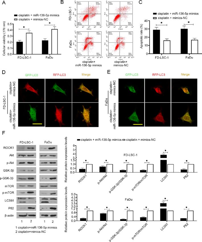
EXPERIMENTAL AND THERAPEUTIC MEDICINE 21: 317, 2021                                            3

cells in each group were scratched with a 200 µl pipette tip and   RRID: AB_2227948; 1:2,000; ProteinTech Group, Inc.) and
washed with serum‑free medium to remove cell debris. After         β‑actin (cat. no. sc‑47778; RRID: AB_2714189; 1:1,000; Santa
scratching, serum‑free medium was used for cell culture and        Cruz Biotechnology, Inc.). After incubating with horseradish
pictures were taken under a light microscope (magnification,       peroxidase‑conjugated goat anti‑rabbit/mouse secondary
ⅹ100; Olympus Corporation) to assess wound closure at 0 and        antibodies (1:5,000; cat. nos. A0208 and A0216; Beyotime
24 h.                                                              Institute of Biotechnology) for 45 min at 37˚C, signals were
                                                                   visualized using enhanced chemiluminescence solution
Invasion assay. Cell invasion was detected using a Transwell       (Beyotime Institute of Biotechnology). The expression
assay. Briefly, Transwell chambers (Corning, Inc.) pre‑coated      levels of protein were normalized to β ‑actin. The value of
with Matrigel were placed in 24‑well plates. Culture medium        each control was adjusted to 1 in each individual repeat of
(MEM medium for FaDu cells and BEBM medium for                     the western blot, therefore the mean value of the control is
FD‑LSC‑1 cells, 800 µl) supplemented with 30% FBS was added        always 1.
to the lower chamber. Cell suspension in 200 µl serum‑free
media was added to the upper chamber at a density of 1.5ⅹ104       Dual‑luciferase reporter assay. The binding site of miR‑136‑5p
cells/well. After incubation in a cell culture incubator at 37˚C   and ROCK1 was predicted using TargetScanHuman 7.2
with 5% CO2 for 24 h, the transwell chambers were washed           (http://www.targetscan.org/vert_72/). miR‑136‑5p was
three times with PBS to remove non‑invading cells. The cells       searched and the multiple genes that miR‑136‑5p may target
on the lower chamber were fixed with 4% paraformaldehyde           were obtained. UTRs were also searched between ROCK1 and
at room temperature for 25 min and then stained with 0.4%          miR‑136‑5p to obtain the targeted binding sequence between
crystal violet solution for 5 min at room temperature. Cells       miR‑136‑5p and ROCK1.
on lower chamber were counted under an inverted light                  For the verification of the relationship between miR‑136‑5p
microscope (magnification, ⅹ200; Olympus Corporation). A           and ROCK1, the wild‑type (wt) ROCK1‑3'‑UTR sequence and
total of five fields were selected for each sample and the mean    the mutant (mut) ROCK1‑3'‑UTR sequence were cloned into the
cell numbers was presented.                                        pmirGLO dual‑luciferase reporter vectors (Genscript Biotech
                                                                   Corporation) to construct pmirGLO‑ROCK1‑3'‑UTR‑wt and
Western blotting analysis. The protein expression levels were      pmirGLO‑ROCK1‑3'‑UTR‑mut plasmids. 293T cells were
detected using western blotting. Briefly, total protein was        harvested and seeded in 12‑well plates. Subsequently, the plas‑
extracted from cells using RIPA lysis buffer (Beijing Solarbio     mids were co‑transfected with miR‑136‑5p mimics/mimic‑NC
Science & Technology Co., Ltd.) and phenylmethanesulfonyl          or miR‑136‑5p inhibitor/inhibitor‑NC into 293T cells using
fluoride protease inhibitor (Beijing Solarbio Science &            Lipofectamine® 2000 reagent. After 48 h of incubation, the
Technology Co., Ltd.). Protein concentration in the lysate was     transfected cells were immediately harvested and assayed
quantified using a BCA protein concentration assay kit (Beijing    for luciferase activity using the dual‑luciferase reporter assay
Solarbio Science & Technology Co., Ltd.). Subsequently,            system (Promega Corporation) according to the manufacturer's
20 µg protein was loaded onto a 10% SDS‑gel and resolved           protocol. Firefly luciferase activity was normalized to Renilla
using SDS‑PAGE. After the protein was transferred to the           luciferase activity.
PVDF membranes (EMD Millipore), the membranes were
incubated with primary antibodies at 4˚C overnight. The            Apoptosis assay. Apoptosis was detected using flow cytom‑
primary antibodies used in the present study were: E‑cadherin      etry analysis. The Annexin V‑ FITC apoptosis detection kit
(cat. no. 20874‑1‑AP, RRID:AB_10697811; 1:10,000;                  (Beyotime Institute of Biotechnology) was used to detect
ProteinTech Group, Inc.), N‑cadherin (cat. no. 22018‑1‑AP,         apoptosis. Cells were harvested and adjusted to a density of
RRID:AB_2813891; 1:5,000; ProteinTech Group, Inc.),                1ⅹ106 cells/tube. Subsequently, cells were treated with 5 µl
vimentin (cat. no. 10366‑1‑AP, RRID:AB_2273020; 1:5,000;           Annexin V‑FITC and 10 µl PI and incubated for 15 min at
ProteinTech Group, Inc.), ROCK1 (cat. no. 21850‑1‑AP,              room temperature in the dark. Finally, cells stained with
RRID:AB_10953526; 1:1,000; ProteinTech Group, Inc.),               Annexin V‑FITC and PI were detected using flow cytometry
Akt (cat. no. AF6259, RRID: AB_2835120; 1:1,000; Affinity          (ACEA Bioscience, Inc.) and analyzed using NovoExpress
Biosciences), phospho‑(p‑)‑Akt (Ser473; cat. no. AF0016,           1.2.5 (ACEA Biosciences, Inc.).
RRID: AB_2810275; 1:1,000; Affinity Biosciences), glycogen
synthase kinase‑3 β (GSK‑3 β) (cat. no. AF5016; RRID:              Autophagy double‑labeled adenovirus infection assay. Cells
AB_2834935; 1:1,000; Affinity Biosciences), p‑GSK‑3 β              were seeded in a 24‑well plate (5x10⁴ cells) and infected with
(Ser9; cat. no. AF2016, RRID: AB_2834439; 1:1,000; Affinity        RFP‑GFP‑LC3‑labeled adenovirus (Hanbio Biotechnology
Biosciences), mTOR (cat. no. AF7803, RRID: AB_2844167;             Co., Ltd.) at a multiplicity of infection of 50. After incubating
1:1,000; Affinity Biosciences), p‑mTOR (S2448; cat.                for 24 h in an incubator at 37˚C supplied with 5% CO2, the
no. AF3308, RRID: AB_2834727; 1:1,000; Affinity                    supernatant of the medium containing the virus solution
Biosciences), microtubule‑associated protein 1 light chain 3       was discarded and replaced with complete medium (MEM
II/I (LC3II/I; cat. no. A7198; 1:1,000; ABclonal Biotech           medium containing 10% FBS for FaDu cells and BEBM
Co., Ltd.), P62 (cat. no. 18420‑1‑AP, RRID: AB_10694431;           medium containing 10% FBS for FD‑LSC‑1 cells). After
1:2,000; ProteinTech Group, Inc.), cleaved caspase‑3               incubating for 24 h in an incubator at 37˚C, images were
(cat. no. AF7022, RRID: AB_2835326; 1:1,000; Affinity              taken using a laser‑scanning confocal microscope equipped
Biosciences), Bax (cat. no. 50599‑2‑lg, RRID: AB_2061561;          with the FV10‑ASW system (magnification, ⅹ400; Olympus
1:5,000; ProteinTech Group, Inc.), Bcl‑2 (cat. no. 12789‑1‑AP;     Corporation).
4 YANG et al: miR-136-5P/ROCK1 AXIS ENHANCES CISPLATIN SENSITIVITY IN HEAD AND NECK CANCER CELLS Figure 1. The expression levels of miR‑136‑5p in LSCC and HPSCC cells. (A) The expression level of miR‑136‑5p in LSCC and HPSCC cells. Following transfection of the cells with (B) miR‑136‑5p mimics, mimics‑NC; or (C) miR‑136‑5p inhibitor or inhibitor‑NC for 24 h, the expression levels of miR‑136‑5p were measured using reverse transcription‑quantitative PCR. Data are presented as the mean ± SD, n=3. *P
EXPERIMENTAL AND THERAPEUTIC MEDICINE 21: 317, 2021                                                            5

Figure 2. miR‑136‑5p suppresses viability, invasion and migration of LSCC and HPSCC cells. (A) After LSCC and HPSCC cells transfected with miR‑136‑5p
mimics, mimics‑NC, miR‑136‑5p inhibitor or inhibitor‑NC for 24 h, cell viability was detected using MTT assays, n=6. Cell migratory abilities were measured
using wound healing assays in (B) FD‑LSC‑1 cells and (C) FaDu cells, scale bar=200 µm. (D) Migratory rates were then quantified. (E) Cell invasion was
observed and (F) measured using transwell assays, scale bar =100 µm. (G) The protein expression levels of E‑cadherin, N‑cadherin and vimentin were mea‑
sured using western blot analysis and normalized to the levels of β‑actin. Data are presented as the mean ± SD, n=3. *P
6            YANG et al: miR-136-5P/ROCK1 AXIS ENHANCES CISPLATIN SENSITIVITY IN HEAD AND NECK CANCER CELLS

Figure 3. miRNA‑136‑5p directly targets ROCK1 in LSCC and HPSCC cells. (A) Binding sites between ROCK1 wt and the mut‑type miR‑136‑5p are shown.
(B) miR‑136‑5p mimics or mimic‑NC and luciferase plasmid containing ROCK1‑wt or ROCK1‑mut transcript were co‑transfected into LSCC and HPSCC
cells, luciferase activity was measured using dual‑luciferase reporter assays. Data are presented as the mean ± SD, n=3. *P
EXPERIMENTAL AND THERAPEUTIC MEDICINE 21: 317, 2021                                                            7

Figure 4. miR‑136‑5p overexpression increases the cisplatin sensitivity of laryngeal squamous cell carcinoma and head and neck squamous cell carcinoma
cells. (A) After cells were transfected with miR‑136‑5p mimics or mimics‑NC and treated with cisplatin (2.6 µM) for 24 h, cell viability was detected using
MTT assays, n=6. (B) Flow cytometric detection of apoptosis using annexin V‑FITC/PI staining and (C) quantification of apoptosis. Autophagy was detected
using an autophagy double‑labeled adenovirus infection assay in (D) FD‑LSC‑1 and (E) FaDu cells, scale bar=50 µm. Free yellow puncta indicates the
autophagosome, free red puncta indicates the autophagolysosome. (F) The protein expression levels of ROCK1, p‑Akt/Akt (Ser473), GSK‑3β, p‑GSK‑3β
(Ser9), p‑mTOR (S2448)/mTOR, LC3II/I and P62 were measured using western blot analysis and normalized to the levels of β‑actin. Data are presented as the
mean ± SD, n=3. *P
8             YANG et al: miR-136-5P/ROCK1 AXIS ENHANCES CISPLATIN SENSITIVITY IN HEAD AND NECK CANCER CELLS

Figure 5. miR‑136‑5p downregulation decreased the cisplatin sensitivity of laryngeal squamous cell carcinoma and head and neck squamous cell carcinoma
cells by targeting ROCK1. (A) After cells were transfected with miR‑136‑5p inhibitor or inhibitor‑NC for 24 h, as well as with cisplatin (2.6 µM) and/or Y27632
(25 µM, ROCK inhibitor) for 24 h, cell viability was detected using MTT assays, n=6. (B) Flow cytometric detection of apoptosis using annexin V‑FITC/PI
staining and (C) quantification of apoptosis. (D) The protein expression levels of ROCK1, p‑GSK‑3β (Ser9)/GSK‑3β, cleaved caspase‑3, Bax and Bcl‑2 were
measured using western blot analysis and normalized to the levels of β ‑actin (D). Data are presented as the mean ± SD, n=3. *P
EXPERIMENTAL AND THERAPEUTIC MEDICINE 21: 317, 2021                                             9

p‑mTOR axis negatively regulates autophagy (56). GSK‑3β is        Competing interests
a major downstream molecule of p‑Akt that inhibits autophagy
by activating mTOR (57). LC3‑I is cleaved and lipidated to        The authors declare that they have no competing interests.
form LC3‑II during autophagosome formation and LC3‑II
is a known autophagosomal marker in mammals (58). P62             References
is a selective autophagy substrate that can be continuously
degraded by autophagy (59). Mathew et al (60) demonstrated         1. Chan JYK, Zhen G and Agrawal N: The role of tumor DNA as
that autophagy inhibited tumorigenesis by interfering                 a diagnostic tool for head and neck squamous cell carcinoma.
                                                                      Semin Cancer Biol 55: 1‑7, 2019.
with the p62 pathway, which is crucial for tumorigenesis.          2. Torre LA, Bray F, Siegel RL, Ferlay J, Lortet‑Tieulent J and
Wu et al (61) reported that autophagy decreases the sensitivity       Jemal A: Global cancer statistics, 2012. CA Cancer J Clin 65:
of lung adenocarcinomas to cisplatin treatment through                87‑108, 2015.
                                                                   3. Solomon B, Young RJ and Rischin D: Head and neck squamous
the activation of the AMPK/mTOR signaling pathway.                    cell carcinoma: Genomics and emerging biomarkers for
The present study showed that miR‑136‑5p overexpression               immunomodulatory cancer treatments. Semin Cancer Biol 52:
combined with cisplatin decreased P62 levels and inhibited            228‑240, 2018.
                                                                   4. G régoi re V, L efebv re J L, Licit ra L a nd Fel ip E;
the Akt/mTOR pathway, a pathway that negatively regulates             EHNS‑ESMO‑ESTRO Guidelines Working Group: Squamous
autophagy (62). The results suggested that miR‑136‑5p                 cell carcinoma of the head and neck: EHNS‑ESMO‑ESTRO
overexpression promoted autophagy, which is conducive                 Clinical Practice Guidelines for diagnosis, treatment and
                                                                      follow‑up. Ann Oncol 21 (Suppl 5): v184‑v186, 2010.
to the inhibition of LSCC and HPSCC development, and               5. Ferlay J, Soerjomataram I, Dikshit R, Eser S, Mathers C,
the increase of cisplatin sensitivity of LSCC and HPSCC               Rebelo M, Parkin DM, Forman D and Bray F: Cancer incidence
cells. Conversely, miR‑136‑5p knockdown decreased the                 and mortality worldwide: Sources, methods and major patterns
                                                                      in GLOBOCAN 2012. Int J Cancer 136: E359‑E386, 2015.
sensitivity to cisplatin in both LSCC and HPSCC cells, while       6. Ferlay J, Shin HR, Bray F, Forman D, Mathers C and Parkin DM:
the inhibition of ROCK1 reversed the effects of miR‑136‑5p            Estimates of worldwide burden of cancer in 2008: GLOBOCAN
knockdown on cisplatin sensitivity, indicating that                   2008. Int J Cancer 127: 2893‑2917, 2010.
                                                                   7. Fong PY, Tan SH, Lim DWT, Tan EH, Ng QS, Sommat K,
miR‑136‑5p may affect the cisplatin sensitivity of LSCC and           Tan DSW and Ang MK: Association of clinical factors with
HPSCC cells by targeting ROCK1. Collectively, the present             survival outcomes in laryngeal squamous cell carcinoma
                                                                      (LSCC). PLoS One 14: e0224665, 2019.
study suggested that miR‑136‑5p renders LSCC and HPSCC             8. Cristina V, Herrera‑Gómez RG, Szturz P, Espeli V and Siano M:
cells more sensitive to cisplatin treatment and miR‑136‑5p            Immunotherapies and future combination strategies for head and
and cisplatin combined promotes chemosensitivity through              neck squamous cell carcinoma. Int J Mol Sci 20: 20, 2019.
                                                                   9. Nowicka Z, Stawiski K, Tomasik B and Fendler W: Extracellular
targeting ROCK1 in LSCC and HPSCC cells. Therefore,                   miRNAs as Biomarkers of Head and Neck Cancer Progression
the miRNA‑136‑5p/ROCK axis may serve as a promising                   and Metastasis. Int J Mol Sci 20: 20, 2019.
                                                                  10. Bushati N and Cohen SM: microRNA functions. Annu Rev Cell
therapeutic target for the treatment of LSCC and HPSCC.               Dev Biol 23: 175‑205, 2007.
                                                                  11. Lee YS and Dutta A: MicroRNAs in cancer. Annu Rev Pathol 4:
Acknowledgements                                                      199‑227, 2009.
                                                                  12. Libbus BL and Johnson LA: The creeping vole, Microtus
                                                                      oregoni: Karyotype and sex‑chromosome differences between
Not applicable.                                                       two geographical populations. Cytogenet Cell Genet 47: 181‑184,
                                                                      1988.
                                                                  13. Zhang Y, Li Y, Han L, Zhang P and Sun S: SUMO1P3 is
Funding                                                               associated clinical progression and facilitates cell migration
                                                                      and invasion through regulating miR‑136 in non‑small cell lung
                                                                      cancer. Biomed Pharmacother 113: 108686, 2019.
No funding was received.                                          14. Zhao H, Liu S, Wang G, Wu X, Ding Y, Guo G, Jiang J and Cui S:
                                                                      Expression of miR‑136 is associated with the primary cisplatin
Availability of data and materials statements                         resistance of human epithelial ovarian cancer. Oncol Rep 33:
                                                                      591‑598, 2015.
                                                                  15. Yu L, Zhou GQ and Li DC: miR‑136 triggers apoptosis in human
The datasets used and/or analyzed during the current study            gastric cancer cells by targeting AEG‑1 and BCL2. Eur Rev Med
are available from the corresponding author on reasonable             Pharmacol Sci 22: 7251‑7256, 2018.
                                                                  16. Zhong Y, Yu C and Qin W: lncRNA SNHG14 promotes inflam‑
request.                                                              matory response induced by cerebral ischemia/reperfusion injury
                                                                      through regulating miR‑136‑5p /ROCK1. Cancer Gene Ther 26:
                                                                      234‑247, 2019.
Authors' contributions                                            17. Zhang W, Shi J, Cheng C and Wang H: CircTIMELESS regulates
                                                                      the proliferation and invasion of lung squamous cell carcinoma
BY and FZ designed the study and wrote the manuscript.                cells via the miR‑136‑5p/ROCK1 axis. J Cell Physiol 235:
                                                                      5962‑5971, 2020.
JZ and WY performed the data collection and confirmed             18. Riento K and Ridley AJ: Rocks: Multifunctional kinases in cell
the authenticity of all the raw data. XJ performed statistical        behaviour. Nat Rev Mol Cell Biol 4: 446‑456, 2003.
analysis. All authors read and approved the final manuscript.     19. Ishizaki T, Maekawa M, Fujisawa K, Okawa K, Iwamatsu A,
                                                                      Fujita A, Watanabe N, Saito Y, Kakizuka A, Morii N, et al: The
                                                                      small GTP‑binding protein Rho binds to and activates a 160
Ethics approval and consent to participate                            kDa Ser/Thr protein kinase homologous to myotonic dystrophy
                                                                      kinase. EMBO J 15: 1885‑1893, 1996.
                                                                  20. Ishizaki T, Naito M, Fujisawa K, Maekawa M, Watanabe N,
Not applicable.                                                       Saito Y and Narumiya S: p160ROCK, a Rho‑associated
                                                                      coiled‑coil forming protein kinase, works downstream of Rho
Patient consent for publication                                       and induces focal adhesions. FEBS Lett 404: 118‑124, 1997.
                                                                  21. del Peso L, Hernández‑Alcoceba R, Embade N, Carnero A,
                                                                      Esteve P, Paje C and Lacal JC: Rho proteins induce metastatic
Not applicable.                                                       properties in vivo. Oncogene 15: 3047‑3057, 1997.
10 YANG et al: miR-136-5P/ROCK1 AXIS ENHANCES CISPLATIN SENSITIVITY IN HEAD AND NECK CANCER CELLS
22. Narumiya S, Tanji M and Ishizaki T: Rho signaling, ROCK 45. Du W, Tang H, Lei Z, Zhu J, Zeng Y, Liu Z and Huang JA:
and mDia1, in transformation, metastasis and invasion. Cancer miR‑335‑5p inhibits TGF‑ β1‑induced epithelial‑mesenchymal
Metastasis Rev 28: 65‑76, 2009. transition in non‑small cell lung cancer via ROCK1. Respir
23. Wong CC, Wong CM, Tung EK, Man K and Ng IO: Rho‑kinase 2 Res 20: 225, 2019.
is frequently overexpressed in hepatocellular carcinoma and 46. Hu C, Zhou H, Liu Y, Huang J, Liu W, Zhang Q, Tang Q, Sheng F,
involved in tumor invasion. Hepatology 49: 1583‑1594, 2009. Li G and Zhang R: ROCK1 promotes migration and invasion of
24. Morgan‑Fisher M, Wewer UM and Yoneda A: Regulation of non small cell lung cancer cells through the PTEN/PI3K/FAK
ROCK activity in cancer. J Histochem Cytochem 61: 185‑198, pathway. Int J Oncol 55: 833‑844, 2019.
2013. 47. Gong H, Zhou L, Khelfat L, Qiu G, Wang Y, Mao K and Chen W:
25. Bishop AL and Hall A: Rho GTPases and their effector proteins. Rho‑associated protein kinase (ROCK) promotes proliferation
Biochem J 348: 241‑255, 2000. and migration of PC‑3 and DU145 prostate cancer cells by
26. Imamura F, Mukai M, Ayaki M and Akedo H: Y‑27632, an targeting LIM kinase 1 (LIMK1) and matrix metalloproteinase‑2
inhibitor of rho‑associated protein kinase, suppresses tumor (MMP‑2). Med Sci Monit 25: 3090‑3099, 2019.
cell invasion via regulation of focal adhesion and focal adhesion 48. Zhang J, He X, Ma Y, Liu Y, Shi H, Guo W and Liu L:
kinase. Jpn J Cancer Res 91: 811‑816, 2000. Overexpression of ROCK1 and ROCK2 inhibits human laryngeal
27. Ohta T, Takahashi T, Shibuya T, Amita M, Henmi N, Takahashi K squamous cell carcinoma. Int J Clin Exp Pathol 8: 244‑251, 2015.
and Kurachi H: Inhibition of the Rho/ROCK pathway enhances 49. Liu Y, Liu J, Wang L, Yang X and Liu X: MicroRNA 195
the efficacy of cisplatin through the blockage of hypoxia‑inducible inhibits cell proliferation, migration and invasion in laryngeal
factor‑1α in human ovarian cancer cells. Cancer Biol Ther 13: squamous cell carcinoma by targeting ROCK1. Mol Med Rep 16:
25‑33, 2012. 7154‑7162, 2017.
28. Thiery JP: Epithelial‑mesenchymal transitions in tumour 50. Carlsson L, Bratman SV, Siu LL and Spreafico A: The cisplatin
progression. Nat Rev Cancer 2: 442‑454, 2002. total dose and concomitant radiation in locoregionally advanced
29. Bhowmick NA, Ghiassi M, Bakin A, Aakre M, Lundquist CA, head and neck cancer: Any recent evidence for dose efficacy?
Engel ME, Arteaga CL and Moses HL: Transforming growth Curr Treat Options Oncol 18: 39, 2017.
factor‑beta1 mediates epithelial to mesenchymal transdifferen‑ 51. Dasari S and Tchounwou PB: Cisplatin in cancer therapy:
tiation through a RhoA‑dependent mechanism. Mol Biol Cell 12: Molecular mechanisms of action. Eur J Pharmacol 740: 364‑378,
27‑36, 2001. 2014.
30. Livak KJ and Schmittgen TD: Analysis of relative gene expression 52. Amable L: Cisplatin resistance and opportunities for precision
data using real‑time quantitative PCR and the 2(‑Delta Delta medicine. Pharmacol Res 106: 27‑36, 2016.
C(T)) Method. Methods 25: 402‑408, 2001. 53. Chen W, Yang Y, Chen B, Lu P, Zhan L, Yu Q, Cao K and Li Q:
31. Cohen GM: Caspases: The executioners of apoptosis. miR‑136 targets E2F1 to reverse cisplatin chemosensitivity in
Biochem J 326: 1‑16, 1997. glioma cells. J Neurooncol 120: 43‑53, 2014.
32. Chipuk JE, Moldoveanu T, Llambi F, Parsons MJ and Green DR: 54. Glick D, Barth S and Macleod KF: Autophagy: Cellular and
The BCL‑2 family reunion. Mol Cell 37: 299‑310, 2010. molecular mechanisms. J Pathol 221: 3‑12, 2010.
33. Zheng J, Ge P, Liu X, Wei J, Wu G and Li X: miR‑136 inhibits 55. Yao Q, Chen J, Lv Y, Wang T, Zhang J, Fan J and Wang L: The
gastric cancer‑specific peritoneal metastasis by targeting significance of expression of autophagy‑related gene Beclin,
HOXC10. Tumour Biol 39: 1010428317706207, 2017. Bcl‑2, and Bax in breast cancer tissues. Tumour Biol 32:
34. Ren H, Qi Y, Yin X and Gao J: miR‑136 targets MIEN1 1163‑1171, 2011.
and involves the metastasis of colon cancer by suppressing 56. Janku F, McConkey DJ, Hong DS and Kurzrock R: Autophagy
epithelial‑to‑mesenchymal transition. OncoTargets Ther 11: as a target for anticancer therapy. Nat Rev Clin Oncol 8: 528‑539,
67‑74, 2017. 2011.
35. Yan M, Li X, Tong D, Han C, Zhao R, He Y and Jin X: miR‑136 57. Azoulay‑Alfaguter I, Elya R, Avrahami L, Katz A and
suppresses tumor invasion and metastasis by targeting RASAL2 Eldar‑Finkelman H: Combined regulation of mTORC1 and
in triple‑negative breast cancer. Oncol Rep 36: 65‑71, 2016. lysosomal acidification by GSK‑3 suppresses autophagy and
36. Yuan Q, Cao G, Li J, Zhang Y and Yang W: MicroRNA‑136 contributes to cancer cell growth. Oncogene 34: 4613‑4623, 2015.
inhibits colon cancer cell proliferation and invasion through 58. Tanida I, Ueno T and Kominami E: LC3 conjugation system in
targeting liver receptor homolog‑1/Wnt signaling. Gene 628: mammalian autophagy. Int J Biochem Cell Biol 36: 2503‑2518,
48‑55, 2017. 2004.
37. Wang Z, Huang C, Zhang A, Lu C and Liu L: Overexpression 59. Bjørkøy G, Lamark T, Brech A, Outzen H, Perander M,
of circRNA_100290 promotes the progression of laryngeal Overvatn A, Stenmark H and Johansen T: p62/SQSTM1 forms
squamous cell carcinoma through the miR‑136‑5p/RAP2C axis. protein aggregates degraded by autophagy and has a protective
Biomed Pharmacother 125: 109874, 2020. effect on huntingtin‑induced cell death. J Cell Biol 171: 603‑614,
38. Chaffer CL and Weinberg RA: A perspective on cancer cell 2005.
metastasis. Science 331: 1559‑1564, 2011. 60. Mathew R, Karp CM, Beaudoin B, Vuong N, Chen G, Chen HY,
39. Cho ES, Kang HE, Kim NH and Yook JI: Therapeutic impli‑ Bray K, Reddy A, Bhanot G, Gelinas C, et al: Autophagy
cations of cancer epithelial‑mesenchymal transition (EMT). suppresses tumorigenesis through elimination of p62. Cell 137:
Arch Pharm Res 42: 14‑24, 2019. 1062‑1075, 2009.
40. Chaffer CL, San Juan BP, Lim E and Weinberg RA: EMT, cell 61. Wu T, Wang MC, Jing L, Liu ZY, Guo H, Liu Y, Bai YY,
plasticity and metastasis. Cancer Metastasis Rev 35: 645‑654, Cheng YZ, Nan KJ and Liang X: Autophagy facilitates lung
2016. adenocarcinoma resistance to cisplatin treatment by activation
41. Kang W, Wang Q, Dai Y, Wang H, Wang M, Wang J, Zhang D, of AMPK/mTOR signaling pathway. Drug Des Devel Ther 9:
Sun P, Qi T, Jin X, et al: Hypomethylation of PlncRNA‑1 6421‑6431, 2015.
promoter enhances bladder cancer progression through the 62. Xu Z, Han X, Ou D, Liu T, Li Z, Jiang G, Liu J and Zhang J:
miR‑136‑5p/Smad3 axis. Cell Death Dis 11: 1038, 2020. Targeting PI3K/AKT/mTOR‑mediated autophagy for tumor
42. Young FE: Efficacy of new tests and the safety of the blood therapy. Appl Microbiol Biotechnol 104: 575‑587, 2020.
supply. Transfusion 30: 4‑5, 1990.
43. Liu S, Goldstein RH, Scepansky EM and Rosenblatt M: This work is licensed under a Creative Commons
Inhibition of rho‑associated kinase signaling prevents breast Attribution-NonCommercial-NoDerivatives 4.0
cancer metastasis to human bone. Cancer Res 69: 8742‑8751, International (CC BY-NC-ND 4.0) License.
2009.
44. Liang H, Zhang C, Guan H, Liu J and Cui Y: lncRNA DANCR
promotes cervical cancer progression by upregulating ROCK1
via sponging miR‑335‑5p. J Cell Physiol 234: 7266‑7278, 2019.You can also read