In Vitro Investigation of the Cytotoxic Effects of Different Detergent-Containing Children's Toothpastes on Human Gingival Epithelial Cells ...
←
→
Page content transcription
If your browser does not render page correctly, please read the page content below
In Vitro Investigation of the Cytotoxic Effects of
Different Detergent-Containing Children's
Toothpastes on Human Gingival Epithelial Cells
Sinem Birant ( sinembirant@iuc.edu.tr )
Istanbul University-Cerrahpasa
Yazgul Duran
Marmara University
Tunc Akkoc
Marmara University
Figen Seymen
Istanbul University
Research Article
Keywords: toothpaste, stem cell, Anneksin V, detergent, SLS
Posted Date: October 6th, 2021
DOI: https://doi.org/10.21203/rs.3.rs-885708/v1
License: This work is licensed under a Creative Commons Attribution 4.0 International License.
Read Full License
Page 1/24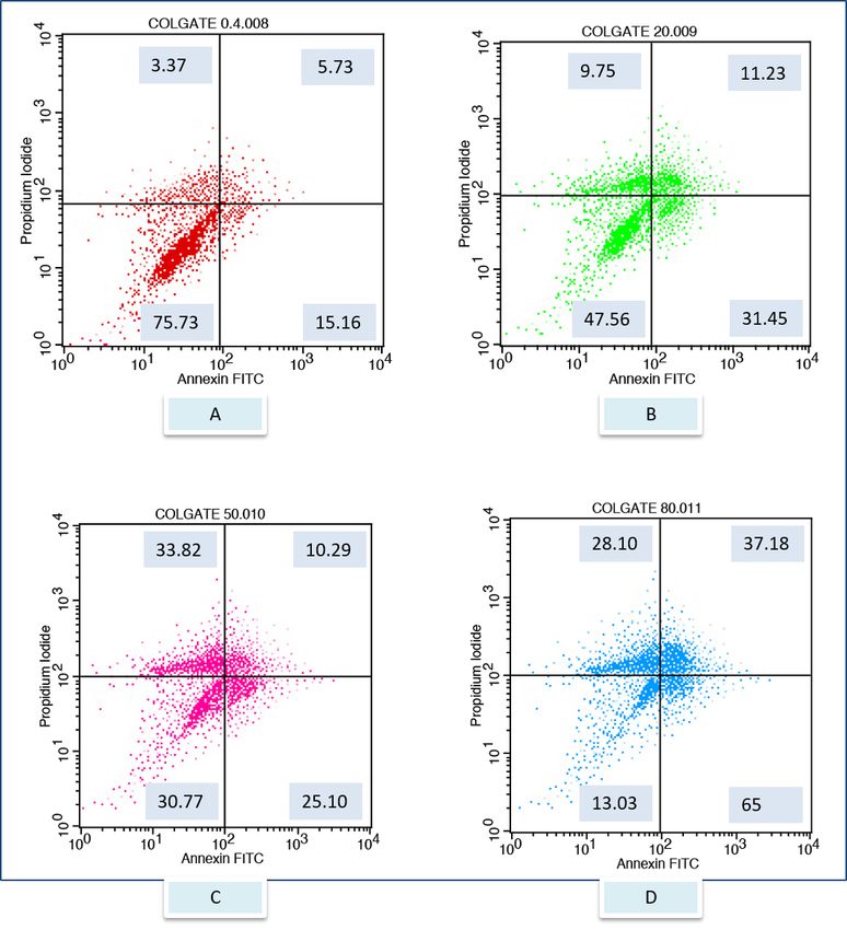
Abstract
Background: This study aimed to evaluate possible cytotoxic effects to gingival epithelial cells exposed
to children toothpastes containing different detergent.

Methods: Tissues required fort he isolation of human gingival epithelial cells were obtained by biopsy
during the extraction of the impacted third molar tooth. Toothpaste solutions of different concentrations
were prepared from five different children’s toothpastes with different detergent contents. Isolated
gingival epithelial cells were stimulated with experimental groups consisting of toothpaste solutions
(Colgate, Sensodyne, Splat, Nenedent, Perlodent) at different concentrations and a control group
consissting of complete Dulbocco’s modified eagle medium. After the experiments, cell viability was
evaluated using flow cytometry. Data analysis were done using One Way ANOVA test and Tukey post-hoc
test.

Results: In all experimental groups, there was a decrease in live cell rates and an increase in dead cell
rates due to increased concentration. The statistically highest live cell ratios were detected in Splat’s
toothpaste solutions after the control group and the group with the lowest viability values was
determined in Colgate group (p
Detergents are substances that reduce surface tension known as surfactants. They have two groups,
hydrophilic and hydrophobic [10,11]. While the long hydrocarbon chain forms the water-repellent
(hydrophobic) part of the molecule, it also provides the molecule with surface active properties. The polar
group forms the water-loving (hydrophilic) part of the molecule and enables it to dissolve in water. The
combination of these polar and apolar groups is defined as the amphiphilic structure. Thanks to the
amphiphilic structure, surfactants can be dissolved in both polar and apolar solvents. While detergents
adhere to water molecules with their polar parts due to these chemical properties, they ensure the removal
of dirt from the environment by holding on to the dirt with their apolar parts [10,11,12,13].

Detergents are classified as anionic, cationic, amphoteric and nonionic detergents according to the ionic
charge of the hydrophilic group they contain. Anionic and amphoteric detergents are frequently used in
toothpaste. Sodium lauryl sulfate, sodium methy cocoyl taurate (adinol), sodium streate (sodium
octadecanoate), sodium lauryl sarcosinate, sodium C12-14 olefin sulfonate, sodium C14-16 olefin
sulfonate from anionic detergents and cocaamidopropyl betaine among amphoteric detergents are
surfactants used frequently in toothpastes. In addition to their foaming and cleansing properties, they are
routinely added to toothpastes due to their antibacterial and plaque inhibition properties [14,15,16,17].

Sodium lauryl sulfate is a detergent that is often used in toothpastes with a ratio of 0.5% to 2%. SLS
prevents the growth of some microorganisms by adsorption to the cell wall, penetration through the cell
wall, interaction with the cell membrane, lipids and proteins, leakage of intracellular components with an
increase in cell permeability and lysis in the cell [18,19]. It has been reported in studies that SLS increases
plaque inhibition, decreases S. Mutans penetration, decreases lactate production, glucosyltransferase
activity and the amount of extracellular polysaccharide created by S. mutans [20,21,22]. Despite these
positive features, some toxic effects of SLS have also been reported. Oral epithelial destruction,
ulcerations and inflammations caused by SLS have been observed in clinical studies. It has been reported
that SLS in the toothpaste denatures the glycoproteins of the mucin layer, causing the barrier function of
the oral mucosa to deteriorate, and the gingiva and buccal mucosa to be more sensitive to irritants such
as exogenous antigens. It has also been stated that SLS may be responsible for a decrease in the
keratinization level of the human oral epithelium. Sodium lauryl sulfate has also been reported to cause
irritation of the oral mucosa in patients with dry mouth and the use of SLS is also associated with
recurrent aphthous ulcers [23, 24,25,26]. Although SLS is the most commonly used surfactant among
toothpastes, surfactants with less side effects such as betaines are also used in toothpastes.
Cocoamidopropyl betaine, an amphoteric detergent, has been reported to have less mucosal irritation and
foaming effect than SLS, and it is more biocompatible [27,28]. The aim of this study was to investigate
the effects of different detergent-containing children's toothpastes on the viability of human primary gum
epithelial cells.

Materials- Methods
The study was approved by the ethics committee of Istanbul University, Faculty of Dentistry (170/2017)
according to Helsinki Declaration guidelines.
                                                   Page 3/24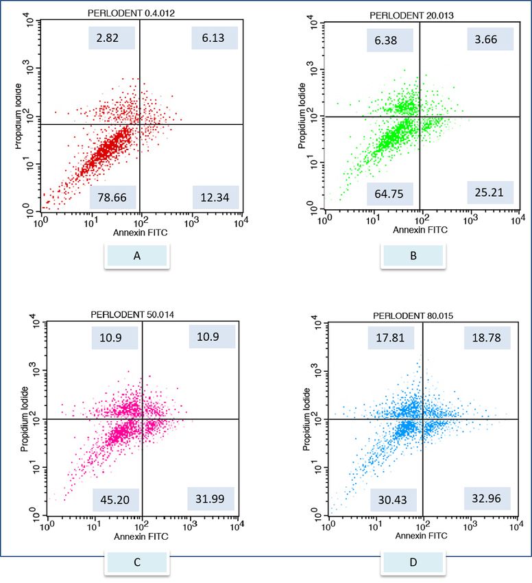
Isolation and charactarization of gingival epithelial cells
(GECs)
5 fully impacted human third molars, which were removed from systemically healthy patients (aged 18-
25 years) were used for tissue biopsy. Gingival tisues surrounding the tooth sockets were collected
immediately after tooth extraction. For the isolation of gingival epithelial cells, the gum tissue was
incubated at 4°C in 0.4% dispase overnight. The epithelium strips were then mechanically separated and
trypsinized in 0.05% trypsin/0.53 mM EDTA (Gibco, Grand Island, NY, USA) at 37°C for 10-15 min. After
strong pipetting, the cell suspension was centrifuged at 700 g for 5 min and the cell pellet was
resusspended in keratinocyte growth medium (Dermalife Basal Medium; Lifeline, Walkersville, MD, USA).
The cells were transferered to T-25 cm2 flask and were placed in the incubator which provided 5%
CO2 environment at 37°C. The keratinocyte growth medium was changed every 2 days and the
proliferation and spreading of the cells on the flask was monitored at regular intervals by invert
miscroscope (EVOS-AMG, Thermo Fisher Scientific, Waltham, MA, USA).

Cells were fixed on the slide using 95%, 70% and 50% alcohol, respectively, at room temperature. Then, the
fixation process was completed by dipping the slide into distilled water. Staining was performed with
hematoxylin (Sigma-Aldrich, St. Louis, MO, USA) for 8 min. After staining, it was washed with distilled
water and the second staining process was started with Eosin (Sigma-Aldrich). After staining with eosin
for 90 seconds, the cells were dehydrated with 95% alcohol and then dipped in xylol 20 times. Microscope
slides were fixed using Permount (Fischer Scientific, Pittsburgh, PA, USA) and epithelial cells were
analyzed by Binocular Research Microscope (Olympus BH2-RFCA) for characterization.

Preparation of toothpaste solutions
The toothpastes used in this study were Colgate6+, Sensodyne Pronamel 6+, Nenedent (4-9), Perlodent
Junior 6+, Splat Juicy. The different detergent contents and other properties of these toothpastes can be
seen in Table 1. Toothpaste solutions of 80%, 50%, 20% and 0.4% concentrations of these toothpastes
used in the study were prepared by the method in our previous study and homogenized extraction liquids
were obtained from toothpastes for cell viability experiments [29] (Birant et al. 2021).

Evaluation of cell viability by flow cytometry
Gingival epithelial cells (5x10⁵ cells) were plated into 48-well plates separately to perform viability
experiments in each concentration of toothpaste solution. The viability experiments were carried out
following the method used in our previous study (Birant et al. 2021). Gingival epithelial cells were
exposed to toothpaste solutions for 2 minutes, , washed with DPBS (Dulbecco’s phosphate buffered
saline) (Gibco, Grand Island, NY, USA) and suspended in serum-free medium. 4 µl of Annexin V (BD
Biosciences, CA, USA) was added to the tubes and the tubes were kept in a dark environment for 10

                                                   Page 4/24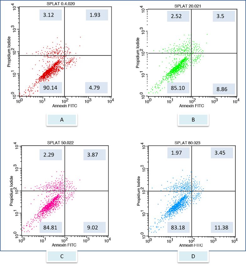
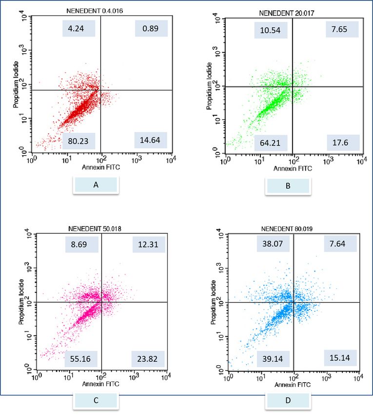
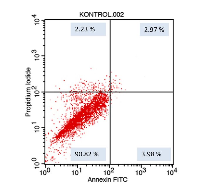
minutes. 200 µl of binding buffer was added and centrifuged at 1500 rpm for 5 min. Tubes were vortexed
by adding 200 µl binding buffer. Then, 10 µl propidium iodide was added to the tubes to read the rates of
viable, necrosis, early and late apoptotic cells in cells exposed to toothpaste solutions. The experiments
with flow cytometry were repeated 5 times, and the average of the results obtained was calculated to
determine the rates of viable, early apototic, late apoptotic and necrotic cells [29]. (Birant et al. 2021).

Statistical Analysis
The obtained datas were analyzed using the IBM SPSS V23 statistical program. While One Way ANOVA
Test eas used for multiple comparisons, Tukey Post-Hoc Test was used for pairwise comparisons.

Results
Isolation and Characterization of Cells
It was observed that the isolated gingival epithelial cells had a cylindrical and cubic morphology by
following their proliferation and reached a confluent structure from the 0th to the 3rd passage (Figure 1).
The microscope image obtained as a result of staining with hematoxylin and eosin for the
characterization of isolated gingival epithelial cells showed that the cells exhibited a cubic morphology
(Figure 2).

Cell viability in cells cultured exposured to the children’s
toothpaste containing the different detergent content
After exposure to the different toothpaste solutions at different toothpaste concentrations, viable and
dead cell ratios were determined graphically according to Annexin-V / PI positive and negativity. Annexin
V (-) and PI (-) live, Annexin V (+) and PI (-) early apoptotic cell, Annexin V (+) and PI (+) late apoptotic cell,
Annexin V (-) and PI (+) considered as a necrotic cell. The flow cytometry graphs of the control group
(CDMEM) in Figure 3, the Splat group in Figure 4, and the Sensodyne group in Figure 5, the Nenedent
group in Figure 6, the Perlodent group in Figure 7 and the Colgate group in Figure 8 show the average
viable, early apototic, late apoptotic and necrotic cell ratios.

The percentages of the cell viability on GECs were represented in Table 2. The lower percentage of viable
cells was observed in Colgate group containing SLS (p
The percentages of dead cell rates on GECs is represented in Table 3,4,5. In the evaluation of early
apoptotic cell rates, a statistically significant difference was found in the comparisons between the
groups at all concentrations except 0.4% (p0.05). The highest early apoptotic cells rates at 20%
concentration was found in the Colgate and Nenedent groups. Early apoptosis rates in the Colgate group
at 50% concentration were found to be statistically significantly higher than all groups (p
constitute the starting point of such studies in biocompatibility and cytotoxicity studies. In this study, in
vitro cell culture tests were preferred to determine the effects of toothpastes on cells.

The cell type used in cell culture studies should be selected in relation to the area of use of the material
whose cytotoxic effects are investigated. Primary cell cultures or continuous cell lines are used in studies
as a biological system in biocompatibility tests. It is stated that continuous cell lines such as L929, 3T3,
HSC-2, MRC-5 can be used in the cytotoxicity assessment tests of materials used in dentistry, since they
can be obtained more easily than primary cell cultures and have rapid reproduction potential. However,
since primary cell cultures are more sensitive than continuous cell lines, they are biological systems that
best reflect the original physiological state, despite the difficulties that arise during the production phase
and the long time to produce [33,35,36,37,38,39]. For this reason, it was preferred to create a primary cell
culture in this study, considering the creation of experimental conditions closer to in vivo
conditions. Gingival epithelial cells were used as a biological system in this study, since the majority of
oral tissues that toothpastes come into contact with during tooth brushing are gum tissues.

In studies for the characterization of gingival epithelial cells, the method of determining epithelial cells
specific CK13 and Vimentin genes by PCR, analysis of phenotypic properties of cells by transmission
electron microscopy, determination of a specific epithelial marker cytokeratin by immunofluorescence
method, staining of cells with Papanicolau staining method and analysis under light microscopy.
methods were used [40]. In this study, the cells, which are easier and quicker to apply than other methods
and are also more cost-effective, were stained with hematoxylin and eosin dyes after fixation with alcohol
on the slide, and the cells were analyzed under light microscopy, and the presence of epithelial cells was
determined.

Many in vitro tests such as MTT, trypan blue exclusion test, micronucleus are used to determine cell
viability. [27,28,41,42,43,44,45,46]. Flow cytometry analysis is frequently recommended in terms of
providing more reliable, faster and more sensitive results than other methods in evaluating cell viability
and cytotoxicity [47]. In addition to determining cell viability, information about different properties of
cells such as immunophenotypic properties, enzyme activities, and specific markers of the cell can be
obtained with this method [48]. In addition, the separation of apoptotic and necrotic cells with this
method is important in terms of different biological responses of these two types of death [49]. In this
study, since gingival cells are labeled with Annexin V and propidium iodide dyes, since they give faster,
more sensitive and reliable results compared to alternative methods used in cell culture studies, it was
ensured that live, early apoptotic, late apoptotic and necrotic cells were determined by flow cytometry
analysis.

In the literature, changes caused by SLS, which is frequently used in toothpaste, on the oral mucosa have
been reported. In addition, in a few studies examining the effects of SLS on cells, it has been stated that
they have a negative effect on cell viability [27,28,50,51,52,53,54,55]. In this study, SLS, sodium lauryl
sarcosinate, sodium C14-16 olefin sulfonate, CAPB containing toothpastes which are reported to be more
biocompatible than SLS, toothpaste without detergent and CDMEM were selected as experimental

                                                   Page 7/24
groups. While determining the concentrations of toothpaste solutions in cell viability experiments, similar
studies have been examined and optimized as 0.4%, 20%, 50% and 80%. In addition, in this study, the
stimulation time of toothpaste solutions with cells was determined as 2 minutes, since the brushing time
was 2 minutes under normal conditions [27,28,41].
When the studies on detergents are examined; Herlofson et al. found a positive relationship between oral
desquamation and SLS in their study [56]. Melsen et al examined the effect of SLS on
monoflurophosphate, and it was stated that SLS reduced the amount of fluoride taken up by the enamel
[57]. Rantanen et al reported that toothpastes containing SLS have an irritating effect on the mucosa
[58]. Shim et al. Investigated the effect of SLS on recurrent aphthous stomatitis and showed that SLS
significantly increased the incidence of ulcers, the duration of ulcers in the mouth, and the pain score [54].
In this study, when the viability rates of different detergent-containing children's toothpaste solutions
onhuman gingival epithelial cells were evaluated, it was seen that the lowest proportion of viable cells
was in toothpaste solutions containing SLS. After the control group, the highest vitality values were
detected in toothpaste without detergent content, followed by toothpaste containing CAPB. The effects of
this study on cell viability Cvikl et al.'s findings in studies examining the effects of adult toothpastes and
children's toothpaste on cells [27,28]. Moore et al. also found that cell viability rates in SLS and betaine
containing toothpastes were lower than the control group. These findings are also similar to the findings
in our study.
In the literature, the effects of toothpastes on cells have been examined only in terms of living cell
proportions. In this study, early apoptotic, late apoptotic and necrotic cell ratios were evaluated as well as
the live cell ratios. In the comparisons between the groups, the Colgate group generally shows the highest
value in terms of early apoptotic, late apoptotic and necrotic cell ratios, while Splat and the control group
generally have similar values in terms of cell death type rates. Considering that SLS increases cellular
permeability by causing denaturation of cellular proteins in this study, we think that the opening of the
pores between cells may cause the release of apoptosis-inducing proteins into the cytosol and ultimately
stimulate apoptosis / necrosis mechanisms. It has been reported that stimulation of apoptosis and
necrosis mechanisms in gingival epithelial cells may prevent periodontal wound healing and prolong the
healing period [59,60,61,62]. In this study, it is thought that the increase in the ratio of apoptotic and
necrotic cells of SLS-containing toothpaste may delay the healing time of periodontal diseases and oral
aphthous ulcers and adversely affect wound healing.
Abbreviations
GEC: gingival epithelial cells; SLS: sodium lauryl sulfate; CDMEM: complete dulbecco’s modified eagles
medium; CABP: cocoamidopropyl betaine; DPBS: dulbecco’s phosphate buffered saline.
Page 8/24Declarations
Authors’ Contributions
Designing the study by Figen Seymen, Sinem Birant; generating the data by Sinem Birant; analyses the
data by Yazgul Duran, Tunc Akkoc; writing the paper by Sinem Birant; approved the final version of this
paper by Sinem Birant, Yazgul Duran, Tunc Akkoc, Figen Seymen.
Funding
This study was supported by the Research Fund of Istanbul University, project no: 26391.
Data Availability of data and materials
Data and materials are available and transparent.
Declarations
Ethics approval:
The study was approved by the ethics committee of Istanbul University, Faculty of Dentistry (170/2017)
following Helsinki Declaration guidelines.
Consent to participate:
Written consents were collected from all the participants before enrollment.
Consent for publication:
All contributing authors agree to the publication of this article. Competing interests The authors declare
no competing interests.
Competing interest:
The authors do not have any conflict of interest.
Page 9/24References
1. Rosan B, Lamont RJ. Dental plaque formation. Microb and Infect. 2000;2:1599–607.
2. Takeshita T, Yasui M, Shibata Y, Furuta M, Saeki Y, Eshima N, et al. Dental plaque development on a
hydroxyapatite disk in young adults observed by using a barcoded pyrosequencing approach. Sci
Report. 2015;5:1–9.
3. Walsiluk A. Fluoride compounds in dental caries prophylaxis in children and adolescents- Review of
polish literature. Przegl Epidemiol. 2017;71:603–11.
4. Claydon NC. Current concepts in toothbrushing and interdental cleaning. Periodontol 2000.
2008;48:10-22.
5. Trubey RJ, Moore SC, Chestnutt IG. Parents reasons for brushing or not brushing their child’s teeth: a
qualitative study. Int J Paediatr Dent. 2014;24(2):104–12.
6. Deery C, Heanue M, Deacon S, Robinson PG, Walmsley AD, Worthington H, et al. The effectiveness of
manual versus powered toothbrushes for dental health: A systematic review. J Dent. 2004;32:197–
211.
7. Wilder RS, Bray KS. Improving periodontal outcomes: merging clinical and behavioral science.
Periodontol 2000. 2016;71:65-81.
8. Welbury R., Duggal M., HoseyT. M. Paediatric dentistry. 3rd ed. Oxford University Press; 2005.
9. Ozkocak CBB, Karaarslan SE, Aytac F. Saliva proteins and their effects on caries. Turkey Dental
Clinics J Sci. 2017;23:56–64.
10. Chambers HF. Miscellaneous antimicrobial agents; disinfectants, antiseptic, sterilants. In: Katzung
BG, editor. Basic&Clinical Pharmacology. 10th ed. Connecticut, Appleton&Lange; 2007. pp. 803–11.
11. Ozyurt M. Aldehyde, peroxygen and peracetic acid and other disinfectants that do not contain
chlorine donating agents and are recommended as instrument disinfectants, their general use and
antimicrobial effectiveness. In: Gunaydın M, Sanic, A, Gurler B, editors. 4th National Sterilization
Disinfection Congress, Congress Book. Scientific Medicine Publishing House, Ankara; 2005. pp. 180–
199.
12. Ananthapadmanabhan KP, Moore DJ, Subramanyan K, Misra M, Meyer F. Cleansing without
compromise: the impact of cleansers on the skin barrier and the technology of mild cleansing.
Dermatol Ther. 2004;17:16–25.
13. Walters KA, Bialik W, Brain KR. The effects of surfactants on penetration across the skin. Int J
Cosmetics Sci. 1993;15:260–70.
14. Shah SK, Niraula TP, Bhattarai A, Chatterjee SK. A comparative study of cationic and anionic
surfactants on the micellar behavior through different composition of methanol-water mixed solvent.
Conductometric Method Bibechan. 2012;8:37–45.
15. Forward GC, James AH, Barnett P, Jackson RJ. Gum health product formulations: what is in them
and why? Periodontol. 2000;15:32–9.
Page 10/2416. Petersen FC, Assev S, Scheie AA. Combined effects of NaF and SLS on acid and polysaccharide
formation of biofilm and planktonic cells. Arch Oral Biol. 2006;51:665–71.
17. Buma R, Maeda T, Kamei M, Kourai H. Pathogenic bacteria carried by companion animals and their
susceptibility to antibacterial agents. Biocontrol Sci. 2006;11: 1–9.
18. Law V, Seow WK. A longitudinal controlled study of factors associated with mutans streptococci
infection and caries lesion initiation in children 21 to 72 months old. Pediatr Dent. 2006;28:58–65.
19. Nordstrom A, Mystikos C, Ramberg P, Birkhed D. Effect on denovo plaque formation of rinsing with
toothpaste slurries and water solutions with a high fluoride concentration (5,000 ppm). Eur J Oral
Sci. 2009;117:563–7.
20. Moran J, Addy M, Newcombe R. The antibacterial effect of toothpastes on the salivary flora. J Clin
Periodontol. 1988;15:193–9.
21. Moran J, Addy M, Newcombe R. Comparison of the effect of toothpastes containing enzymes or
antimicrobial compounds with a conventional fluoride toothpaste on the development of plaque and
gingivitis. J Clin Periodontol. 1989;16:295–9.
22. Evans A, Leishman SJ, Walsh LJ. Inhibitory effects of children’s toothpastes on Streptococcus
mutans, Streptococcus sanguinis and Lactobacillus acidophilus. Eur Arch Paediatr Dent.
2015;16:219–26.
23. Macdonald JM, Tobin CA, Burkemper NM, Hurley MY. Oral Leukoedema with mucosal desquamation
caused by toothpaste containing sodium lauryl sulfate. Case Let. 2015;97:4–5.
24. Neppelberg E, Costea DE, Vintermyr OK, Johannessen AC. Dual effects of sodium lauryl sulphate on
human oral epithelial structure. Experiment Dermatol. 2007;16:574–9.
25. Siegel IA, Gordon HP. Surfactant-induced alterations of permeability of rabbit oral mucosa in vitro.
Exp Mol Pathol. 1986;44:132–7.
26. Ahlfors EE, Lyberg T. Contact sensitivity reactions in the oral mucosa. Acta Odontol Scand.
2001;59:248–54.
27. Cvikl B, Lussi A, Moritz A, Gruber R. The in vitro impact of toothpaste extracts on cell viability. Eur
Oral Sci. 2015;123:179–85.
28. Cvikl B, Lussi A, Moritz A, Gruber R. Dentifrices for children differentially affect cell viability in vitro.
Clin Oral Invest. 2017;21:453–61.
29. Birant S, Duran Y, Gokalp M, Akkoc T, Seymen F. Effects of different-containing children’s toothpastes
on the viability, osteogenic and chondrogenic differentiation of human dental periodontal ligament
stem cells and gingival stem cells in vitro. Tissue and Cell 2021,72:1–12.
30. Lawrence LM., Farquharson A, Brown RS, Vatanka HO. Oral tissue irritants in toothpaste: a case
report. J Clin Pediatr Dent. 2013;38:75–8.
31. Herlofson BB, Barkvoll P. Oral mucosal desquamation caused by two toothpaste detergents in an
experimental model. Eur J Oral Sci. 1996;104:21–6.
Page 11/2432. Skaare AB, Rolla G, Barkvoll P. The influence of triclosan, zinc or propylene glycol on oral mucosa
exposed to sodium lauryl sulphate. Eur J Oral Sci. 1997;105:527–33.
33. Craig RG, Powers JM, Sakaguchı RL. Craig’s Restorative Dental Materials.12th ed. St. Louis: Mosby;
2006.
34. Schmalz G. Use of cell cultures for toxicity testing of dental materials- advantages and limitations. J
Dent. 1994;22:6–11.
35. Arenholt-Bindslev D, Bleeg H. Characterization of two types of human oral fibroblast with a potential
application to cellular toxicity studies; tooth pulp fibroblasts and buccal mucosa fibroblasts. Int
Endod J. 1990;23:84–91.
36. Illeperuma RP, Park YJ, Kim JM, Bae JY, Che ZM, Son HK, et al.. Immortalized gingival fibroblasts as a
cytotoxicity test model for dental materials. J Mater Sci Mater Med. 2012;23:753–62.
37. International Organization for Standardization. Dentistry- Biological evaluation of medical devices.
Tests for in vitro cytotoxicity. ISO 10993-5. 2009. https://www.iso.org/standard/36406.html.
Accessed 13 Feb.2019
38. Schmalz G. Concepts in biocompatibility testing of dental restorative materials. Clin Oral
Invest.1997;1:154–62.
39. Tuncer S, Demirci M. Biocompatibility Evaluations of Dental Materials. J Atatürk Univ
Dentist.2011;21:141–9.
40. Russo FB, Pignatari GC, Fernandes IR, Dias JLRM, Beltrao-Braga PCB. Epithelial cells from oral
mucosa: How to cultivate them? Cytotechnol. 2016;68:2105–14.
41. Ghapanchi J, Kamali F, Moattari A, Poorshahidi S, Shahin E, Rezazadeh F, et al. In vitro Comparison
of cytotoxic and antibacterial effects of 16 commercial toothpastes. J Int Oral Health. 2015;7:39–43.
42. Fiori J, Teti G, Gotti R, Mazzotti G, Falconi M. Cytotoxic activity of guaiazulene on gingival fibroblasts
and influence of light exposure on guaiazzulene-induced cell death. Toxico In Vitro. 2011;25:64–72.
43. Eyuboglu GB, Yesilyurt C, Erturk M. Evaluation of cytotoxicity of dentin desensitizing products. Oper
Dentist. 2015;40:503–14.
44. Kalil Bussadori S, Marcilio Santos E, Cardoso Guedes C, Jansiski Motta L, Santos Fernandes KP,
Mesquita-Ferrari RA, et al. Cytotoxicity assessment of casein phosphopeptide-amorphous calcium
phosphate (CPP-ACP) paste. ConScient Saude. 2010;9:354–9.
45. Fernandes JPS, Mello Moura ACV, Marques MM, Nicoletti MA. Cytotoxicity evaluation of Curcuma
zedoaria Roscoe fluid extract used in oral hygiene products. Acta Odontol Scand. 2012;70:610–4.
46. Olgun Erdemir E, Sengun A, Ulker M. Cytotoxicity of mouthrinses on epithelial cells by micronucleus
test. Eur J Dent. 2007;1:80–5.
47. Zhou H, Shen Y, Wang Z, Li L, Zheng Y, Hakkinen L, et al. ve ark. In Vıtro Cytotoxicity Evaluation of a
Novel Root Repair Material. J Endod. 2013;39:478–83.
48. Kanev MO, Gokalp Muranlı FD. Flow cytometry and usage areas. J SAÜ Fen Bil. 2016;20:33–8.
Page 12/2449. Coskun G, Ozgur H. Molecular Mechanism of Apoptosis and Necrosis. Arch Med Rev J.
2011;20:145–58.
50. Salzer S, Rosema NAM, Maertin ECJ, Slot DE, Timmer CJ, Dorfer CE, et al. The effectiveness of
dentifrices without and with sodium lauryl sulfate on plaque, gingivitis and gingival abrasion- a
randomized clinical trial. Clin Oral Invet. 2016;20:443–50.
51. Healy CM, Paterson M, Joyston-Bechal S, Williams DM, Thornhill MH. The effect of a sodium lauryl
sulfate-free dentifrice on patients with recurrent oral ulceration. Oral Dis. 1999;5:39–43.
52. Barkvoll P, Rolla G, Svendsen AK. Interaction between chlor- hexidine digluconate and sodium lauryl
sulfate in vivo. J Clin Periodontol. 1989;16:593–5.
53. Allen AL, Hawley CE, Cutright DE, Seibert JS. An investigation of the clinical and histologic effects of
selected dentifrices on human palatal mucosa. J Periodontol. 1975;46:102–12.
54. Shim YJ, Choi JH, Ahn HJ, Kwon JS. Effect of sodium lauryl sulfate on recurrent aphthous
stomatitis: a randomized controlled clinical trial. Oral Dis. 2012;18:655–60.
55. Moore C, Addy M, Moran J. Toothpaste detergents: a potential source of oral soft tissue damage? Int
J Dent Hyg. 2008;6:193–8.
56. Herlofson BB, Brodin P, Aars H. Increased Human Gingival Blood Flow Induced by Sodium Lauryl
Sulfate. J Clin Periodontol. 1996;123:1004–7.
57. Melsen B, Rolla G. Reduced Clinical effect of monofluorophosphate in the presence of sodium lauryl
sulphate. Caries Res, 1983;17:549–53.
58. Rantanen I, Jutila K, Nicander I, Tenovuo J, Soderling E. The effects of two sodium lauryl sulphate-
containing toothpastes with and without betaine on human oral mucosa in vivo. Swed Dent J.
2003;27:31–4.
59. Semlali A, Chakir J, Goulet JP, Chmielwski W, Rouabhia M. Whole cigarette smoke promets human
gingival epithelial cell apoptosis and inhibits cell repair processes. J Periodont Res. 2011;46: 533–
41.
60. Rouabhia M. Interactions between host and oral commensal microorganisms are key events in
health and disease status. Can J Infect Dis. 2002;13: 47–51.
61. Weindl G, Wagener J, Schaller M. Epithelial cells and innate antifungal defense. J Dent Res. 2010;89:
666–75.
62. Bahri R, Saidane-Mosbahi D, Rouabhia M. Candida famata modulates toll-like receptor, beta-
defensin, and proinflammatory cytokine expression by normal human epithelial cells. J Cell Physiol.
2010;222: 209–18.
Tables
Table 1
Composition of materials evaluated
Page 13/24Materials Composition Manufacturer
Colgate 6+ Sorbitol, aqua, hydrated silica, PEG-12, Sodium Lauryl Sulfate, Colgate
cellulose gum, sodium saccharin, sodium fluoride (1450 ppm F-), Palmolive
aroma,hydroxypropyl methylcellulose, menthol, glycerin, cinnamal, Company,
eugenol, limonene, CI 77891, CI 42090 Belgium
Nenedent Aqua, hydrated silica, glycerin, xylitol, propylene glycol, xanthan gum, Dentinox,
Kids (4-9 titanium dioxide, aroma, Sodium Lauryl Sarcosinate, disodium EDTA, Berlin,
aged) sodiummonofluorophophate (500 ppm F-), sodium chloride Germany
Perlodent Aqua, sorbitol, hydrated silica, propylene glycol, tetrapotassium Rossmann,
Junior 6+ pyrophosphate, xanthan gum, Sodium C14-16 Olefin Germany
Sulfonate, aroma, titanium dioxide, sodium fluoride (1450 ppm F-),
sodium saccharin, phenoxyethanol, ethylhexyl glycerin
Sensodyne Aqua, sorbitol, hydrated silica, glycerin, PEG-6, Cocamidopropyl Glaxo Smith
Pronamel Betaine, xanthan gum, aroma, sodium fluoride(1450 ppm F-), , sodium Kline, ABD
6+ saccharin, sucralose, titanium dioxide, sodium hydroxide, limonene
Splat Aqua*, dicalcium phosphate dihydrate*,hydrogenated starch
Juicy hydrolsate*, glycerin*, hydroxyapatite, cellulose gum*, aroma, xanthan
gum*, potassium thiocyanate, lactoferrin*, lactoperoxidase*, glucose
oxidase*, glucose pentaacetate, aloe barbadensis leaf extract*,
sodium mthylparaben, hydrolyzed casein*, glycyrrhiza glabra root
extract* (*natural origins)
Complete %10 oranında FBS (Fötal sığır serumu), %1 penisilin/ streptomycin Gibco, Grand
DMEM ilave edilmiş DMEM (Dulbecco’s Modified Eagles Medium) Island, USA
(CDMEM)
Table 2
Comparison of viability of gingival epithelial cells between groups
Page 14/24Concentration
0,40% 20% 50% 80%
Colgate 6+ 75.74 ± 3.18a 47.56 ± 3.49a 30.77 ± 4.26a 13.04 ± 2.98a
Splat Juicy 90.14 ± 0.95d 85.1 ± 1.77b 84.82 ± 1.6b 83.19 ± 1.88b
Sensodyne Pronamel 6+ 84.66 ± 1.58c 78.8 ± 1.16c 67.47 ± 1.68c 57.63 ± 0.83c
Nenedent Kids 80.23 ± 0.93b 64.21 ± 0.91d 55.17 ± 1.2d 39.15 ± 0.91d
Perlodent Junior 6+ 78.66 ± 1.84ab 64.75 ± 0.91d 45.21 ± 1.81e 30.43 ± 4.05e
CDMEM 90.82 ± 1.04d 9.,82 ± 1.04e 90.82 ± 1.04f 90.82 ± 1.04f
p 0.000* 0.000* 0.000* 0.000*
a-b-c-d-e-f: There is no difference between the groups with the same
One Way ANOVA Test ; Tukey post–hoc Test
* Significant p value at 0.05 level
Table 3
Comparison of early apoptotic cell rates ingingival epithelial cells between groups
Page 15/24Concentration
0,40% 20% 50% 80%
Colgate 6+ 3.38 ± 2.01 9.75 ± 3.55cd 33.83 ± 2.81d 28.11 ± 3.07b
Splat Juicy 3.12 ± 1.48 2.52 ± 1.14a 2.29 ± 0.78a 1.97 ± 0.4a
Sensodyne Pronamel 6+ 1.85 ± 0.53 4.52 ± 0.67ab 12.09 ± 1.11c 9.73 ± 0.57c
Nenedent Kids 4.24 ± 0.98 10.55 ± 1.79d 8.69 ± 1.36b 38.08 ± 1.27d
Perlodent Junior 6+ 2.83 ± 0.67 6.39 ± 1.14bc 10.9 ± 0.99bc 17.82 ± 3.44e
CDMEM 2.24 ± 1.26 2.24 ± 1.26a 2.24 ± 1.26a 2.24 ± 1.26a
p 0.078 0.000* 0.000* 0.000*
a-b-c-d-e-f: There is no difference between the groups with the same
One Way ANOVA Test ; Tukey post–hoc Test
* Significant p value at 0.05 level
Table 4
Comparison of late apoptotic cell rates ingingival epithelial cells between groups
Page 16/24Concentration
0,40% 20% 50% 80%
Colgate 6+ 5.73 ± 1.66c 11.23 ± 3.2c 10.29 ± 2.55b 37.19 ± 2.68e
Splat Juicy 1.93 ± 0.32ab 3.51 ± 0.65a 3.87 ± 0.96a 3.46 ± 0.97ab
Sensodyne Pronamel 6+ 0.70 ± 0.37a 1.5 ± 0.37a 5.25 ± 0.64a 11.96 ± 1.02c
Nenedent Kids 0.89 ± 0.26a 7.65 ± 1b 12.31 ± 0.53b 7.65 ± 0.6bc
Perlodent Junior 6+ 6.13 ± 1.01c 3.66 ± 0.64a 10.9 ± 1.19b 18.78 ± 4.84d
CDMEM 2.97 ± 0.86b 2.97 ± 0.86a 2.97 ± 0.86a 2.97 ± 0.86a
p 0.000* 0.000* 0.000* 0.000*
a-b-c-d-e-f: There is no difference between the groups with the same
One Way ANOVA Test ; Tukey post–hoc Test
* Significant p value at 0.05 level
Table 5
Comparison of necrotic cell rates ingingival epithelial cells between groups
Concentration
0,40% 20% 50% 80%
Colgate 6+ 15.16 ± 2.4c 31.46 ± 4e 25.11 ± 2.91d 21.67 ± 2.84c
Splat Juicy 4.79 ± 0.54a 8.86 ± 0.87b 9.03 ± 1.03b 11.39 ± 1.53b
Sensodyne Pronamel 6+ 12.79 ± 0.96bc 15.18 ± 1.09c 15.19 ± 1.28c 20.69 ± 0.91c
Nenedent Kids 14.65 ± 0.61bc 17.6 ± 1.1c 23.83 ± 0.7d 15.15 ± 0.92b
Perlodent Junior 6+ 12.35 ± 0.68b 25.21 ± 1.3d 31.99 ± 1.2e 32.97 ± 3.99d
CDMEM 3.98 ± 1.09a 3.98 ± 1.09a 3.98 ± 1.09a 3.98 ± 1.09a
p 0.000* 0.000* 0.000* 0.000*
a-b-c-d-e-f: There is no difference between the groups with the same
One Way ANOVA Test ; Tukey post–hoc Test
* Significant p value at 0.05 level
Page 17/24Figures
Figure 1
Light microscope images of GECs in P0, P1, P2 and P3 passages
Figure 2
Determination of characterization of GECs by Binocular Research Microscope (Olympus BH2-RFCA) 400X
Gingival epithelial cells, Binocular Research Microscope (Olympus BH2-RFCA) 400X
Page 18/24Figure 3
Flow cytometry graph related to the effect of CDMEM control group on gingival epithelial cells (x: Annexin
V FITC, y: PIPE)
Page 19/24Figure 4
Flow cytometry graph related to the effect of Splat toothpaste solutions on gum epithelial cells (x:
Annexin V FITC, y: PIPE) A) Splat 0.4% B) Splat 20% C) Splat 50% D) Splat 80%
Page 20/24Figure 5
Flow cytometry graph related to the effect of Sensodyne toothpaste solutions on gum epithelial cells (x:
Annexin V FITC, y: PIPE) A) Sensodyne 0.4% B) Sensodyne 20% C) Sensodyne 50% D) Sensodyne 80%
Page 21/24Figure 6
Flow cytometry graph related to the effect of nenedent toothpaste solutions on gum epithelial cells (x:
Annexin V FITC, y: PIPE) A) Nenedent 0.4% B) Nenedent 20% C) Nenedent 50% D) Nenedent 80%
Page 22/24Figure 7
Flow cytometry graph related to the effect of perlodent toothpaste solutions on gum epithelial cells (x:
Annexin V FITC, y: PIPE) A) Perlodent 0.4% B) Perlodent 20% C) Perlodent 50% D) Perlodent 80%
Page 23/24Figure 8
Flow cytometry graph related to the effect of Colgate toothpaste solutions on gum epithelial cells (x:
Annexin V FITC, y: PIPE) A) Colgate 0.4% B) Colgate 20% C) Colgate 50% D) Colgate 80%
Page 24/24You can also read