Different MDSC Activity of G-CSF/Dexamethasone Mobilized Neutrophils: Benefits to the Patient?
←
→
Page content transcription
If your browser does not render page correctly, please read the page content below
ORIGINAL RESEARCH
                                                                                                                                           published: 21 July 2020
                                                                                                                                    doi: 10.3389/fonc.2020.01110

                                              Different MDSC Activity of
                                              G-CSF/Dexamethasone Mobilized
                                              Neutrophils: Benefits to the Patient?
                                              Cathelijn E. M. Aarts 1*, Ida H. Hiemstra 1 , Charita Furumaya 1 , Robin van Bruggen 1 and
                                              Taco W. Kuijpers 1,2
                                              1
                                               Department of Blood Cell Research, Sanquin Research, Amsterdam University Medical Center (AUMC), University of
                                              Amsterdam, Amsterdam, Netherlands, 2 Department of Pediatric Immunology, Rheumatology & Infectious Diseases, Emma
                                              Children’s Hospital, AUMC, University of Amsterdam, Amsterdam, Netherlands

                                              Human neutrophils exert a well-known role as efficient effector cells to kill pathogenic
                                              micro-organisms. Apart from their role in innate immunity, neutrophils also have the
                                              capacity to suppress T cell-mediated immune responses as so-called granulocyte-
                                              myeloid-derived suppressor cells (g-MDSCs), impacting the clinical outcome of various
                                              disease settings such as cancer. Patients undergoing chemotherapy because of
                                              an underlying malignancy can develop prolonged bone marrow suppression and
                                              are prone to serious infections because of severe neutropenia. Concentrates of
                                              granulocytes for transfusion (GTX) constitute a therapeutic tool and rescue treatment
                                              to fight off these serious bacterial and fungal infections when antimicrobial therapy is
                            Edited by:        ineffective. GTX neutrophils are mobilized by overnight G-CSF and/or Dexamethasone
                Panagiota S. Filippou,
  Teesside University, United Kingdom
                                              stimulation of healthy donors. Although the phenotype of these mobilized neutrophils
                        Reviewed by:
                                              differs from the circulating neutrophils under normal conditions, their anti-microbial
                      Nelita Du Plessis,      function is still intact. In contrast to the unaltered antimicrobial effector functions,
 Stellenbosch University, South Africa        G-CSF/Dexamethasone-mobilized neutrophils were found to lack suppression of the
                         Rolf Kiessling,
     Karolinska Institutet (KI), Sweden       T cell proliferation, whereas G-CSF-mobilized or Dexamethasone-mobilized neutrophils
                  *Correspondence:            could still suppress the T cell proliferation upon cell activation equally well as control
                  Cathelijn E. M. Aarts       neutrophils. Although the mechanism of how G-CSF/Dex mobilization may silence
                   c.aarts@sanquin.nl
                                              the g-MDSC activity of neutrophils without downregulating the antimicrobial activity
                  Specialty section:
                                              is presently unclear, their combined use in patients in the treatment of underlying
        This article was submitted to         malignancies may be beneficial—irrespective of the number of circulating neutrophils.
     Molecular and Cellular Oncology,
                                              These findings also indicate that MDSC activity does not fully overlap with the
               a section of the journal
                Frontiers in Oncology         antimicrobial activity of human neutrophils and offers the opportunity to elucidate the
           Received: 15 March 2020            feature(s) unique to their T-cell suppressive activity.
            Accepted: 03 June 2020
                                              Keywords: neutrophils, MDSC activity, granulocyte transfusions, GTXs, mobilized-neutrophils
            Published: 21 July 2020
                             Citation:
Aarts CEM, Hiemstra IH, Furumaya C,
      van Bruggen R and Kuijpers TW
                                              INTRODUCTION
     (2020) Different MDSC Activity of
    G-CSF/Dexamethasone Mobilized
                                              Patients who undergo chemotherapy are prone to develop neutropenia and are thereby susceptible
 Neutrophils: Benefits to the Patient?        to serious bacterial and fungal infections (1). In addition to antimicrobial therapy, granulocyte
                Front. Oncol. 10:1110.        transfusions (GTX) can be a therapeutic option to improve the clinical outcome in case of a
       doi: 10.3389/fonc.2020.01110           deteriorating clinical condition because of the lack of efficacy of antimicrobial agents only (2–4).

Frontiers in Oncology | www.frontiersin.org                                       1                                            July 2020 | Volume 10 | Article 1110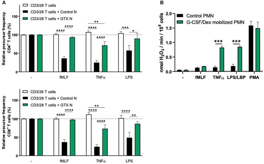
Aarts et al.                                                                                    MDSC Activity of G-CSF/Dex-Mobilized Neutrophils

In the past we and others have extensively described the                 isolation kit of Miltenyi-Biotec (Bergisch Gladbach, Germany)
combined administration of G-CSF and Dexamethasone to                    according to the manufacturer’s instructions. Neutrophils were
healthy donors in order to generate sufficient numbers of cells          obtained from the pellet fraction after erythrocyte lysis with
for these GTX products. These mobilized GTX neutrophils show             hypotonic ammonium chloride solution at 4◦ C as previously
a changed phenotype but a completely intact ability to respond,          described (14).
migrate and kill invading pathogens (5).
    Next to their role of efficient innate immunity killers
of micro-organisms, neutrophils are also recognized to be                T Cell Proliferation Assay
involved in modulation of adaptive immune responses in various           Purified T cells were labeled with CFSE (Molecular probes,
disease settings including cancer (6–9). Immature and mature             Life Technologies, Carlsbad, CA, USA) and cultured in 96-well
neutrophils were reported to have the capacity to suppress T cell-       flat bottom plates (Nunclon Delta Surface, Thermo Scientific,
mediated immune responses as so-called granulocyte-myeloid-              Waltham, MA, USA) for 4–6 days at 37◦ C in IMDM medium
derived suppressor cells (g-MDSCs), and thereby affect the               (Gibco, Life Technologies, Carlsbad, CA, USA), supplemented
clinical outcome of cancer patients. In fact, in cancer patients         with 10% (v/v) fetal calf serum (Bodinco, Alkmaar, The
the presence of increased neutrophil counts in the circulation           Netherlands), 104 U/mL penicillin, 10 ng/mL streptomycin,
is directly related with a bad prognosis (9). While the function         200 mM glutamine, and 0.00035% (v/v) β-mercaptoethanol
of g-MDSCs has been investigated in depth and in murine                  (Sigma-Aldrich, Saint Louis, MO, USA). To induce proliferation,
experimental models in particular, the characterization of human         the T cells were stimulated by anti-CD3 (clone 1XE [IgE
g-MDSC activity is still controversial. Lectin-type Oxidized             isotype] hybridoma supernatant, 1:1,000, Sanquin, Amsterdam,
LDL receptor 1 (LOX-1) has been suggested to be a marker                 The Netherlands) and anti-CD28 (clone 15E8 [IgG1 isotype] at
to discriminate g-MDSCs from circulating human mature                    5 µg/mL, Sanquin) monoclonal antibodies (moAbs; at 20,000 T
neutrophils and would therefore allow for better distinction             cells/well). Neutrophils from blood, collected from the pellet
without the use of a gradient (low-density g-MDSCs versus high-          fraction after density centrifugation, were added in a 1:3
density mature neutrophils) (10). However we have found in a             ratio (60,000 neutrophils/well), in the presence or absence
recent study that activated mature neutrophils also express LOX-         of neutrophil-activating stimuli: fMLF (1 µM, Sigma), TNFα
1 (11), questioning the fact if LOX-1 is indeed a suitable g-MDSC        (10 ng/mL, Peprotech EC, London, UK) or LPS (20 ng/mL, E. coli
marker. We have recently demonstrated that mature neutrophils            055:B5, Sigma).
(i.e., high-density) from healthy donors can exert MDSC activity             After 4–6 days, the cells were harvested from the culture
(i.e., suppress immune responses) but only upon cell activation          plates and stained with APC-labeled anti-CD4 (clone SK3, BD
(11–13), which correlates to the LOX-1 expression. Moreover, the         Biosciences, San Jose, CA, USA) and PerCPCy5.5-labeled anti-
mechanisms involved in the MDSC activity greatly overlapped              CD8 (clone SK1, Biolegend, San Diego, CA, USA) antibodies.
with the toxic antimicrobial effector functions of neutrophils,          The T cell proliferation was assessed by measuring the CFSE
being dependent on cell-cell contact (adhesion), production of           dilution of CD4+ and CD8+ T cells via flow cytometry.
reactive oxygen species (ROS) and release of their granular
content (degranulation) (11).
    In this study we investigated whether neutrophils obtained           ROS Production
upon overnight mobilization of neutrophils into the bloodstream          NADPH oxidase activity was assessed as the release of hydrogen
in healthy GTX donors may have a potentially relevant impact as          peroxide, determined by the Amplex Red method (Molecular
MDSCs in the treatment of oncology patients.                             Probes, Life Technologies, Carlsbad, CA, USA) by neutrophils
                                                                         (1x10⁶ /mL) stimulated with: fMLF (1 µM), TNFα (10 ng/mL),
MATERIALS AND METHODS                                                    LPS (20 ng/mL) + LPS-binding protein (LBP) (50 ng/mL, R&D
                                                                         Systems, Minneapolis, MN, USA) or PMA (100 ng/mL, Sigma) in
Study Approval                                                           the presence of Amplex Red (0.5 µM) and horseradish peroxidase
Heparinized peripheral blood samples were collected from                 (1 U/mL). Fluorescence was measured at 30-s intervals for 4 h
healthy granulocyte transfusion donors 1 day after combined              with the HTS7000+ plate reader (Tecan, Zurich, Switzerland).
G-CSF (600 µg, subcutaneously) and dexamethasone (8 mg,                  Maximal slope of hydrogen peroxide release was assessed over a
orally) treatment (G-CSF/Dex), or upon their preference with G-          2-min interval.
CSF or dexamethasone alone, as described previously (5). Blood
samples were collected after obtaining informed consent and in
accordance with the Declaration of Helsinki.                             Antibodies and Flow Cytometry
                                                                         The following directly conjugated antibodies were used for
Blood Cell Isolation                                                     flow cytometry analysis: PB-labeled anti-CD11b (clone ICRF44,
Neutrophils and peripheral blood mononuclear cells (PBMC)                BD Biosciences) and PECy7-labeled anti-CD16 (clone 3G8,
were isolated from whole blood by gradient centrifugation using          BD Biosciences).
isotonic Percoll (Pharmacia, Uppsala, Sweden) with a specific               Flow cytometry data were acquired using Canto II flow
density of 1.076 g/mL. T cells were isolated from the PBMC               cytometer (BD Biosciences) and analyzed using FlowJo software
fraction by magnetic-activated cell sorting with the Pan T cell          (Tree Star, USA).

Frontiers in Oncology | www.frontiersin.org                          2                                        July 2020 | Volume 10 | Article 1110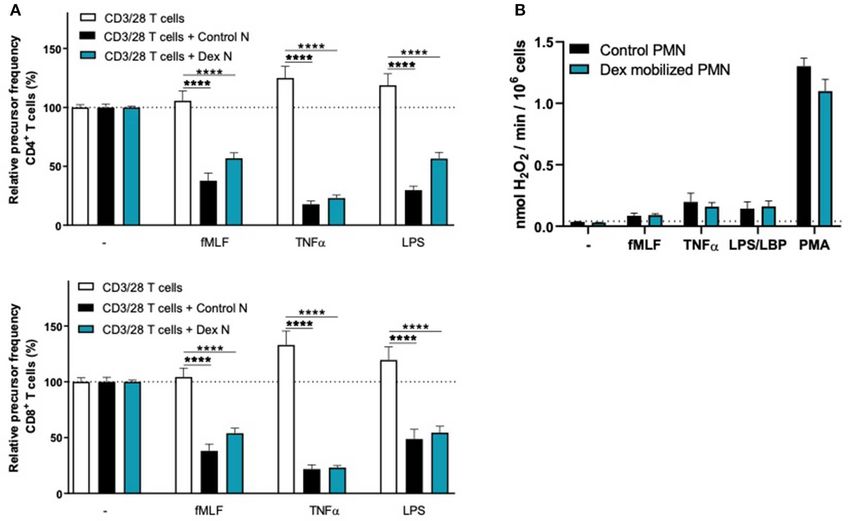
Aarts et al.                                                                                                        MDSC Activity of G-CSF/Dex-Mobilized Neutrophils

  FIGURE 1 | G-CSF/Dex-mobilized donors have an increased amount of neutrophils including immature and mature neutrophils. (A) Absolute neutrophil count of
  peripheral blood from G-CSF/Dex-treated donors before and after administration n = 5. (B) Surface marker expression of CD11b and CD16 was measured by flow
  cytometry analysis of neutrophils from blood of healthy donors (left panel), neutrophil progenitors from bone marrow (center panel) and G-CSF/Dex-mobilized
  neutrophils. The four indicated neutrophil progenitor populations are (pro)myelocytes (1, CD11bNEG CD16NEG ), metamyelocytes (2, CD11bPOS CD16NEG ), band cells (3,
  CD11bPOS CD16DIM ) and segmented cells (4, CD11bPOS CD16POS ). Shown are representative FACS analysis images (n = 3).

Statistics                                                                                To investigate the MDSC activity (i.e., suppression of immune
Statistical analysis was performed with GraphPad Prism version                        responses) of these G-CSF/Dex-mobilized neutrophils, we now
8 for Windows (GraphPad Software, San Diego, CA, USA).                                performed additional T cell proliferation assays. In our previous
Data were evaluated by one-way ANOVA or unpaired two-tailed                           study (11), where we have optimized our T cell proliferation
student’s t-test. The results are presented as the mean ± SEM.                        assay, we have studied the mechanism behind the suppressive
Data were considered significant when p < 0.05.                                       activity of activated mature neutrophils in more depth. Here we
                                                                                      found that neutrophils exert their suppressive activity in the first
                                                                                      hours/day of the cell culture, after which the suppressed T cells
RESULTS                                                                               are no longer prone to T cell stimulation. Furthermore, the most
                                                                                      optimal read-out of the T cell proliferation by CFSE dilution was
G-CSF/Dex Mobilized Neutrophils Are Not                                               between 4 and 6 days of cell culture.
Able to Suppress the T Cell Proliferation                                                 Neutrophils from G-CSF/Dex donors or from healthy controls
We received blood from healthy granulocyte transfusion                                were cultured simultaneously for 5 days in the presence
donors routinely treated with the combination of G-CSF                                of isolated CFSE-labeled T cells from an unrelated healthy
and dexamethasone to test whether the mobilization of                                 donor and were left unstimulated or activated with either
neutrophils into the bloodstream resulted in a change of                              fMLF, TNFα, or LPS. Just as previously described, T cell
MDSC activity.                                                                        proliferation was induced by the strong and uniform activation
   One day after G-CSF/Dex administration, the absolute                               by the combination of monoclonal anti-CD3 and anti-CD28
neutrophil count in the peripheral blood was ∼30 times increased                      antibodies and quantified as relative “recursor frequency”:
compared to the neutrophil count before administration                                i.e., percentage of naïve cells in the initial population that
(Figure 1A). The rapid increase in blood neutrophil numbers                           underwent one or more divisions upon anti-CD3/anti-CD28
induced by G-CSF/Dex resulted from the predominant release of                         antibodies (17). The precursor frequency was then normalized
mature (∼80%) and some immature (∼20%) neutrophils from                               for the condition of anti-CD3/CD28-stimulated T cells and non-
the bone marrow into the circulation (Figure 1B). Neutrophil                          activated neutrophils.
progenitor cells can be divided in four different developmental                           We observed that G-CSF/Dex-mobilized neutrophils were
stages, namely (pro)myelocytes, metamyelocytes, band cells and                        not able to suppress the T cell proliferation of CD4+ or
segmented neutrophils based on the expression of cell surface                         CD8+ T cells, neither under resting conditions nor upon
markers CD11b and CD16 (15, 16), which were all present                               their activation (Figure 2A). Only the neutrophils from healthy
in the G-CSF/Dex-mobilized neutrophil fraction (Figure 1B).                           controls were able to suppress T cell proliferation upon proper
Apart from the release of the reserve pool of neutrophils from                        activation. One of the main effector mechanism in which
the bone marrow, also the demargination of neutrophils from the                       activated neutrophils suppress the T cell proliferation is the
(lung) vasculature as well as activation of neutrophils due to the                    production of reactive oxygen species (ROS) (11, 18–20). The G-
overnight G-CSF/Dex may contribute to a change in phenotype                           CSF/Dex-mobilized neutrophils showed normal ROS production
and function of these GTX neutrophils (5). Although the exact                         upon fMLF stimulation and even to a larger extent upon TNFα
contribution of each of these processes remains unclear, G-                           or LPS/LBP stimulation, when compared to neutrophils from
CSF/Dex-mobilized neutrophils have a completely intact ability                        healthy donors (Figure 2B). These data indicate that the lack of
to respond to signs of infection, migrate toward an ongoing                           MDSC activity of G-CSF/Dex-mobilized neutrophils cannot be
infection and kill invading pathogens as we had previously                            ascribed to an impaired respiratory burst. As previously shown,
studied in great detail (5).                                                          also degranulation and adhesion properties were unremarkable

Frontiers in Oncology | www.frontiersin.org                                       3                                                July 2020 | Volume 10 | Article 1110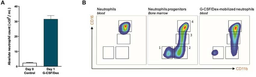
Aarts et al. MDSC Activity of G-CSF/Dex-Mobilized Neutrophils
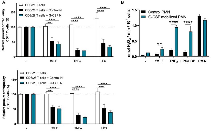
FIGURE 2 | G-CSF/Dex-mobilized neutrophils cannot suppress T cell proliferation despite normal/increased ROS production. (A) Purified CFSE-labeled T cells from
healthy donors (n = 4) were cultured in absence (white bars) or presence of mature neutrophils from control donors (black bars, n = 4), or G-CSF/Dex-mobilized
neutrophils (green bars, n = 3). T cells were stimulated with anti-CD3 and anti-CD28 antibodies. Neutrophils were kept unstimulated or activated with the indicated
stimuli. After 4 or 5 days, cells were harvested and analyzed by flow cytometry for CFSE dilution among CD4+ (upper graph) and CD8+ (lower graph) T cells. Error
bars indicate SEM; the statistical analysis one-way ANOVA was used. (B) G-CSF/Dex-mobilized neutrophils or control neutrophils were stimulated with the indicated
stimuli and production of H2 O2 was determined by measuring Amplex Red conversion into fluorescent Resorufin (n = 3–4). Error bars indicate SEM; the statistical
analysis unpaired two-tailed t-test was used. *p < 0.05, **p < 0.01, ***p < 0.001, ****p < 0.0001.
and very similar to those of normal neutrophil from healthy Dex-mobilized neutrophils were both able to suppress the T
controls without prior mobilization for GTX products (5, 21). cell proliferation of CD4+ and CD8+ T cells upon activation,
comparable to neutrophils from healthy controls (Figures 4A,
Both G-CSF- and Dex-Mobilized 5A). Also the activation of the NADPH oxidase complex
Neutrophils Can Suppress the T Cell required for ROS production was intact. Whereas, the G-CSF-
Proliferation mobilized neutrophils showed a higher level of ROS production
To investigate whether the lack of MDSC activity by G-CSF/Dex- upon fMLF, TNFα, or LPS/LBP stimulation (Figure 4B),
mobilized neutrophils is caused by the G-CSF or Dex component, similar to G-CSF/Dex-mobilized neutrophils (Figure 2B).
we isolated neutrophils 1 day after administration from healthy Dex-mobilized neutrophils showed a normal ROS production,
donors who had received only G-CSF or dexamethasone. As comparable to neutrophils from non-mobilized healthy
we had observed with the G-CSF/Dex donors, the absolute donors (Figure 5B).
neutrophil counts of either G-CSF- or Dex-treated donors Collectively our data suggest that the MDSC activity is
were increased in the peripheral blood compared to the only absent when neutrophils are mobilized with both G-CSF
numbers of circulating neutrophils prior to the administration and Dexamethasone.
of mobilizing agent, i.e., around nine and three times higher,
respectively (Figure 3A). Although the increase in circulating
neutrophils was not as high as in G-CSF/Dex-treated donors, DISCUSSION
also the number of immature neutrophils released into the
blood stream were lower in case of the use of G-CSF or Dex Myeloid-derived suppressor cells have been described as a
only. A small population of CD11bPOS CD16DIM cells was heterogeneous subset of immature myeloid cells, defined by their
present in the G-CSF-mobilized neutrophil fraction next to the capacity to suppress T cell activation and proliferation. Apart
mature neutrophils (CD11bPOS CD16POS ), whereas the Dex- from their malignant transformation, tumor cells also create a
mobilized neutrophil fraction only comprised phenotypically chronic state of inflammation. In cancer, aberrant emergency
mature neutrophils (Figure 3B). The G-CSF-mobilized and myelopoiesis, which is defined as the early exit of progenitor
Frontiers in Oncology | www.frontiersin.org 4 July 2020 | Volume 10 | Article 1110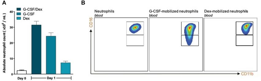
Aarts et al.                                                                                                         MDSC Activity of G-CSF/Dex-Mobilized Neutrophils

  FIGURE 3 | G-CSF- and Dex-mobilized donors have an increased amount of neutrophils consisting mostly of mature neutrophils. (A) Absolute neutrophil count of
  peripheral blood of healthy donors (white bar, n = 6) or from G-CSF/Dex-treated, G-CSF-treated or Dex/treated donors 1 day after administration (n = 3). (B) Surface
  marker expression of CD11b and CD16 was measured by flow cytometry analysis of neutrophils from blood of healthy donors (left panel), G-CSF-mobilized
  neutrophils (center panel) and Dex-mobilized neutrophils. Shown are representative FACS analysis images (n = 3).

  FIGURE 4 | G-CSF-mobilized neutrophils can suppress the T cell proliferation and have an elevated respiratory burst. (A) Purified CFSE-labeled T cells from healthy
  donors (n = 6) were cultured in absence (white bars) or presence of mature neutrophils from control donors (black bars, n = 6), or G-CSF-mobilized neutrophils (blue
  bars, n = 3). T cells were stimulated with anti-CD3 and anti-CD28 ntibodies. Neutrophils were kept unstimulated or activated with the indicated stimuli. After 4 or 5
  days, cells were harvested and analyzed by flow cytometry for CFSE dilution among CD4+ (upper graph) and CD8+ (lower graph) T cells. Error bars indicate SEM; the
  statistical analysis one-way ANOVA was used. (B) G-CSF-mobilized neutrophils or control neutrophils were stimulated with the indicated stimuli and production of
  H2 O2 was determined by measuring Amplex Red conversion into fluorescent Resorufin (n = 3–6). Error bars indicate SEM; the statistical analysis unpaired two-tailed
  t-test was used. **p < 0.01, ***p < 0.001, ****p < 0.0001.

neutrophils from bone marrow, is driven by tumor cell-                                 cancer patients with G-CSF for neutropenia could affect the
derived and/or locally tissue-induced factors including colony                         patients negatively in terms of g-MDSC enrichment (22, 27).
stimulating factors such as GM-CSF, G-CSF and M-CSF (22, 23).                          However, as we have recently reported by studying bone marrow
These factors are thought to contribute to the release of immature                     fractions of myeloid cells in different stages of their development,
neutrophil-like cells that represent a unique immature g-MDSC                          immature neutrophils are not capable of producing ROS (28).
subpopulation (22). The presence and tumor infiltration of                             The formation of these toxic metabolites have been shown in
MDSCs have been associated with poor prognosis (24–26).                                several studies to be one of the main effector mechanisms in
Hence, an important issue was raised as to whether treating                            the suppression of T cells, i.e., MDSC activity (11, 18–20). In

Frontiers in Oncology | www.frontiersin.org                                        5                                                July 2020 | Volume 10 | Article 1110
Aarts et al.                                                                                                          MDSC Activity of G-CSF/Dex-Mobilized Neutrophils

  FIGURE 5 | Dex-mobilized neutrophils can suppress the T cell proliferation and have normal respiratory burst. (A) Purified CFSE-labeled T cells from healthy donors (n
  = 4) were cultured in absence (white bars) or presence of mature neutrophils from control donors (black bars, n = 4), or Dex-mobilized neutrophils (blue bars, n = 3).
  T cells were stimulated with anti-CD3 and anti-CD28 antibodies. Neutrophils were kept unstimulated or activated with the indicated stimuli. After 4 or 5 days, cells
  were harvested and analyzed by flow cytometry for CFSE dilution among CD4+ (upper graph) and CD8+ (lower graph) T cells. Error bars indicate SEM; the statistical
  analysis one-way ANOVA was used. (B) Dex-mobilized neutrophils or control neutrophils were stimulated with the indicated stimuli and production of H2 O2 was
  determined by measuring Amplex Red conversion into fluorescent Resorufin (n = 3–6). Error bars indicate SEM; the statistical analysis unpaired two-tailed t-test was
  used. ****p < 0.0001.

line with these observations, we have previously demonstrated                           and cellular ATP depletion of the T cells. In this study we
that the immature neutrophils in bone marrow fractions from                             have not assessed other suppressive activities than the most
healthy control individuals were not able to suppress the T                             relevant function by which MDSC activity is defined, i.e., T
cell proliferation upon activation. In contrast, MDSC activity                          cell proliferation. In our previous study (11), g-MDSC activity
in these bone marrow samples was induced in case of the                                 suppressing the T cell proliferation was found to coincide with
most mature neutrophils being fully differentiated, as indicated                        the lack of cytokine production, making it less likely that a strong
by morphology and expression of surface markers (28). Our                               induction of T regulatory cells (Tregs) as additional means of
previous findings on bone marrow derived myeloid progenitors                            suppressive activity would contribute as a result of the direct
question the presence of a subset of highly effective granulocyte-                      MDSC activity per se. We have also not extended our studies
related MDSCs that can be released into the circulation to fulfill                      to possible alternative modes of T cell suppression that might
instantaneously strong T cell suppressive activity in humans (28).                      be independent of cell-cell contact and may be based on soluble
Although we cannot exclude the presence of such a bone marrow                           factors otherwise (29), although such factors have limited impact
subset in case of the presence of cancer that may chronically                           in our mixed cell culture, as previously demonstrated when kept
induce the development of such a subset of MDSCs, we could not                          separated by a permeable filter (11).
detect such spontaneously active MDSCs in chemotherapy-naïve                               The reason underlying the inability of G-CSF/Dex mobilized
patients newly diagnosed with Head-Neck Cancer or Mamma                                 neutrophils to perform MDSC activity is as yet unclear. We may
Carcinoma (11). Still, a myeloid progenitor MDSC may be                                 speculate on the sequential steps of MDSC activity following
released to “home” to the tumor microenvironment to develop                             initial cell-cell-interactions to eliminate T cells by ROS and
locally in a strong suppressor cell but supportive data are as yet                      degranulation, which may be facilitated by trogocytosis, i.e.,
not available to the best of our knowledge.                                             the uptake of membrane fragments from T cells by activated
   In our previous study (11), MDSC activity of neutrophils                             neutrophils. Neutrophil trogocytosis does occur at an early stage
in cancer patients and controls was found to be very similar                            during the multi-step process of exerting its full g-MDSC activity,
and depended completely on prior activation. The process of                             and may be an initial, necessary but not sufficient step in
MDSC activity was defined by a the damaged small T cell subset                          this process. Neutrophils from chronic granulomatous disease
undergoing cell death as indicated by morphological alterations                         (CGD) patients, unable to generate ROS, do not show MDSC

Frontiers in Oncology | www.frontiersin.org                                         6                                                July 2020 | Volume 10 | Article 1110
Aarts et al.                                                                                                           MDSC Activity of G-CSF/Dex-Mobilized Neutrophils

activity while the extent of trogocytosis was indistinguishable                          while leaving the effector mechanisms of neutrophils against
from that of control neutrophils (11). The fact that both ROS                            microbial pathogens unaffected, and show that mature G-
and degranulation are required while being spared in case of                             CSF/Dex-mobilized neutrophils indeed meet such conditions (5).
G-CSF/Dex mobilized neutrophils leaves us with an as yet                                 Although GTX products are rarely used in practice, they can
unidentified process that seems to be selectively involved in the                        be life-saving. The fact that G-CSF/Dex-mobilized products are
initiation of g-MDSC activity.                                                           without MDSC activity would be an additional positive safety
   There is sufficient data to support the active role of neutrophil                     issue for using these products in case they are needed. Moreover,
MDSC activity in-vivo, for instance, in the ovarian cancer                               these products may help to clarify the mechanisms in place to
microenvironment (12). MDSC activity of the neutrophils is                               modulate g-MDSC activity specifically without downregulating
actively induced by as yet not fully identified substances within                        the antimicrobial activity.
the ascites fluid of these patients. Similar results were obtained
when pleural fluid of patients with local metastases were tested                         DATA AVAILABILITY STATEMENT
(12), supporting the in-vivo relevance of neutrophil-mediated
MDSC activity. Therefore, G-CSF-mobilized neutrophils could                              The raw data supporting the conclusions of this article will be
have a pro-tumor response when entering the tumor milieu,                                made available by the authors, without undue reservation, to any
as we show here, and treating cancer patients with G-CSF                                 qualified researcher.
alone for neutropenia may be an important issue to reconsider
unless dexamethasone can be used simultaneously to reduce the                            ETHICS STATEMENT
inherent MDSC activity.
   The relevance to further elucidate g-MDSC activity and the                            The studies involving human participants were reviewed and
mechanism by which the combined use of G-CSF and Dex may                                 approved by Sanquin Research Institutional Ethical Committee.
selectively silence this activity bares important relevance to the                       The patients/participants provided their written informed
use of checkpoint inhibitors as well as use of tumor-infiltrating                        consent to participate in this study.
lymphocytes (TILs) as novel forms of effective immunotherapy to
treat cancer (30–32). In cancer patients the presence of increased                       AUTHOR CONTRIBUTIONS
neutrophil counts in the circulation is directly related with a
poor prognosis (9). Our data show that G-CSF/Dex-mobilized                               TK and CA are the principle investigators, who conceived, and
neutrophils lack most of their T cell damaging MDSC activity.                            designed the study. CA, IH, and CF performed the experiments.
Thus, G-CSF/Dex treatment may be a way to silence neutrophils                            RB contributed to the design of the study. CA and IH initiated
within the tumor environment and thereby protect TILs from                               many of the experiments and performed the analysis. CA wrote
local damage, and hence help to improve the development                                  the manuscript together with TK. All authors contributed to the
of more effective anti-cancer immunotherapies. Our current                               article and approved the submitted version.
studies are focusing on differences in cell-cell contact, signal
transduction in both neutrophils and T cells as well as proteomics                       FUNDING
approaches to find out which toxic mechanisms may be impaired
such that T cells may stay unimpaired.                                                   This work was supported by Sanquin Blood Supply Product and
   In this study, we explored whether g-MDSC activity of                                 Process Development Cellular Products Fund (PPOC 2089 &
neutrophils can be selectively inhibited when treating cancer,                           1873) and the Louise Vehmeijer Foundation.

REFERENCES                                                                                6. Kolaczkowska E, Kubes P. Neutrophil recruitment and function in health and
                                                                                             inflammation. Nat Rev Immunol. (2013) 13:159–75. doi: 10.1038/nri3399
 1. Bodey G, Bueltmann B, Duguid W, Gibbs D, Hanak H, Hotchi M, et al.                    7. Ostrand-Rosenberg S, Sinha P. Myeloid-derived suppressor cells:
    Fungal infections in cancer patients: an international autopsy survey. Eur J             linking inflammation and cancer. J Immunol. (2009) 182:4499–
    Clin Microbiol Infect Dis. (1992) 11:99–109. doi: 10.1007/BF01967060                     506. doi: 10.4049/jimmunol.0802740
 2. Bishton M, Chopra R. The role of granulocyte transfusions                             8. Treffers LW, Hiemstra IH, Kuijpers TW, van den Berg TK,
    in neutropenic patients. Br J Haematol. (2004) 127:501–                                  Matlung HL. Neutrophils in cancer. Immunol Rev. (2016)
    8. doi: 10.1111/j.1365-2141.2004.05221.x                                                 273:312–28. doi: 10.1111/imr.12444
 3. Demla A, Madsen LT, Dains J. Effectiveness of granulocyte transfusions in             9. Solito S, Marigo I, Pinton L, Damuzzo V, Mandruzzato S, Bronte V. Myeloid-
    neutropenic adult oncology patients: a comprehensive review of the literature.           derived suppressor cell heterogeneity in human cancers. Ann N Y Acad Sci.
    J Adv Pract Oncol. (2016) 7:410–7. doi: 10.6004/jadpro.2016.7.4.4                        (2014) 1319:47–65. doi: 10.1111/nyas.12469
 4. Grigull L, Pulver N, Goudeva L, Sykora KW, Linderkamp C, Beilken                     10. Condamine T, Dominguez GA, Youn JI, Kossenkov AV, Mony S, Alicea-
    A, et al. G-CSF mobilised granulocyte transfusions in 32 paediatric                      Torres K, et al. Lectin-type oxidized LDL receptor-1 distinguishes population
    patients with neutropenic sepsis. Support Care Cancer. (2006) 14:910–                    of human polymorphonuclear myeloid-derived suppressor cells in cancer
    6. doi: 10.1007/s00520-006-0041-x                                                        patients. Sci Immunol. (2016) 1:aaf8943. doi: 10.1126/sciimmunol.aaf8943
 5. Drewniak A, Boelens JJ, Vrielink H, Tool AT, Bruin MC, van den Heuvel-               11. Aarts CEM, Hiemstra IH, Béguin EP, Hoogendijk AJ, Bouchmal S, van
    Eibrink M, et al. Granulocyte concentrates: prolonged functional capacity                Houdt M, et al. Activated neutrophils exert myeloid-derived suppressor
    during storage in the presence of phenotypic changes. Haematologica. (2008)              cell activity damaging T cells beyond repair. Blood Adv. (2019) 3:3562–
    93:1058–67. doi: 10.3324/haematol.12489                                                  74. doi: 10.1182/bloodadvances.2019031609

Frontiers in Oncology | www.frontiersin.org                                          7                                                July 2020 | Volume 10 | Article 1110
Aarts et al. MDSC Activity of G-CSF/Dex-Mobilized Neutrophils
12. Singel KL, Emmons TR, Khan ANH, Mayor PC, Shen S, patients with newly diagnosed glioblastoma. Neuro Oncol. (2011) 13:591–
Wong JT, et al. Mature neutrophils suppress T cell immunity 9. doi: 10.1093/neuonc/nor042
in ovarian cancer microenvironment. JCI Insight. (2019) 24. Keskinov AA, Shurin MR. Myeloid regulatory cells in tumor
4:e122311. doi: 10.1172/jci.insight.122311 spreading and metastasis. Immunobiology. (2015) 220:236–
13. Singel KL, Grzankowski KS, Khan ANMNH, Grimm MJ, D’Auria, 42. doi: 10.1016/j.imbio.2014.07.017
AC, et al. Mitochondrial DNA in the tumour microenvironment 25. Solito S, Falisi E, Diaz-Montero CM, Doni A, Pinton L, Rosato A, et al.
activates neutrophils and is associated with worse outcomes in A human promyelocytic-like population is responsible for the immune
patients with advanced epithelial ovarian cancer. Br J Cancer. (2019) suppression mediated by myeloid-derived suppressor cells. Blood. (2011)
120:207–17. doi: 10.1038/s41416-018-0339-8 118:2254–65. doi: 10.1182/blood-2010-12-325753
14. Gazendam RP, van Hamme JL, Tool AT, van Houdt M, Verkuijlen PJ, Herbst 26. Umansky V, Blattner C, Gebhardt C, Utikal J. The role of myeloid-derived
M, et al. Two independent killing mechanisms of Candida albicans by human suppressor cells (MDSC) in cancer progression. Vaccines (Basel). (2016)
neutrophils: evidence from innate immunity defects. Blood. (2014) 124:590– 4:36. doi: 10.3390/vaccines4040036
7. doi: 10.1182/blood-2014-01-551473 27. Luyckx A, Schouppe E, Rutgeerts O, Lenaerts C, Fevery S, Devos T, et al.
15. Borregaard N, Sørensen OE, Theilgaard-Mönch K. Neutrophil granules: G-CSF stem cell mobilization in human donors induces polymorphonuclear
a library of innate immunity proteins. Trends Immunol. (2007) 28:340– and mononuclear myeloid-derived suppressor cells. Clin Immunol. (2012)
5. doi: 10.1016/j.it.2007.06.002 143:83–7. doi: 10.1016/j.clim.2012.01.011
16. Hoogendijk AJ, Pourfarzad F, Aarts CEM, Tool ATJ, Hiemstra IH, Grassi L, 28. Aarts CEM, Hiemstra IH, Tool ATJ, van den Berg TK, Mul E, van Bruggen R,
et al. Dynamic transcriptome-proteome correlation networks reveal human et al. Neutrophils as suppressors of T cell proliferation: does age matter? Front
myeloid differentiation and neutrophil-specific programming. Cell Rep. Immunol. (2019) 10:2144. doi: 10.3389/fimmu.2019.02144
(2019) 29:2505–19.e4. doi: 10.1016/j.celrep.2019.10.082 29. Granot Z. Neutrophils as a therapeutic target in cancer. Front Immunol. (2019)
17. van Leeuwen EM, de Bree GJ, Remmerswaal EB, Yong SL, Tesselaar K, ten 10:1710. doi: 10.3389/fimmu.2019.01710
Berge IJ, et al. IL-7 receptor alpha chain expression distinguishes functional 30. Azoury SC, Straughan DM, Shukla V. Immune checkpoint inhibitors for
subsets of virus-specific human CD8+ T cells. Blood. (2005) 106:2091– cancer therapy: clinical efficacy and safety. Curr Cancer Drug Targets. (2015)
8. doi: 10.1182/blood-2005-02-0449 15:452–62. doi: 10.2174/156800961506150805145120
18. Kramer PA, Prichard L, Chacko B, Ravi S, Overton ET, Heath SL, et al. 31. Darvin P, Toor SM, Sasidharan Nair V, Elkord E. Immune checkpoint
Inhibition of the lymphocyte metabolic switch by the oxidative burst of human inhibitors: recent progress and potential biomarkers. Exp Mol Med. (2018)
neutrophils. Clin Sci (Lond). (2015) 129:489–504. doi: 10.1042/CS20140852 50:1–11. doi: 10.1038/s12276-018-0191-1
19. Pillay J, Kamp VM, van Hoffen E, Visser T, Tak T, Lammers JW, et al. A subset 32. Rohaan MW, Wilgenhof S, Haanen JBAG. Adoptive cellular
of neutrophils in human systemic inflammation inhibits T cell responses therapies: the current landscape. Virchows Arch. (2019) 474:449–
through Mac-1. J Clin Invest. (2012) 122:327–36. doi: 10.1172/JCI57990 61. doi: 10.1007/s00428-018-2484-0
20. Schmielau J, Finn OJ. Activated granulocytes and granulocyte-derived
hydrogen peroxide are the underlying mechanism of suppression of t-cell Conflict of Interest: The authors declare that the research was conducted in the
function in advanced cancer patients. Cancer Res. (2001) 61:4756–60. absence of any commercial or financial relationships that could be construed as a
21. Drewniak A, van Raam BJ, Geissler J, Tool AT, Mook OR, van den Berg TK, potential conflict of interest.
et al. Changes in gene expression of granulocytes during in vivo granulocyte
colony-stimulating factor/dexamethasone mobilization for transfusion Copyright © 2020 Aarts, Hiemstra, Furumaya, van Bruggen and Kuijpers. This is an
purposes. Blood. (2009) 113:5979–98. doi: 10.1182/blood-2008-10-182147 open-access article distributed under the terms of the Creative Commons Attribution
22. Bergenfelz C, Leandersson K. The generation and identity of License (CC BY). The use, distribution or reproduction in other forums is permitted,
human myeloid-derived suppressor cells. Front Oncol. (2020) provided the original author(s) and the copyright owner(s) are credited and that the
10:109. doi: 10.3389/fonc.2020.00109 original publication in this journal is cited, in accordance with accepted academic
23. Raychaudhuri B, Rayman P, Ireland J, Ko J, Rini B, Borden EC, practice. No use, distribution or reproduction is permitted which does not comply
et al. Myeloid-derived suppressor cell accumulation and function in with these terms.
Frontiers in Oncology | www.frontiersin.org 8 July 2020 | Volume 10 | Article 1110You can also read