IFN γ and IL 18 in conditioned media of parasite infected host and IL 21 silenced colorectal cancer cells
←
→
Page content transcription
If your browser does not render page correctly, please read the page content below
EXPERIMENTAL AND THERAPEUTIC MEDICINE 21: 103, 2021

         IFN‑γ and IL‑18 in conditioned media of parasite‑infected
              host and IL‑21‑silenced colorectal cancer cells
   PENG GE1, CHING YI ONG2, ABDALLA ESHTIYAG ABDALKAREEM2,3, BOON YIN KHOO2 and BO YUAN1

                   1
                    Department of General Surgery, Xi'an Central Hospital, Xi'an, Shaanxi 710003, P.R. China;
             2
              Institute for Research in Molecular Medicine (INFORMM), Universiti Sains Malaysia, Penang 11800,
                             Malaysia; 3Tropical Medicine Research Institute, Khartoum 11111, Sudan

                                       Received September 15, 2019; Accepted August 11, 2020

                                                        DOI: 10.3892/etm.2020.9535

Abstract. The presence of certain soluble factors may provide         and proliferating cell nuclear antigen mRNA expression was
a possible selective advantage for a parasite to gradually            altered in host cells incubated with various levels of IFN‑γ and
modify cell proliferation in neighbouring cells, which may            IL‑18, as well as in IL‑21‑silenced HCT116 cells compared
result in chronic diseases. These soluble factors present in the      with the respective controls. In conclusion, the current study
conditioned medium also allow the parasite to invade rapidly          provided preliminary evidence on the fundamental molecular
into more host cells. The present study aimed to determine            mechanisms of host‑parasite interactions that result in chronic
the levels of a group of type 1 T helper (Th1) cytokines in the       diseases, which may aid in the treatment of these diseases in
conditioned media of host cells infected with parasites and in        the relevant endemic regions.
IL‑21‑silenced colorectal cancer cells. The conditioned media
of human foreskin fibroblasts (HFFs) parasitized with the RH          Introduction
and ME49 strains of Toxoplasma gondii for 10 days were
prepared, and subsequently the levels of the Th1 cytokines in         Toxoplasmosis is caused by the protozoan parasite
the conditioned media were determined by ELISA. HFFs were             Toxoplasma gondii, where >60% of populations are infected
incubated with the growth media containing selected soluble           with these parasites (1,2). The infection often occurs in areas of
factors, and cell proliferation markers were subsequently             lower altitudes, hot and humid climates (2). Our previous study
analysed by reverse transcription‑quantitative PCR. The               demonstrated that a low dose of the virulent type I RH strain
mRNA expression level of cell proliferation markers was also          of Toxoplasma gondii (100 parasites) rapidly caused a lethal
examined in IL‑21‑silenced HCT116 cells, where the levels of          infection in mice within 4 days (3). By contrast, mortality with
soluble factors in the conditioned media were also determined         the avirulent type II ME49 strain of T. gondii occurred with
as aforementioned. The results of the present study demon‑            a higher initial dose (1x10⁵ parasites) at 6‑8 days post‑infec‑
strated that HFFs parasitized with ME49 released elevated             tion (3). The T. gondii strain RH exhibits high virulence in
levels of IFN‑γ and lower levels of IL‑18 into the conditioned        animals, but mice have been demonstrated to survive infec‑
medium compared with the controls. These phenomena were               tion with the low virulence ME49 strain of T. gondii (4). It is
not observed in the conditioned medium of HFFs parasitized            hypothesised that parasitized host cells release soluble factors
with RH. Similar levels of these soluble factors were also            into the conditioned medium following T. gondii infection to
detected in the conditioned medium of IL‑21‑silenced HCT116           regulate the parasite invasion into the host cells. However, the
cells. The results of the present study also revealed that Ki67       differences of these soluble factors in the conditioned media
                                                                      of host cells parasitized with RH and ME49 remain unclear.
                                                                      Different soluble factors are believed to be secreted into the
                                                                      conditioned medium by host cells parasitized with various
                                                                      strains of T. gondii, as these factors provide a possible selective
Correspondence to: Dr Bo Yuan, Department of General                  advantage for the parasite to rapidly invade host cells (5). A
Surgery, Xi'an Central Hospital, 161 Xiwu Road, Xincheng, Xi'an,      previous study has demonstrated that the ability to develop
Shaanxi 710003, P.R. China                                            chronicity of infection is dependent on type 1 T helper (Th1)
E‑mail: yuanbodandanbaba@sina.com
                                                                      cells (6). In addition, overstimulation of Th1 cytokines has
Abbreviations: Th1, type 1 T helper; HFFs, human foreskin             been associated with the acute virulence of T. gondii (3).
fibroblasts; siRNA, small interfering RNA; PCNA, proliferating cell   Therefore, soluble Th1 cytokines are likely to constitute the
nuclear antigen; cDNA, complementary DNA; OS, overall survival        factors that underlie the differences between the conditioned
                                                                      media of host cells parasitized with RH and ME49.
Key words: interferon‑γ, interleukin‑18, host‑parasite interaction,       The present study investigated the levels of soluble Th1
gene silencing, colorectal cancer, cell proliferation marker          cytokines in the conditioned media of host cells parasitized
                                                                      with the RH and ME49 strains of T. gondii at different time
                                                                      intervals. The current study also compared the levels of soluble
2 GE et al: CYTOKINES OF PARASITE-INFECTED AND IL-21 SILENCED CELLS
factors in the conditioned media of IL‑21‑silenced colorectal at room temperature. The unbound substances were removed
cancer cells and the impact of the soluble factors on the mRNA with a 1X PBS washing buffer, according to the manufacturer's
expression level of cell proliferation markers in both the host instructions. An enzyme‑linked antibody specific for each Th1
cells post parasitic infection and colorectal cancer cells post cytokine was added to the wells and incubated for 1 h at room
silencing of the IL‑21 gene. The IL‑21 gene was silenced in a temperature. Following another washing step, a substrate solu‑
colorectal cancer cell line, as it has been indicated to reduce tion was added to the wells for colour development. The colour
the proliferation of the cells, and it may also be used to study intensity was measured using a plate reader (Tecan Group,
the molecular mechanisms of host‑parasite interactions that Ltd.) at 450 nm, and the colour development was proportional
cause chronic diseases with respect to colorectal cancer (7). to the amount of the Th1 cytokines present in the samples. The
The present study provided useful information on the funda‑ cytokine level was calculated as the ratio of the experimental
mental molecular mechanisms of the host‑parasite interactions value (pg/ml) relative to the value in non‑infected host cells.
that may aid in early diagnosis, novel prescription drugs The statistically significant difference relative to day 2 ratio
and cost‑effective strategies for the treatment of infectious was considered.
disease‑associated colorectal cancer in the future.
Incubation of HFFs with media containing specific soluble
Materials and methods factors. Briefly, HFFs were seeded at a density of 100 cells/ml
into T‑25 cell culture flasks (Thermo Fisher Scientific, Inc.)
Preparation of conditioned media of host cells parasitized with and maintained as aforementioned. When HFFs were
RH and ME49. The RH and ME49 strains of T. gondii were a ~80% confluent, the growth media were removed from the
kind gift from Prof. Rahmah Noordin, INFORMM, Universiti culture flasks. The HFF feeder layer was then exposed to
Sains Malaysia. The stocks were stored in liquid nitrogen. The growth medium containing 1 ng/ml human IFN‑γ research
parasites were cultured and propagated in vitro and adjusted grade (cat. no.: 130‑096‑873), 1 ng/ml Abnova human IL‑18
to a concentration of 100 parasites/ml. The parasite numbers recombinant protein (cat. no. P3632) or growth medium only
were estimated using a Neubauer haemocytometer chamber for 4‑10 days in triplicate (day 2 was excluded to reduce the
(Electron Microscopy Sciences, Inc.). Human foreskin fibro‑ use of cytokines and growth media). It is expected that an
blasts (HFFs), which were originally purchased from the alteration in HFF proliferation was observed when the HFFs
American Type Culture Collection (cat no. CRL‑2522) and were incubated in growth media containing IFN‑γ and IL‑18
maintained in the laboratory, were used as the host cells and compared with growth medium alone. The incubated HFFs
cultured in DMEM supplemented with 10% FBS, 100 U/ml were subsequently harvested for analysis of mRNA expression,
penicillin and 100 mg/ml streptomycin (all from Thermo Fisher as described in the following section.
Scientific, Inc.). HFFs were used as the host for parasites since
they are not differentiated, which allows the parasites to prop‑ Analysis of the mRNA expression levels of cell proliferation
agate rapidly (8). The growth medium of HFFs was changed markers in HFFs. Total RNA was extracted from HFFs
every 2‑3 days, and the cells were cultured at 37˚C in a humidi‑ incubated with specific soluble factors for 4‑10 days using a
fied atmosphere with 5% CO2. HFFs were seeded at a density commercially available RNeasy Mini Kit (Qiagen, Inc.) for
of 1,000 cells/ml in T‑75 cell culture flasks (Thermo Fisher total RNA extraction. RNA (1.0 µg) was reverse‑transcribed
Scientific, Inc.). The cell number was also estimated using a into cDNA at 60˚C using a commercially available RevertAid
haemocytometer chamber. When HFFs were 80% confluent, First Strand cDNA Synthesis kit (Thermo Fisher Scientific,
RH or ME49 (100 parasites/ml) were added to the HFF feeder Inc.), which was subsequently used for the analysis of cell
layer. The co‑culture was incubated for 2‑10 days. The growth proliferation markers at the mRNA expression level by
medium was removed from the co‑culture at different time quantitative PCR (qPCR). Primers for Ki67 and proliferating
intervals; every 2 days post‑infection and used as the condi‑ cell nuclear antigen (PCNA) were designed using Primer
tioned medium. The collected conditioned media, as well as Express v2.0, and qPCR was performed using ABI 7500 Fast
the control media from non‑parasitized HFFs (culture super‑ Sequence Detection System (both from Applied Biosystems;
natants of HFFs only), were passed through a 0.22‑mm filter Thermo Fisher Scientific, Inc.). The primer sequences used
(Thermo Fisher Scientific, Inc.) to separate the parasites, host were as follows: Ki67 forward, 5'‑CCAACTGTTG GTCTC
cells and other cell debris. The conditioned media collected at GCGTAAG ‑3' and reverse, 5'‑ATCTGTCCAG CTGTAGTG
various time intervals were used for the immunoassays of Th1 CCCA‑3'; PCNA forward, 5'‑GGCACTCAAG GACCTCAT
cytokines. CAAC‑3' and reverse, 5'‑GTGAGCTGCACCAAAGAGACG
T‑3'; Transforming growth factor‑ α forward, 5'‑CAGACC
Immunoassay of Th1 cytokines in the conditioned media TTCC TAC TTG GCC TGTAA‑3' and reverse, 5'‑GACG GA
of parasite‑infected host. Immunoassays for human IL‑1β GTT C TT GAC AGA GTT T TG‑3'; Chemokine C‑C motif
(cat no. KA0356), IL‑10 (cat no. KA0125), IL‑12p40 ligand 5 forward, 5'‑AGCC TCT CCCACAGGTACCAT‑3'
(cat no. KA0178), IL‑18 (cat no. KA0561), IFN‑γ (cat no. 3045) and reverse, 5'‑GGC AGT AGC A AT GAG GAT GAC A‑3';
and TNF‑α (cat no. P3453) were performed using commer‑ Epidermal growth factor forward, 5'‑TGTGGTTCTCAGATT
cially available ELISA kits (Abnova). According to the GGGCTATG‑3' and reverse 5'‑GATGAGGGCTTCAGCATG
manufacturer's instructions, microtiter plates were pre‑coated CT‑3'; and GAPDH forward, 5'‑CAAGTTCAACGGCACAGT
with antibodies specific for the Th1 cytokines. The collected CAAG‑3' and reverse, 5'‑CTCCTGGAAGATGGTGATTGG
conditioned media (~200 µl) were added to the respective T‑3'. GAPDH was used as the internal control. A total reac‑
wells and allowed to react with the bound antibody for 2.5 h tion volume of 25 µl was used, which included SYBR® GreenEXPERIMENTAL AND THERAPEUTIC MEDICINE 21: 103, 2021 3
Master Mix (Applied Biosystems; Thermo Fisher Scientific, mRNA level, whereas the conditioned media collected from
Inc.), 900 nM of each primer and 5 µl cDNA (25 ng). The the IL‑21‑silenced cells were subjected to immunoassay of
following thermocycling conditions were used: 95˚C for cytokines.
10 min, followed by 40 cycles of 95˚C for 15 sec and 60˚C for
1 min. Fold‑changes in gene expression were calculated using Analysis of cell proliferation markers at mRNA level and
the 2‑ΔΔCq method (9). The relative expression of each target cytokines in conditioned media of IL‑21‑silenced cells by qPCR
gene in the samples of the host cells incubated with growth and ELISA. Total RNA was extracted from IL‑21‑silenced
media containing soluble factors was normalised to that of HCT116 cells at 1‑3 days post‑transfection. A total of 1.0 µg
the target gene in the samples of the host cells incubated with RNA was reverse‑transcribed into cDNA, which was used for
growth media alone (control). The expression level of each the analysis of cell proliferation marker expression at the mRNA
gene in fold‑change was calculated relative to day 4 gene level by qPCR. The same primers for the detection of Ki67
expression level. and PCNA were used as aforementioned, and the fold‑changes
in gene expression were calculated relative to those of the
Systematic review of the associations between IL‑18, IFN‑ γ control cells (day 1), as aformentioned. On the other hand,
and IL‑21. The following procedure was used for the system‑ the conditioned media collected from IL‑21‑silenced HCT116
atic review: i) The research question ‘what is the relationship cells at 1‑3 days post‑transfection were added to the wells and
between IL‑18, IFN‑ γ and IL‑21 in human diseases’ was allowed to react with the bound antibodies on the microtiter
used as the topic of the analysis; ii) the systematic reference plates that were pre‑coated with antibodies specific for IL‑18
management software EndNote 9.0.1 (Clarivate Analytics) and IFN‑ γ for 2.5 h at room temperature. The unbound
was used to extensively search the published studies on this substances were removed with a wash buffer, according to
topic worldwide; iii) the studies associated with this topic were the manufacturer's instructions, and ELISA was subsequently
screened in the PubMed databases at the National Library of performed as aforementioned. The cytokine level was calcu‑
Medicine (NLM; https://pubmed.ncbi.nlm.nih.gov/) to iden‑ lated as the ratio of the experimental value in the silenced
tify evidence for subsequent examination; iv) the studies on cells (pg/ml) relative to the value in control cells. Statistically
the associations between IL‑18, IFN‑γ and IL‑21 in infectious significant differences were considered relative to day 1 ratio.
disease and colorectal cancer research in an endemic region
were extracted to answer the research question; v) the contents Statistical analysis. Data are presented as the mean ± SD. All
of each article were analysed, and the evidence was synthesised statistical analyses were performed with one‑way ANOVA
to determine bias with qualitative statistics tools, for example followed by Tukey's post hoc test using GraphPad v6.01
PCR and Immunoassays; and vi) the conclusion was returned software (GraphPad Software, Inc.). The experiments
into a report to support the analysis. were repeated at least twice in three technical replicates to
ensure reproducibility. P4 GE et al: CYTOKINES OF PARASITE-INFECTED AND IL-21 SILENCED CELLS
Figure 2. mRNA expression levels of cell proliferation markers in host cells
incubated with growth media containing IL‑18 and IFN‑γ for different time
intervals. (A) Ki67 and (B) PCNA. Host cells incubated with growth medium
were used as a control. The target mRNA expression was normalised to
the expression of GAPDH in the same sample. At least two independent
experiments were performed. Data are presented as the mean ± SD from
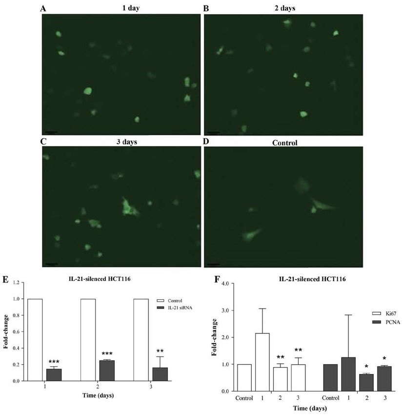
triplicate cultures. *PEXPERIMENTAL AND THERAPEUTIC MEDICINE 21: 103, 2021 5 Figure 3. mRNA expression levels of cell proliferation markers in IL‑21‑silenced HCT116 cells. (A‑D) Efficacy of IL‑21 gene silencing for 1, 2 and 3 days under an inverted fluorescence microscope (magnification of x100 was used). The control image depicts the fluorescence of non‑silenced cells. (E) Differences in IL‑21 mRNA expression in cells transfected with IL‑21 small interfering RNA and control (non‑transfected) cells detected by reverse transcription‑quantitative PCR. (F) mRNA expression was normalised to the expression of GAPDH in each sample. At least two independent experiments were performed. Data are presented as the mean ± SD from triplicate cultures. *P
6                                   GE et al: CYTOKINES OF PARASITE-INFECTED AND IL-21 SILENCED CELLS

                                                                                  Discussion

                                                                                  The present study on Th1 immune responses following
                                                                                  parasitic infection of host cells revealed that IL‑18 and IFN‑γ
                                                                                  likely represented the growth factors that differentiated a
                                                                                  possible selective advantage and disadvantage for RH and
                                                                                  ME49 strains of T. gondii to invade into surrounding host
                                                                                  cells. These factors may modify the proliferation of the host
                                                                                  cells, which was observed as a reduction in Ki67 and PCNA
                                                                                  mRNA expression levels in the IL‑18‑ and IFN‑γ‑stimulated
                                                                                  host cells at the end of the experiments. These soluble factors
                                                                                  may also allow the parasites, such as ME49, to slowly invade
                                                                                  into a higher number of surrounding host cells, resulting in
                                                                                  chronic diseases. Similarly, reduction in the mRNA expression
                                                                                  level of the cell proliferation markers Ki67 and PCNA was also
                                                                                  observed in the IL‑21‑silenced HCT116 cells in the present
                                                                                  study, which indicated that IL‑21 may regulate the prolif‑
                                                                                  eration of diseased cells. Different levels of IL‑18 and IFN‑γ
                                                                                  were also detected in the conditioned media of IL‑21‑silenced
                                                                                  HCT116 cells. By studying HFFs (host cells) and HCT116 cells
                                                                                  (diseased cells), the present study aimed to elucidate the funda‑
                                                                                  mental molecular mechanisms of the host‑parasite interactions
                                                                                  that subsequently cause chronic diseases, such as colorectal
                                                                                  cancer, and examine whether these two cell types released the
                                                                                  same soluble factors in the conditioned media. The collective
                                                                                  information may be useful for future treatment of infec‑
                                                                                  tious disease‑associated colorectal cancer, and IL‑21, IL‑18
Figure 4. Soluble factor levels in conditioned media of IL‑21‑silenced            and IFN‑γ may be used to develop tools for early diagnosis,
HCT116 cells. (A) IL‑18 and (B) IFN‑γ. Conditioned media of non‑parasit‑
                                                                                  novel prescription drugs and cost‑effective strategies for the
ized host cells were used as a control. The cytokine level was calculated as
the ratio of the experimental value (pg/ml) relative to that of the control. At   treatment of these diseases.
least two independent experiments were performed. Data are presented as the           IL‑18 and IFN‑γ are Th1‑type cytokines that are produced
mean ± SD from triplicate cultures. **P
EXPERIMENTAL AND THERAPEUTIC MEDICINE 21: 103, 2021 7
Table I. Summary of the associations between IL‑18 and IFN‑γ with IL‑21 observed in the present and previous studies.
Cytokine
‑‑‑‑‑‑‑‑‑‑‑‑‑‑‑‑‑‑‑‑‑‑‑‑‑‑‑‑‑‑‑‑‑‑‑‑‑‑‑‑‑‑‑‑‑‑‑‑‑‑‑‑‑‑‑‑‑‑
Day IL‑18a IFN‑γa IL‑21b Comment
2 + + ‑ Early chronic parasitic infection
4 + + ‑ Early chronic parasitic infection
6 + ++ + IFN‑γ and cancerous factor levels start to be induced in host cells with chronic
parasitic infection
8 ‑ +++ ++ IFN‑γ and cancerous factor levels continue to be induced when IL‑18 is decreased
in the conditioned media of host cells with chronic parasite infection
10 ‑ +++ +++ IFN‑γ is maintained at a high level, and the cancerous factor continues to be
induced when IL‑18 is decreased in the conditioned media of host cells with
chronic parasite infection
a
Cytokines in chronic infection determined in the present study; bhypothesised cytokine levels based on a previous study (19). ‑, indicates low
expression level, +, medium expression level; ++, high expression level; +++, very high expression level.
phenomenon was not observed in the type I parasitic infection Ki67 and PCNA in IL‑18‑ and IFN‑γ‑stimulated host cells,
with the RH strain. which was also observed in IL‑21‑silenced HCT116 cells.
A previous study has demonstrated that IL‑18 and IL‑21 in IL‑18 is a uniquely pleiotropic member of the IL‑1 family, and
different combinations enhance IFN‑γ production in human it is synthesised as a 24 kDa precursor protein and cleaved into
NK and T cells (19). The results of the present study revealed an 18 kDa mature form by caspase‑1 (25). The level of IL‑18
that infection with ME49 reduced the levels of IL‑18, but was reduced in the conditioned media of host cells parasitized
increased those of IFN‑γ in the conditioned media compared with ME49 for 8‑10 days in the present study. Stimulation
with those in the control cells. The overview of the associa‑ of the host cells with IL‑18 induced an optimal level of cell
tion between IL‑18 and IFN‑g with IL‑21 is summarised in proliferation markers dependent on the time following stimu‑
Table I, whereby a low expression level of IL‑21 is hypothe‑ lation (bell shaped curve), which was reflected in the mRNA
sised at early chronic parasitic infection when both IL‑18 and expression levels of Ki67 and PCNA. However, prolonged
IFN‑γ are maintained at moderate levels. When the infection incubation of the host cells with IL‑18 also reduced cell prolif‑
time is prolonged, IL‑21 continues to be induced, while IL‑18 eration marker expression. These results were consistent with
and IFN‑ γ are decreased and maintained at a high level, those of a previous study indicating that IL‑18 exerted both
respectively. IL‑18 is required for IFN‑γ gene activation in cancer‑promoting and cancer‑suppressing functions (26). IL‑18
both bacterial and viral infections (20‑23). Therefore, a low has been revealed to promote the proliferation and invasion of
level of IL‑18 in the conditioned media of parasite‑infected pancreatic cancer cells, and higher IL‑18 levels in pancreatic
host cells may be insufficient to induce high levels of IFN‑γ. cancer tissues were associated with a shorter overall survival
The presence of other factors in the conditioned media, (OS), increased invasion and metastasis, compared with patients
e.g. IL‑21, likely contributes to high levels of IFN‑γ and to with lower IL‑18 levels (26). However, in the same study, higher
the development of cancerous cells. This phenomenon may plasma levels of IL‑18 were associated with longer OS. IL‑18
also explain the relationship between parasitic infection and has been demonstrated to exhibit antitumourtumour activity
colorectal cancer. IL‑21 was the focus of the present study, in preclinical models and increase the serum concentrations
as a high level of IL‑21 expression has been detected in of IFN‑γ, granulocyte macrophage colony‑stimulating factor
colorectal cancer cells, such as HT29 and HCT116 (7). In our and soluble Fas ligand (27). By contrast, the role of IFN‑γ was
previous study, a high level of IL‑21 was also detected in the demonstrated to be more direct and straightforward in the
serum samples of patients diagnosed with colorectal cancer present study. IFN‑γ was induced in the conditioned media of
with a history of parasitic infections (24). IL‑18 or IL‑21 the host cells parasitized with ME49, and stimulation of the host
alone have been demonstrated to represent weak inducers of with IFN‑γ reduced cell proliferation markers. These results
IFN‑γ production, but the combination of IL‑18 and IL‑21 suggested that IFN‑γ may reduce cell proliferation markers,
has been reported to induce notable activation of IFN‑γ gene which was also observed in IL‑21‑silenced HCT116 cells in
expression (19). Therefore, it was hypothesised that IL‑21 is the present study. A previous study reported that intravesical
another soluble factor that may be present in the conditioned instillations of 0.7 mg IFN‑γ produced a significant cytostatic
media, and it was examined in the present study whether effect on superficial bladder cancer cells, which was evidenced
IL‑21 silencing resulted in the reduction of cell proliferation by the decrease in growth fractions, measured using antigens
that may be associated with the levels of IL‑18 and IFN‑γ in of PCNA and Ki67 (28). IFN‑γ has been also demonstrated
the conditioned media. to sustain the expression of PCNA and the G1/S regulator
In the present study, IL‑18 and IFN‑γ were demonstrated retinoblastoma proteins, including cyclin D1, cyclin E and
to alter the mRNA expression of the cell proliferation markers cdk2, and maintain low p27 levels (29). However, the effects8 GE et al: CYTOKINES OF PARASITE-INFECTED AND IL-21 SILENCED CELLS
of prolonged IFN‑γ stimulation on Ki67 and PCNA expression Availability of data and materials
were only observed at day 10 in the current study. A more
direct method, such as flow cytometry or western blotting, to The datasets used and/or analysed during the current study
evaluate the cell cycle‑associated proteins should be utilised in are available from the corresponding author on reasonable
future research to produce a stronger evidence for this hypoth‑ request.
esis. The results of the analysis of the mRNA expression
levels of cell proliferation markers in IL‑21‑silenced HCT116 Authors' contributions
cells indicated that IL‑21 likely regulated the proliferation of
HCT116 cells. The IL‑21‑silenced HCT116 cells may be used PG, BYK and BY made substantial contributions to the
as a model to ensure whether the modification of diseased cell design of the present study. CYO and AEA participated in all
proliferation released the soluble factors as the aforementioned experiments under technical support provided by BYK. BYK
investigation outlines. interpreted the results, drafted and revised the manuscript. All
A previous study has revealed that IL‑18‑deficient mice authors read and approved the final manuscript.
were highly resistant to chronic T. muris infection, and
in vivo treatment of normal mice with recombinant IL‑18 Ethics approval and consent to participate
suppressed IL‑4 and IL‑13 secretion. However, the treatment
did not affect the level of IFN‑γ in the mice (6). The present Not applicable.
in vitro study did not observe an association between IL‑18
and ME49 infection resistance. However, the levels of IL‑18 Patient consent for publication
and IFN‑γ exhibited opposing trends during ME49 infection
for 10 days, which supported the hypothesis that IL‑18 does Not applicable.
not function as an IFN‑γ‑inducing cytokine during chronic
infections, but serves other roles, such as direct regulation Competing interests
of Th2 cytokines (30). Another study has demonstrated that
CD8+ T cells and IFN‑γ were required for the host immunity The authors declare that they have no competing interests.
to RH and ME49 (31). However, these differences may reflect
stage‑specific (tachyzoite vs. bradyzoite) or strain‑specific References
(RH vs. ME49) requirements for the host immunity, which
remain unclear, primarily since the majority of toxoplasma 1. Ferra B, Holec‑Gąsior L and Grąźlewska W: Toxoplasma gondii
recombinant antigens in the serodiagnosis of toxoplasmosis
strains have been indicated to be virulent during secondary in domestic and farm animals. Animals (Basel) 10: 1245, 2020.
infections (32). Additional studies should be conducted in the 2. Galeh T M, Sa r vi S, Montazer i M, Moosazadeh M,
future to provide stronger evidence for the validation of the Nakhaei M, Shariatzadeh SA and Daryani A: Global status of
Toxoplasma gondii seroprevalence in rodents: A systematic
hypothesis of the present study. To the best of our knowl‑ review and meta‑analysis. Front Vet Sci 7: 461, 2020.
edge, Schistosomiasis attracts little attention and support 3. Mordue DG, Monroy F, La‑Regina M, Dinarello CA and
worldwide owing to the geographical barriers and certain Sibley LD: Acute toxoplasmosis leads to lethal overproduction of
Th1 cytokines. J Immunol 167: 4574‑4584, 2001.
political issues (33). The disease may also be considered 4. Gavrilescu LC and Denkers EY: IFN‑gamma overproduction
as unimportant as it primarily occurs in individuals living and high level apoptosis are associated with high but not low
in poor, rural communities and in endemic regions (33). virulence Toxoplasma gondii infection. J Immunol 167: 902‑909,
2001.
Moreover, current studies focus mainly on pandemic issues 5. Hakimi MA, Olias P and Sibley LD: Toxoplasma effectors
rather than neglected diseases. targeting host signaling and transcription. Clin Microbiol
In conclusion, the results of the present study may elucidate Rev 30: 615‑645, 2017.
6. Helmby H, Takeda K, Akira S and Grencisa RK: Interleukin
the fundamental molecular mechanisms of host‑parasite (Il)‑18 promotes the development of chronic gastrointestinal
interactions that cause chronic diseases. The results may also helminth infection by downregulating IL‑13. J Exp Med 194:
provide useful information for future studies on groups of 355‑364, 2001.
7. Abdalkareem EA, Ong CY, Lim BH and Khoo BY: Neutralising
genes that regulate Th cell responses during colorectal cancer FGF4 protein in conditioned medium of IL‑21‑silenced HCT116
and parasitic infection. cells restores the invasiveness of the colorectal cancer cells.
Cytotechnology 70: 1363‑1374, 2018.
8. Khan A and Grigg ME: Toxoplasma gondii: Laboratory mainte‑
Acknowledgements nance and growth. Curr Protoc Microbiol 44: 20C.1.1‑20C.1.17,
2017.
The authors would like to thank Professor Rahmah Noordin 9. Livak KJ and Schmittgen TD: Analysis of relative gene expres‑
sion data using real‑time quantitative PCR and the 2(‑Delta Delta
of Institute for Research in Molecular Medicine (INFORMM), C(T)) method. Methods 25: 402‑408, 2001.
Universiti Sains Malaysia for providing the RH and ME49 10. Eshtiyag A, Lim BH and Khoo BY: Silencing of IL21 in HT29
strains of T. gondii for the present study. and HCT116 cells to determine its role in the proliferation
of colorectal cancer associated with Schistosoma mansoni
infection. Australian J Basic Applied Sci 9: 39‑44, 2015.
Funding 11. Eberl M, Beck E, Coulson PS, Okamura H, Wilson RA and
Mountford AP: IL‑18 potentiates the adjuvant properties of IL‑12
in the induction of a strong Th1 type immune response against a
The present study was funded by the Universiti Sains Malaysia recombinant antigen. Vaccine 18: 2002‑2008, 2000.
Short‑term Grant Scheme (grant no. 304/CIPPM/6311018) 12. Chang CY, Lee J, Kim EY, Park HJ, Kwon CH, Joh JW and
and Fundamental Research Grant Scheme Fasa 1/2017 Kim SJ: Intratumoral delivery of IL‑18 naked DNA induces
T‑cell activation and Th1 response in a mouse hepatic cancer
(grant no. 203/CIPPM/6711599). model. BMC Cancer 7: 87, 2007.EXPERIMENTAL AND THERAPEUTIC MEDICINE 21: 103, 2021 9
13. Srinivasan A, Salazar‑Gonzalez RM, Jarcho M, Sandau MM, 24. Abdalkareem EA, Tan GC, Abdalla HS, Lim BH and Khoo BY:
Lefrancois L and McSorley SJ: Innate immune activation of Identification of specific proteins in colorectal cancer patients
CD4 T cells in salmonella‑infected mice is dependent on IL‑18. with Schistosoma mansoni infection as a possible biomarker for
J Immunol 178: 6342‑6349, 2007. the treatment of this infection. Asian Pac J Trop Dis 4 (Suppl):
14. Spolski R and Leonard WJ: Interleukin‑21: Basic biology S720‑S724, 2014.
and implications for cancer and autoimmunity. Annu Rev 25. Kuppala MB, Syed SB, Bandaru S, Varre S, Akka J and
Immunol 26: 57‑79, 2008. Mundulru HP: Immunotherapeutic approach for better manage‑
15. Ozaki K, Spolski R, Feng CG, Qi CF, Cheng J, Sher A, ment of cancer‑role of IL‑18. Asian Pac J Cancer Prev 13:
Morse HC III, Liu C, Schwartzberg PL and Leonard WJ: A 5353‑5361, 2012.
critical role for IL‑21 in regulating immunoglobulin production. 26. Guo X, Zheng L, Jiang J, Zhao Y, Wang X, Shen M, Zhu F,
Science 298: 1630‑1634, 2002. Tian R, Shi C, Xu M, et al: Blocking NF‑κ B is essential for the
16. Pot C, Jin H, Awasthi A, Liu SM, Lai CY, Madan R, Sharpe AH, immunotherapeutic effect of recombinant IL‑18 in pancreatic
Karp CL, Miaw SC, Ho IC and Kuchroo VK: Cutting edge: cancer. Clin Cancer Res 22: 5939‑5950, 2016.
IL‑27 induces the transcription factor c‑Maf, cytokine IL‑21, 27. Wigginton JM, Lee JK, Wiltrout TA, Alvord WG, Hixon JA,
and the costimulatory receptor ICOS that coordinately act Subleski J, Back TC and Wiltrout RH: Synergistic engagement
together to promote differentiation of IL‑10‑producing Tr1 cells. of an ineffective endogenous anti‑tumor immune response and
J Immunol 183: 797‑801, 2009. induction of IFN‑gamma and Fas‑ligand‑dependent tumor
17. Spolski R, Kim HP, Zhu W, Levy DE and Leonard WJ: eradication by combined administration of IL‑18 and IL‑2.
IL‑21 mediates suppressive effects via its induction of IL‑10. J Immunol 169: 4467‑4474, 2002.
J Immunol 182: 2859‑2867, 2009. 28. Stavropoulos NE, Ioachim E, Pappa L, Hastazeris K and
18. Stumhofer JS, Silver JS and Hunter CA: IL‑21 is required for Agnantis NJ: Antiproliferative activity of interferon gamma in
optimal antibody production and T cell responses during chronic superficial bladder cancer. Anticancer Res 19: 4529‑4533, 1999.
Toxoplasma gondii infection. PLoS One 8: e62889, 2013. 29. Chew LJ, King WC, Kennedy A and Gallo V: Interferon‑gamma
19. Strengell M, Matikainen S, Sirén J, Lehtonen A, Foster D, inhibits cell cycle exit in differentiating oligodendrocyte progen‑
Julkunen I and Sareneva T: IL‑21 in synergy with IL‑15 or IL‑18 itor cells. Glia 52: 127‑143, 2005.
enhances IFN‑gamma production in human NK and T cells. 30. Yasuda K, Nakanishi K and Tsutsui H: Interleukin‑18 in health
J Immunol 170: 5464‑5469, 2003. and disease. Int J Mol Sci 20: 649, 2019.
20. Sareneva T, Matikainen S, Kurimoto M and Julkunen I: Influenza 31. Gigley JP, Bhadra R and Khan IA: CD8 T cells and
A virus‑induced IFN‑alpha and IL‑18 synergistically enhance Toxoplasma gondii: A new paradigm. J Parasitol Res 2011:
IFN‑gamma gene expression in human T cells. J Immunol 160: 243796, 2011.
6032‑6038, 1998. 32. Jensen KDC, Camejo A, Melo MB, Cordeiro C, Julien L,
21. Nakanishi K, Yoshimoto T, Tsutsui H and Okamura H: Grotenbreg GM, Frickel EM, Ploegh HL, Young L and Saeij JP:
Interleukin‑18 regulates both Th1 and Th2 responses. Ann Rev Toxoplasma gondii superinfection and virulence during
Immunol 19: 423‑474, 2001. secondary infection correlate with the exact ROP5/ROP18 allelic
22. Pirhonen J, Sareneva T, Kurimoto M, Julkunen I and combination. mBio 6: e02280, 2015.
Matikainen S: Virus infection activates IL‑1 beta and IL‑18 33. Abdalkareem EA and Yin KB: A current perspective of
production in human macrophages by a caspase‑1‑dependent schistosomiasis in association with colorectal carcinogenesis.
pathway. J Immunol 162: 7322‑7329, 1999. Open Infect Dis J 11: 7‑12, 2019.
23. Pien GC, Satoskar AR, Takeda K, Akira S and Biron CA:
Cutting edge: Selective IL‑18 requirements for induction of This work is licensed under a Creative Commons
compartmental IFN‑gamma responses during viral infection. Attribution-NonCommercial-NoDerivatives 4.0
J Immunol 165: 4787‑4791, 2000. International (CC BY-NC-ND 4.0) License.You can also read