Downregulation of CD44 Inhibits Proliferation, Invasion and Migration of Osteosarcoma Cells by Regulating the Expression of Cathepsin S
←
→
Page content transcription
If your browser does not render page correctly, please read the page content below

Downregulation of CD44 Inhibits Proliferation,
Invasion and Migration of Osteosarcoma Cells by
Regulating the Expression of Cathepsin S
Lingwei Kong ( konglingwei0408@126.com )
Chengde Medical University Affiliated Hospital
Hairu Ji
Chengde Medical College: Chengde Medical University
Xintian Gan
Chengde Medical University Affiliated Hospital
Sheng Cao
Chengde Medical University Affiliated Hospital
Zhehong Li
Chengde Medical University Affiliated Hospital
Yu Jin
Chengde Medical University Affiliated Hospital
Research Article
Keywords: Osteosarcoma, CD44, cathepsin S, proliferation, migration, invasion
Posted Date: October 4th, 2021
DOI: https://doi.org/10.21203/rs.3.rs-943715/v1
License: This work is licensed under a Creative Commons Attribution 4.0 International License.
Read Full License
Page 1/16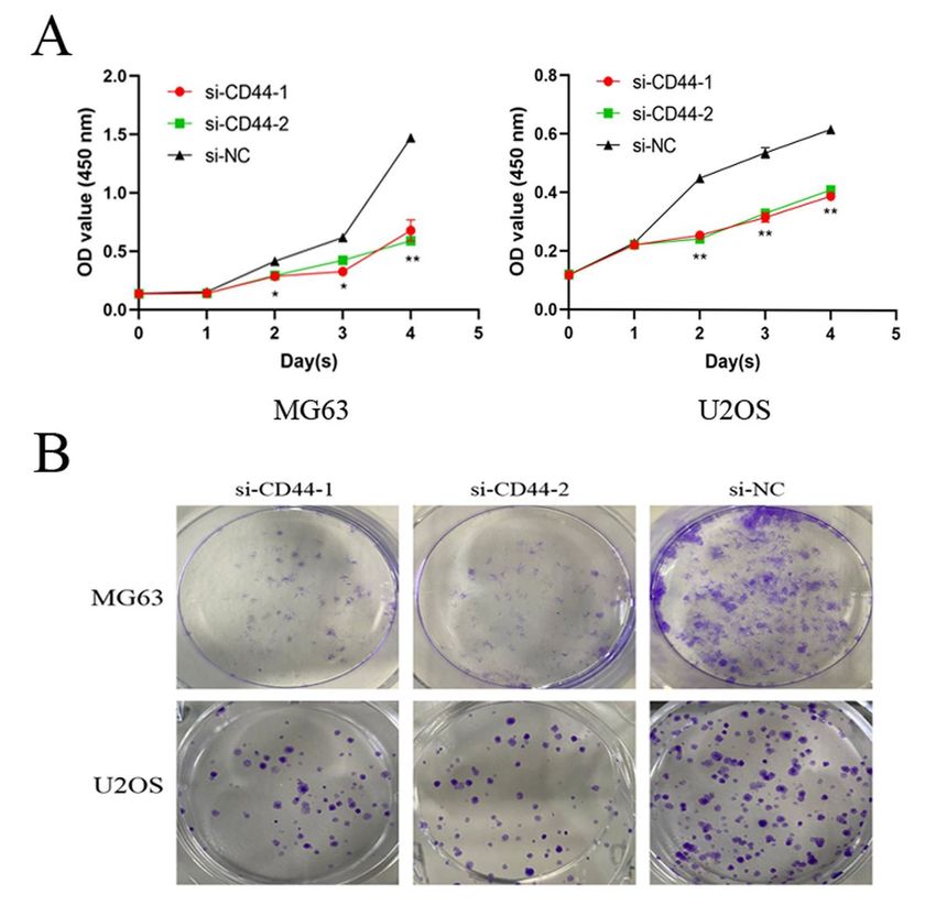
Abstract
Background

Osteosarcoma (OS) is a malignant bone tumour of mesenchymal origin. These tumours are
characterised by rich vascularisation, therefore promoting rapid proliferation and facilitating metastasis.
CD44 has been reported to be involved in OS, but its role and molecular mechanisms in the pathogenesis
of the disease are not fully determined.

Methods

In this study, we investigated the antitumor effect of CD44 on the development of OS and further explored
the molecular mechanisms. The expression of CD44, cathepsin S and MMP-9 was detected by Western
blot (WB) and reverse transcription-polymerase chain reaction (RT-qPCR) in different cell lines (MG63,
U2OS OS and hFOB 1.19). To elucidate the role of CD44 in OS, MG63 and U2OS cells were treated with
small interference RNA (siRNA) to knock down CD44, and the knockdown efficiency was validated with
GFP and RT-qPCR. Furthermore, cell proliferation was assayed using Cell Counting Kit‑8 (CCK-8) and
colony formation assays, and cell migration and invasion were assayed by transwell and wound-healing
assays.

Results

We found that CD44 expression in the MG63 and U2OS OS cell lines was markedly increased compared
to that of the human osteoblast hFOB 1.19 cell line. Knockdown of CD44 inhibited proliferation,
migration, and invasion of MG63 and U2OS cells, possibly by regulating the expression of cathepsin S in
OS.

Conclusion

Taken together, our data reinforced the evidence that CD44 knockdown inhibited cell proliferation,
migration, and invasion of OS cells accompanied by altered expression of cathepsin S. These findings
offer new clues for OS development and progression, suggesting CD44 as a potential therapeutic target
for OS.

Introduction
Osteosarcoma (OS) is the most common primary bone tumour, mainly occurring in children and
adolescents, and the third most frequent in adults, following chondrosarcoma and chordoma. The overall
incidence of OS is 3.4 per million cases per year worldwide [1], and the principal cause of death in
patients suffering from OS is pulmonary metastasis [2]. Osteosarcoma is a primary bone cancer
characterised by cancer cells that produce calcified osteoid extracellular matrix and inducing lung
metastases with a high frequency. Despite recent advances in treating osteosarcoma with a combination
of chemotherapy and surgery, the 5-year survival rate remains low, and the prognosis for patients is
                                                  Page 2/16
poor [3]. The cellular and molecular mechanisms underlying the progression of osteosarcoma, including
the rate of cancer cell proliferation, the formation of metastatic lesions and the development of drug
resistance, remain unclear [4].

Cluster of differentiation 44 (CD44) is a complex transmembrane adhesion glycoprotein considered an
essential bridge molecule as it links the extracellular matrix and intracellular skeletal proteins and
participates in intracellular signal transduction, affecting cell deformation or movement through
cytoskeletal changes [5]. Numerous studies have reported that CD44 not only participates in normal
cellular functions but also plays pivotal roles in pathological processes [6]. For example, CD44-RhoA-YAP
signalling mediates mechanics-induced fibroblast activation, and targeting this pathway could ameliorate
crystalline silica-induced silicosis and provide a potential therapeutic strategy to mitigate fibrosis [7]. It is
noteworthy that CD44 expression was found upregulated in different tumours [8-10], promoting cancer
cell invasion and migration [11, 12]. However, the role and molecular mechanisms of CD44 in the
development and progression of OS remain uncertain.

The present study analysed the CD44 expression pattern in OS cell lines using reverse
transcription‑quantitative PCR (RT‑qPCR) and Western blot (WB). Furthermore, loss‑of‑function
experiments were performed to investigate the biological roles of CD44 in OS. The results revealed that
CD44 expression was upregulated in OS cell lines. In addition, in vitro assays revealed that CD44
downregulation inhibited cell proliferation, migration, and invasion, probably by regulating cathepsin S.
These findings suggest that CD44 functions as an oncogene and future research may contribute to the
development of new tools for the diagnosis and treatment of OS.

Materials And Methods
Cell culture

The human OS MG63 and U2OS cell lines were purchased from the Cell Bank of Shanghai Institute of
Cell Biology (Shanghai, China) and cultured in modified Eagle’s medium (MEM, Gibco) supplemented with
10% fetal bovine serum (FBS, Gibco) at 37 ˚C with 5% CO2.

The normal human osteoblastic cell line hFOB 1.19 was purchased from the Cell Bank of Shanghai
Institute of Cell Biology (Shanghai, China) and maintained in D-MEM/F-12 (Gibco) supplemented with
10% FBS (Gibco) and 0.3 mg/mL Geneticin (G418; Gibco) at 37 ˚C with 5% CO2.

Small interference RNA transfection

Small interference RNA (siRNA) for transfection were purchased from Ribobio (Guangzhou, China).
Transfections (50 nM final concentration of siRNA) were performed using Invitrogen Lipofectamine 2000
(Thermo Fisher Scientific) following the protocols of the manufacturer. Three different siRNAs (si-CD44-
1,si-CD44-2, si-CD44-3) were tested, and si-CD44-1 and si-CD44-2 were selected for subsequent
experiments. A control siRNA (si-NC) was used in all the experiments.

                                                    Page 3/16
RT‑qPCR analysis
MG63 and U2OS cells were treated with si-CD44 or si-NC for 24 h, and total RNA was extracted from the
OS cell lines and the normal human osteoblastic cell line hFOB 1.19 using Trizol (Supersmart, China).
Next, 2 μg of RNA was used to synthesise the complementary DNA (cDNA) by reverse transcriptase
(ABclonal, China). The resulting complementary cDNA was used for PCR analysis. The relative levels of
genes were detected by RT‑qPCR using SYBR Premix Ex Taq™ (ABclonal, China). The PCR cycling
conditions were 95 ˚C for 5 min, followed by denaturation for 10 sec at 95 ˚C and extension for 20 sec at
60 ˚C for 40 cycles. GAPDH was used as an internal loading control. All reactions were performed in
triplicates. Fold changes were calculated using the 2‑ΔΔCq method. The primers were as follows: CD44
forward, 5'‑GAGCAGCACTTCAGGAGGTT‑3' and reverse, 5'‑TGGTTGCTGTCTCAGTTGCT-3'; cathepsin
S forward, 5'‑GCAGTGGCACAGTTGCATAA‑3' and reverse, 5'‑AGCACCACAAGAACCCATGT-3';
GAPDH forward, 5'‑GTCTCCTCTGACTTCAACAGCG‑3' and reverse, 5'‑ACCACCCTGTTGCTGTAGCCAA-3'.
Western blot analysis
MG63 and U2OS cells were treated with si-CD44 or si-NC for 48 h, and total proteins were extracted using
RIPA buffer containing protease inhibitor cocktail. Protein concentrations were determined using the BCA
Protein Assay (Multi sciences). Proteins (30 µg/lane) were separated by 10% SDS‑PAGE and transferred
to PVDF membranes. The membranes were blocked with 5% non‑fat milk for 2 h at room temperature
(RT). Next, the membranes were incubated with anti-CD44 (1:2000, ABclonal, China), cathepsin
S (1:2000, Affinity, China), and MMP-9 (1:2000, ABclonal, China) at 4 ˚C overnight. Subsequently, the
appropriate horseradish peroxidase (HRP)-linked secondary antibodies (1:5000, Sera care) were used to
visualise the immunoreactivity. GAPDH was used as an internal control. The intensity of each band was
measured with Image J.
Cell Counting Kit‑8 (CCK‑8) assay
MG63 and U2OS cells were treated with si-CD44 or si-NC for 24 h. Cells were prepared into suspension
and MG-63, and U2OS cells were seeded in 96-well plates at a density of 1x10³ cells per well and
incubated in a humidified incubator at 37 ˚C for 24, 48, 72 h and 96 h. Subsequently, the cells were
incubated with 10 µl CCK‑8 solution for another 1 h at 37 ˚C. Optical density (OD) was determined at a
wavelength of 450 nm.
Colony formation assay
MG63 and U2OS cells were treated with CD44 siRNA or negative control for 24 h. Cells were then
resuspended and seeded in 6-well plates at a density of 500 cells per well and cultured for 15 days.
Subsequently, cells were fixed with pre-cooled methanol for 30 min at RT and stained with 0.1% crystal
violet for 20 min at RT, washed twice with PBS and twice with double distilled water. The colonies were
counted and analysed under a light microscope.
Page 4/16Wound-healing assay
To evaluate the role of CD44 in OS cell migration, MG63 and U2OS cells were transfected with si-CD44 or
si-NC for 24 h. Cells were resuspended and seeded in 6-well plates at a density of 1×10⁶ cells per well,
and 2 ml of culture medium supplemented with 10% FBS was added. Cells were grown to 90%
confluence, and then a uniform and consistent wound was scraped on the bottom of the 6-well plate with
a 200 μL plastic pipette tip (time set as 0 h). PBS was used to remove floating cells. Subsequently, cells
were incubated in fresh complete medium (1% FBS) for 0, 24 and 48 h and the number of migrated cells
were observed and counted under a light microscope.
Transwell assay
Migration and invasion abilities of MG‑63 and U2OS cells were measured using a transwell assay. The
Matrigel was incubated at 37˚C for 5 h before testing. OS cells were transfected with si-CD44 or si-NC for
24 h. 1x10⁵ transfected cells were resuspended in serum‑free medium and seeded in the upper chamber
with or without Matrigel (BD Biosciences) for the invasion and migration assays, respectively.
Subsequently, medium containing 20% FBS was added to the lower chambers. Following a 24 h
incubation, the cells from the upper compartments were scraped off with cotton swabs, while the cells
that migrated to or invaded the lower surface of the membrane were fixed with pre-cooled methanol at RT
for 20 min and stained with 0.1% crystal violet at RT for 20 min. The stained cells were counted in five
random fields off view under a light microscope at x200 magnification, and all experiments were repeated
three times.
Statistical analysis
The results are presented as the mean ± SD. Statistical analyses were performed using SPSS 23.0 (IBM
Corp, USA) and GraphPad Prism 9.0 (La Jolla, CA, USA) software. ANOVA test was applied to compare
differences among multiple groups. P < 0.05 was considered to indicate a statistically significant
difference.
Results
1. CD44 is upregulated in OS cell lines. The present study first examined CD44 expression levels in the
MG63, U2OS, and hFOB 1.19 cell lines by WB and RT-qPCR. Compared with the hFOB 1.19 cell line,
CD44 expression was markedly upregulated in the OS cell lines (Fig. 1A-C).
2. CD44 knockdown in MG63 and U2OS cells in vitro. MG63 and U2OS cells were transfected with si-
CD44 or si-NC for 24 h, and the transfection efficiency was detected using fluorescence
microscopy (Fig. 2A). CD44 mRNA and protein expression was quantified by RT-PCR and Western
blot, respectively, after CD44 knockdown in MG63 and U2OS cells. As shown in Figure 2B and C, the
results revealed that the siRNA transfection decreased CD44 expression, but as expected, no
significant difference was observed between the si‑NC and control groups. For subsequent
Page 5/16experiments, two (siCD44-1, siCD44-2) of the three CD44 siRNA with high transfection efficiency were
selected.
3. CD44 knockdown inhibited the proliferation of MG63 and U2OS cells. To assess the role of CD44 in
MG63 and U2OS cell proliferation, siRNA was transfected to silence CD44 expression. Subsequently,
cell proliferation was assessed using CCK‑8 and colony formation assays. As demonstrated by the
result of the CCK‑8 assay, cell growth was suppressed in CD44-silenced MG63 and U2OS cells
compared with the si‑NC-transfected cells (Fig. 3A). In addition, the colony formation ability of
si‑CD44‑transfected cells was decreased (Fig. 3B). These results revealed that downregulation of
CD44 markedly decreased the proliferation of MG63 and U2OS cells.
4. CD44 knockdown inhibited the migration and invasion of MG63 and U2OS cells. To investigate the
role of CD44 in the migration and invasion of OS cells, the wound-healing assay were used at 0, 12,
or 24 h after transfection. The result of the wound-healing assay showed that the migration
distances of cells transfected with si-NC was compared to the migration distances in CD44-silenced
cells (Fig. 4A). The result of the transwell migration and invasion assay showed that the number of
control si-NC-transfected cells was more than the number of CD44-silenced cells (Fig. 4B).
Furthermore, Western blot was applied to evaluate the matrix metalloproteinase MMP‑9 protein
levels. As shown in Figure 4C, silencing of CD44 decreased MMP‑9 expression in MG‑63 and U2OS
cells compared with the si-NC group. Therefore, the results suggested that the migration and
invasion abilities of MG‑63 and U2OS cells were suppressed following CD44 knockdown.
5. CD44 knockdown downregulated the expression of cathepsin S in MG63 and U2OS cells. To further
confirm the underlying mechanism of CD44 in OS, we detected the mRNA and protein expression of
cathepsin S by RT-PCR and Western blot after CD44 knockdown in MG63 and U2OS cells. The mRNA
and protein levels of cathepsin S in the CD44-silenced OS cells were markedly reduced compared
with the control cells (si-NC) at 24 and 48 h after transfection (p < 0.01) (Fig. 5A–5B). These data
indicated that CD44 exerted its effects in OS in part by regulating cathepsin S.
Discussion
The current treatment for osteosarcoma is surgical resection and combined neoadjuvant chemotherapy,
which has increased the 5-year overall survival rate for osteosarcoma patients from less than 20% to
more than 60% [13]. However, survival rates for patients with metastatic or recurrent osteosarcoma have
remained virtually unchanged over the past 30 years, with an overall 5-year survival rate as low as 20% [3,
14]. The molecular mechanisms underlying the development of OS have not been fully explored.
Therefore, it is crucial to elucidate the predictive markers of OS and their potential regulatory
mechanisms.
CD44, also known as homing cell adhesion molecule, is a cell surface transmembrane glycoprotein
molecule involved in cell-cell and cell-extracellular matrix communication. In humans, CD44 proteins are
encoded by a highly conserved gene located on the short arm of chromosome 11 (11p13) [15]. CD44 can
affect cell growth, proliferation and motility through changes in the cytoskeleton, as well as being
Page 6/16involved in intracellular signal transduction [16]. Therefore, CD44 is a critical bridging molecule between
the extracellular matrix and intracellular skeletal proteins.
CD44 expression is elevated in a wide range of malignant tumours [17], such as colon tumours [18],
ovarian clear cell carcinoma [19], and glioblastoma [20]. Overexpression of CD44 splice mesenchymal
isoform in OS cells induced EMT and invasion, followed by the gain of stem-like characteristics and
chemoresistance [21]. In gastric cancer, CD44V6 regulates the transformation of normal mucosal
epithelial cells into tumour cells and is associated with gastric cancer differentiation, lymph node
metastasis, and pathological staging [22]. CD44 potentiates AKT activation to induce phosphorylation
and nuclear translocation of Mdm2, which terminates the p53 genomic surveillance response. This
allows DNA-damaged hepatocytes to escape p53-induced death and go on to become HCC
progenitors [23]. We confirmed that CD44 is highly expressed in osteosarcoma cell lines compared to
hFOB 1.19 human osteoblasts. Hence, we next evaluated the effects of CD44 on the proliferation,
invasion, and migration of osteosarcoma cells.
We found that downregulation of CD44 inhibited the proliferation, invasion, and migration ability of
osteosarcoma cells MG63 and U2OS, which is consistent with the reported role of CD44 in most
tumours [8, 10, 17], suggesting that CD44 may be a pro-oncogene in osteosarcoma.
To know more about the molecular mechanisms, we next investigated the expression of cathepsin S. Like
matrix metalloproteinases, members of the histone family have been associated with metastasis and
cancer recurrence [24]. Cathepsin S (CTSS), a member of the histone family of enzymes, is highly
expressed in renal clear cell carcinoma [25], hepatocellular carcinoma [26], cervical cancer [27], lung
cancer [28] and other tumours and is an essential regulator of tumour growth and invasion. Suppression
of cell migration and invasion by modulation of Ca2+-dependent downstream effectors after CTSS
inhibition [29]. The expression of CTSS was regulated by PI3K/Akt and Ras/Raf/MAPK signalling
pathways, is a candidate target for blocking the metastasis of breast and oral cancers [30, 31]. However,
it is not clear what role cathepsin S plays in osteosarcoma. Previous experiments found that cathepsin S
was highly expressed in osteosarcoma cells and that silencing inhibited the invasion and metastasis of
osteosarcoma cells.
Previous studies have reported that CD44 can promote tumour stem cell properties in triple-negative
breast cancer by regulating the PI3K/AKT pathway [32]. Interaction of hyaluronan and CD44 enhances
neutrophil phagocytosis and IL-8 production via the p38 and ERK1/2-MAPK signalling pathways [33].
Activation of the receptor complex CD74/CD44 can lead to activation of the ERK1/2, PI3K-Akt signalling
cascade, NFκB and AMP-activated protein kinase (AMPK) pathways [34]. Therefore, we speculate that
CD44 may exert its biological effects on osteosarcoma cells through the regulation of cathepsin S. The
experimental results showed that cathepsin S expression was downregulated after CD44 silencing
compared to the si-NC group. Therefore, we suggest that CD44 may exert regulation of osteosarcoma cell
proliferation, invasion, and metastasis through the regulation of cathepsin S. However, the specific
Page 7/16regulatory mechanisms of the two still need to be further explored, and this will be the focus of our
subsequent work.
Conclusion
This study shows that inhibition of CD44 attenuates cell proliferation, migration, and invasion, possibly
by regulating the expression of cathepsin S in OS cells. These findings suggest that CD44 may be an
oncogenic factor in the progression of OS and may be a promising molecular marker for the diagnosis
and treatment of OS.
Abbreviations
OS: Osteosarcoma; CD44: cluster of differentiation 44; CCK8: Cell Counting Kit‑8; MMP9: Matrix
metalloproteinase 9; WB: Western blot; RT-qPCR: reverse transcription-polymerase chain reaction; siRNA:
small interference RNA
Declarations
Acknowledgements
Thank you for medical science research project in Hebei province (20200352, 20200378) and natural
science research youth fund for higher education in Hebei province (QN2020107) support.
Authors’ contributions
Lingwei Kong was responsible for manuscript writing and experiment conducting; Hairu Ji and Xintian
Gan contributed to data collection and analysis; Sheng Cao collected the literature and explained the
results; Yu Jin designed this study and reviewed this article. The authors read and approved the final
manuscript.
Funding
This work was supported by medical science research project in Hebei province (20200352, 20200378)
and natural science research youth fund for higher education in Hebei province (QN2020107).
Availability of data and materials
The datasets supporting the conclusions of this article are included within the article.
Declarations
Ethics approval and consent to participate Not applicable.
Consent for publication
Page 8/16Not applicable.
Competing interests
The authors report no declarations of interest.
References
[1] A.C. da Costa, F. Santa-Cruz, L.A.R. Mattos, M.A. Rego Aquino, C.R. Martins, A.A. Bandeira Ferraz, J.L.
Figueiredo, Cathepsin S as a target in gastric cancer, Mol Clin Oncol 12(2) (2020) 99-103.
[2] M.F. Heymann, F. Lézot, D. Heymann, The contribution of immune infiltrates and the local
microenvironment in the pathogenesis of osteosarcoma, Cellular immunology 343 (2019) 103711.
[3] L. Marchandet, M. Lallier, C. Charrier, M. Baud'huin, B. Ory, F. Lamoureux, Mechanisms of Resistance to
Conventional Therapies for Osteosarcoma, Cancers (Basel) 13(4) (2021).
[4] K.H. Lu, R.C. Lin, J.S. Yang, W.E. Yang, R.J. Reiter, S.F. Yang, Molecular and Cellular Mechanisms of
Melatonin in Osteosarcoma, Cells 8(12) (2019).
[5] G.T. Lim, D.G. You, H.S. Han, H. Lee, S. Shin, B.H. Oh, E.K.P. Kumar, W. Um, C.H. Kim, S. Han, S. Lee, S.
Lim, H.Y. Yoon, K. Kim, I.C. Kwon, D.G. Jo, Y.W. Cho, J.H. Park, Bioorthogonally surface-edited extracellular
vesicles based on metabolic glycoengineering for CD44-mediated targeting of inflammatory diseases,
Journal of extracellular vesicles 10(5) (2021) e12077.
[6] Y. Dong, A.A. Arif, J. Guo, Z. Ha, S.S.M. Lee-Sayer, G.F.T. Poon, M. Dosanjh, C.D. Roskelley, T. Huan, P.
Johnson, CD44 Loss Disrupts Lung Lipid Surfactant Homeostasis and Exacerbates Oxidized Lipid-
Induced Lung Inflammation, Front Immunol 11 (2020) 29.
[7] S. Li, C. Li, Y. Zhang, X. He, X. Chen, X. Zeng, F. Liu, Y. Chen, J. Chen, Targeting Mechanics-Induced
Fibroblast Activation through CD44-RhoA-YAP Pathway Ameliorates Crystalline Silica-Induced Silicosis,
Theranostics 9(17) (2019) 4993-5008.
[8] R. Pothuraju, S. Rachagani, S.R. Krishn, S. Chaudhary, R.K. Nimmakayala, J.A. Siddiqui, K. Ganguly, I.
Lakshmanan, J.L. Cox, K. Mallya, S. Kaur, S.K. Batra, Molecular implications of MUC5AC-CD44 axis in
colorectal cancer progression and chemoresistance, Mol Cancer 19(1) (2020) 37.
[9] K.E. Gomez, F. Wu, S.B. Keysar, J.J. Morton, B. Miller, T.S. Chimed, P.N. Le, C. Nieto, F.N. Chowdhury, A.
Tyagi, T.R. Lyons, C.D. Young, H. Zhou, H.L. Somerset, X.J. Wang, A. Jimeno, Cancer Cell CD44 Mediates
Macrophage/Monocyte-Driven Regulation of Head and Neck Cancer Stem Cells, Cancer Res 80(19)
(2020) 4185-4198.
[10] X. Liu, R. Taftaf, M. Kawaguchi, Y.F. Chang, W. Chen, D. Entenberg, Y. Zhang, L. Gerratana, S. Huang,
D.B. Patel, E. Tsui, V. Adorno-Cruz, S.M. Chirieleison, Y. Cao, A.S. Harney, S. Patel, A. Patsialou, Y. Shen, S.
Page 9/16Avril, H.L. Gilmore, J.D. Lathia, D.W. Abbott, M. Cristofanilli, J.S. Condeelis, H. Liu, Homophilic CD44
Interactions Mediate Tumor Cell Aggregation and Polyclonal Metastasis in Patient-Derived Breast Cancer
Models, Cancer discovery 9(1) (2019) 96-113.
[11] H. Cao, J. Xiao, M.E. Reeves, K. Payne, C.S. Chen, D.J. Baylink, G. Marcucci, Y. Xu, Discovery of
proangiogenic CD44+mesenchymal cancer stem cells in an acute myeloid leukemia patient's bone
marrow, J Hematol Oncol 13(1) (2020) 63.
[12] T. Kong, R. Ahn, K. Yang, X. Zhu, Z. Fu, G. Morin, R. Bramley, N.C. Cliffe, Y. Xue, H. Kuasne, Q. Li, S.
Jung, A.V. Gonzalez, S. Camilleri-Broet, M.C. Guiot, M. Park, J. Ursini-Siegel, S. Huang, CD44 Promotes PD-
L1 Expression and Its Tumor-Intrinsic Function in Breast and Lung Cancers, Cancer Res 80(3) (2020) 444-
457.
[13] C. Zheng, F. Tang, L. Min, F. Hornicek, Z. Duan, C. Tu, PTEN in osteosarcoma: Recent advances and
the therapeutic potential, Biochim Biophys Acta Rev Cancer 1874(2) (2020) 188405.
[14] J. Cui, D. Dean, F.J. Hornicek, Z. Chen, Z. Duan, The role of extracelluar matrix in osteosarcoma
progression and metastasis, Journal of experimental & clinical cancer research : CR 39(1) (2020) 178.
[15] A. Ouhtit, B. Rizeq, H.A. Saleh, M.M. Rahman, H. Zayed, Novel CD44-downstream signaling pathways
mediating breast tumor invasion, International journal of biological sciences 14(13) (2018) 1782-1790.
[16] P. Govindaraju, L. Todd, S. Shetye, J. Monslow, E. Puré, CD44-dependent inflammation, fibrogenesis,
and collagenolysis regulates extracellular matrix remodeling and tensile strength during cutaneous
wound healing, Matrix biology : journal of the International Society for Matrix Biology 75-76 (2019) 314-
330.
[17] C. Chen, S. Zhao, A. Karnad, J.W. Freeman, The biology and role of CD44 in cancer progression:
therapeutic implications, J Hematol Oncol 11(1) (2018) 64.
[18] S.P.J. Joosten, M. Spaargaren, H. Clevers, S.T. Pals, Hepatocyte growth factor/MET and CD44 in
colorectal cancer: partners in tumorigenesis and therapy resistance, Biochim Biophys Acta Rev Cancer
1874(2) (2020) 188437.
[19] Y. Yamada, T. Miyamoto, H. Kashima, H. Kobara, R. Asaka, H. Ando, S. Higuchi, K. Ida, T. Shiozawa,
Lipocalin 2 attenuates iron-related oxidative stress and prolongs the survival of ovarian clear cell
carcinoma cells by up-regulating the CD44 variant, Free radical research 50(4) (2016) 414-25.
[20] T.T. Lah, M. Novak, B. Breznik, Brain malignancies: Glioblastoma and brain metastases, Semin
Cancer Biol 60 (2020) 262-273.
[21] R. Bhattacharya, T. Mitra, S. Ray Chaudhuri, S.S. Roy, Mesenchymal splice isoform of CD44 (CD44s)
promotes EMT/invasion and imparts stem-like properties to ovarian cancer cells, J Cell Biochem 119(4)
(2018) 3373-3383.
Page 10/16[22] S. Liang, H.L. Li, G.Y. Han, J.H. Cui, CD44V6 regulates gastric carcinoma occurrence and development
through up-regulating VEGF expression, European review for medical and pharmacological sciences
21(22) (2017) 5121-5128.
[23] D. Dhar, L. Antonucci, H. Nakagawa, J.Y. Kim, E. Glitzner, S. Caruso, S. Shalapour, L. Yang, M.A.
Valasek, S. Lee, K. Minnich, E. Seki, J. Tuckermann, M. Sibilia, J. Zucman-Rossi, M. Karin, Liver Cancer
Initiation Requires p53 Inhibition by CD44-Enhanced Growth Factor Signaling, Cancer cell 33(6) (2018)
1061-1077.e6.
[24] A. Steimle, H. Kalbacher, A. Maurer, B. Beifuss, A. Bender, A. Schafer, R. Muller, I.B. Autenrieth, J.S.
Frick, A novel approach for reliable detection of cathepsin S activities in mouse antigen presenting cells, J
Immunol Methods 432 (2016) 87-94.
[25] E. Song, W. Song, M. Ren, L. Xing, W. Ni, Y. Li, M. Gong, M. Zhao, X. Ma, X. Zhang, R. An, Identification
of potential crucial genes associated with carcinogenesis of clear cell renal cell carcinoma, J Cell
Biochem 119(7) (2018) 5163-5174.
[26] X. Wang, L. Xiong, G. Yu, D. Li, T. Peng, D. Luo, J. Xu, Cathepsin S silencing induces apoptosis of
human hepatocellular carcinoma cells, American journal of translational research 7(1) (2015) 100-10.
[27] M.C. Hsin, Y.H. Hsieh, P.H. Wang, J.L. Ko, I.L. Hsin, S.F. Yang, Hispolon suppresses metastasis via
autophagic degradation of cathepsin S in cervical cancer cells, Cell Death Dis 8(10) (2017) e3089.
[28] R. Brown, S. Nath, A. Lora, G. Samaha, Z. Elgamal, R. Kaiser, C. Taggart, S. Weldon, P. Geraghty,
Cathepsin S: investigating an old player in lung disease pathogenesis, comorbidities, and potential
therapeutics, Respir Res 21(1) (2020) 111.
[29] H.H. Lin, S.J. Chen, M.R. Shen, Y.T. Huang, H.P. Hsieh, S.Y. Lin, C.C. Lin, W.W. Chang, J.Y. Chang,
Lysosomal cysteine protease cathepsin S is involved in cancer cell motility by regulating store-operated
Ca(2+) entry, Biochim Biophys Acta Mol Cell Res 1866(12) (2019) 118517.
[30] M.J. Hsieh, C.W. Lin, M.K. Chen, S.Y. Chien, Y.S. Lo, Y.C. Chuang, Y.T. Hsi, C.C. Lin, J.C. Chen, S.F. Yang,
Inhibition of cathepsin S confers sensitivity to methyl protodioscin in oral cancer cells via activation of
p38 MAPK/JNK signaling pathways, Scientific reports 7 (2017) 45039.
[31] J. Gautam, Y.K. Bae, J.A. Kim, Up-regulation of cathepsin S expression by HSP90 and 5-HT7 receptor-
dependent serotonin signaling correlates with triple negativity of human breast cancer, Breast Cancer Res
Treat 161(1) (2017) 29-40.
[32] J. Bai, W.B. Chen, X.Y. Zhang, X.N. Kang, L.J. Jin, H. Zhang, Z.Y. Wang, HIF-2α regulates CD44 to
promote cancer stem cell activation in triple-negative breast cancer via PI3K/AKT/mTOR signaling, World
journal of stem cells 12(1) (2020) 87-99.
Page 11/16[33] C.H. Lu, C.H. Lin, K.J. Li, C.Y. Shen, C.H. Wu, Y.M. Kuo, T.S. Lin, C.L. Yu, S.C. Hsieh, Intermediate
Molecular Mass Hyaluronan and CD44 Receptor Interactions Enhance Neutrophil Phagocytosis and IL-8
Production via p38- and ERK1/2-MAPK Signalling Pathways, Inflammation 40(5) (2017) 1782-1793.
[34] H. Su, N. Na, X. Zhang, Y. Zhao, The biological function and significance of CD74 in immune
diseases, Inflamm Res 66(3) (2017) 209-216.
Figures
Figure 1
CD44 is upregulated in OS cell lines. (A) CD44 mRNA levels in MG63, U2OS, and hFOB 1.19 cell lines. **P
< 0.01 vs. hFOB group. (B) and (C) CD44 protein levels in MG63, U2OS, and hFOB 1.19 cell lines. **P <
0.01 vs. hFOB group.
Page 12/16Figure 2
CD44 knockdown in MG63 and U2OS cells in vitro. (A) Transfection efficiency of MG‑63 and U2OS cells
was assessed by fluorescence microscopy. GFP, green fluorescent protein. Magnification, x200. (B)
Reverse transcription‑quantitative PCR analysis was used to assess the mRNA expression levels of CD44
in MG‑63 and U2OS cells after transfection for 24h. **P < 0.01 vs. si-NC group. (C) Western blot was used
to assess CD44 expression levels in MG‑63 and U2OS cells 48 h after transfection.
Page 13/16Figure 3
CD44 knockdown inhibited the proliferation of MG63 and U2OS cells. (A) Cell proliferation and (B) colony
formation in MG 63 and U2OS cells. All data are presented as the mean ± SD of n = 3 experiments (*P <
0.05, **P < 0.01 vs. si-NC group).
Page 14/16Figure 4
CD44 knockdown inhibited the migration and invasion of MG63 and U2OS cells. (A) A wound-healing
assay was performed to detect the migration of MG 63 and U2OS cells. (B) Transwell assay was
performed to detect migration and invasion of MG 63 and U2OS cells. Magnification, x200. (C)
Expression levels of migration and invasion related proteins (MMP 9) were detected by Western blotting.
Page 15/16Figure 5
CD44 knockdown downregulated the expression of cathepsin S in MG63 and U2OS cells. (A) mRNA
expression levels of CD44, cathepsin S, and β-actin were detected by RT-qPCR. (B) Protein expression
levels of CD44, cathepsin S, and β-actin were detected by Western blotting. **P < 0.01 vs. control group.
Page 16/16You can also read