Cellular and Molecular Characteristics of Established Childhood Soft-Tissue Sarcoma Cell Lines
←
→
Page content transcription
If your browser does not render page correctly, please read the page content below
Journal of
 Cancer Research and Therapeutic Oncology
  Review                                                                                                                 Open Access

 Cellular and Molecular Characteristics of Established Childhood Soft-Tissue
 Sarcoma Cell Lines
 Tohru Sugimoto1,2,3,4,*, Hiroshi Kuroda1,3,4,5, Yasumishi Kuwahara3, Yoshiki Katsumi3, Akiko Misawa3, Hiroshi
 Moritake4, Hajime Hosoi3
 1
  Shiga-ken Saiseikai Nursing School, Saiseikai Imperial Gift Foundation Inc., Ritto, Shiga, Japan
 2
  Saiseikai Shiga Hospital, Saiseikai Imperial Gift Foundation Inc., Ritto, Shiga, Japan
 3
  Department of Pediatrics, Kyoto Prefectural University of Medicine, Kyoto, Japan
 4
  Department of Pediatrics, University of Miyazaki, Miyazaki, Japan
 5
  Department of Pediatrics, Kyoto City Hospital, Kyoto, Japan
 *Corresponding author: Tohru Sugimoto, Shiga-ken Saiseikai Nursing School, Saiseikai Imperial Gift Foundation Inc., Ritto,
 Shiga, Shiga 520-3046, Japan. Tel:+81-77-553-7002; Fax: +81-77-553-7246; E-mail: tosugimo@koto.kpu-m.ac.jp

 Received Date: July 22, 2019 Accepted Date: September 20, 2019 Published Date: September 23, 2019

 Citation: Tohru Sugimoto (2019) Cellular and Molecular Characteristics of Established Childhood Soft-Tissue Sarcoma Cell
 Lines. J Cancer Res Therap Oncol 7: 1-12.

     Abstract

               From 1984 to 2013, we established 29 childhood tumor cell lines, including 13 soft-tissue-sarcoma cell lines. Here,
     we provide an overview of these soft-tissue sarcoma cell lines, their origins, characteristics, and highlight their potential as
     valuable research tools for fundamental research and development of new treatments. The cell lines were established from
     three patients with rhabdomyosarcoma (RMS), five patients with Ewing sarcoma family tumors, four patients with rhabdoid
     tumors, and one patient with clear cell sarcoma of soft parts. In particular, we demonstrate the potential of the MRT cell
     lines in preclinical in vivo and in vitro studies to evaluate molecular target therapy using gefitinib and trastuzumab activated
     antibody-dependent cellular cytotoxicity. Moreover, the clear cell sarcoma cell line, MP-CCS-SY, was established from a
     metastatic tumor in the left Achilles tendon of a 17-year-old girl, which represents a rare cell line for this cancer, and will help
     to gain a better understanding of the molecular biology of this malignancy and serve as a useful tool for developing boron
     neutron capture therapy.

     Keywords: Soft-tissue-sarcomas; Rhabdomyosarcoma; Ewing sarcoma family tumor; Malignant rhabdoid tumor; Clear cell
     sarcoma of soft parts; Clinical characteristics.

     Abbreviations: RMS: rhabdomyosarcoma; PNET: peripheral primitive neuroectodermal tumor; ESFT: Ewing sarcoma fam-
     ily of tumor; MRT: Malignant rhabdoid tumor; CCS: clear cell sarcoma of soft parts

 ©2019 The Authors. Published by the JScholar under the terms of the Crea-
 tive Commons Attribution License http://creativecommons.org/licenses/
 by/3.0/, which permits unrestricted use, provided the original author and
 source are credited.

JScholar Publishers                                                                          J Cancer Res Therap Oncol 2019 | Vol 7: 202
2
Introduction                                                                 In the last 30 years, from 1984 to 2013, we have estab-
                                                                   lished 29 tumor cell lines from children, including 13 soft-tis-
          Soft-tissue sarcomas originate from immature embry-      sue sarcoma cell lines at three Institutes (SCMS, KP and MP).
onal mesoderm or mesenchymal tissues, and can therefore po-        Here, we established from children with [1] rhabdomyosarcomas
tentially develop from any anatomical site. Recent advances in     (RMS), [2] Ewing sarcoma family tumors (EW), [3] malignant
progressive therapy have led to a high cure rate for soft-tissue   rhabdoid tumors (MRT), and [4] clear cell sarcoma of soft parts
sarcomas with combinations of chemotherapy, surgery, radiation     (CCS) (Table 1). The goal of this review is to further highlight
therapy, and supportive therapy. In addition, chromosomal anal-    the utility and potential of these soft-tissue sarcoma cell lines to
ysis and more recent advanced molecular biological techniques      improve understanding of basic and clinical aspects of pediatric
that can identify chromosomal aberrations have improved both       oncology.
the diagnosis and prognosis of the disease, along with insight
into the underlying mechanism of tumor biology.

                              Age/gen-                                                                 Out-
  No          Cell line                       Primary tumor             Stage    Culture Sample                      References
                                der                                                                    come
                                                        Rhadomyosarcoma

   1       SCMC-RM2            10y/F         Rt-abdominal wall           IV          Bone marrow        Dead             11

   2       KP-RMS-DH           14y/M        Lt-mandibular cavity         IV          Bone marrow        Dead        Unpublished

   3       KP-RMS-KH            14/F              Perinea               IV       Primary tumor          Dead        Unpublished

                                                   Ewing sarcoma family tumor

   1        KP-EW-YI           16y/M              Rt-pelvis              II      Primary tumor          Dead             17

   2        KP-EW-MS           16y/F             Rt-breech              IV           Bone marrow        Dead             17

   3      MP-ASKIN-SA          13y/M              Lt-chest               II      Primary tumor          Dead             18

   4       KP-PNET-TO          15y/M              Lt-kidney             IV              Lt-iliac        Dead        Unpublished

   5        KP-EW-AK           15y/M             Lt-scapula             IV       Primary tumor          Dead        Unpublished

                                                    Malignant rhabdoid tumor

   1       KP-MRT-NS           2m/F              Lt-kidney               II            Ascites          Dead          19, 20, 24

   2       KP-MRT-RY           1m/M              Lt-kidney               I       Primary tumor          Dead        19, 20, 22, 23

   3       MP-MRT-AN           3m/F                 liver               IV      Peripheral blood        Dead          20, 21, 24

   4       KP-MRT-YM           5m/M             Rt-chest wall            I       Primary tumor          Alive         20, 22, 23

                                                  Clear cell sarcoma of soft parts

                                                                                     Lt-popliteall
   1       MP-CCS-SY           13y/F             Lt-Achillis             II                             Dead           26, 28
                                                                                         fossa

Table 1: Our established soft-tissue-sarcoma cell lines from children

 JScholar Publishers                                                                      J Cancer Res Therap Oncol 2019 | Vol 7: 202
3
Establishment of 13 soft-tissue sarcoma cell                            nosis of RMS in these patients (Figure 1).

lines
          The idea for establishing a cell line was born in 1983,
when one of the authors (TS) was a clinical research fellow at
Cell Culture Laboratory, Department of Molecular Biology, Ros-
well Park Memorial Institute (Buffalo, NY, USA), as establish-
ment of tumor cell lines was considered to be an important mark
of a researcher at that time. Here, we focus on the cell lines estab-
lished for soft-tissue sarcomas with the ultimate goal to unravel
novel diagnostic or therapeutic methods.

         Tumor samples from patients for cell culture were ob-
tained by biopsy, operation, or autopsy and were finely minced
with a scalpel and cultured. Mononuclear cell fractions from the
bone marrow or peripheral metastatic cells were prepared by Fi-
coll-Hypaque centrifugation. The cells were cultured in RPMI
                                                                        Figure. 1 Expression of MyoD1 in rhabdomyosarcoma, neuro-
1640 medium containing penicillin (100 U/ml), streptomycin
                                                                        blastoma, and other tumor cell lines
(100 µg/ml), and 15% heated-inactivated fetal calf serum at 37℃
                                                                        PAX3/FKHR chimera product in alveolar RMS cell lines
in a 5% CO2 atmosphere. The medium was replaced every 3–4
days. Cell lines were considered to be established when they were                  One of the molecular characteristics of alveolar RMS is
cultured for more than 60 passages over a 2-year period [1].            the translocation t (2;13) (q35;q14), which results in the PAX3/
                                                                        FKHR chimera product, whereas the translocation t (1;13) (p36;
RMS                                                                     q14) results in the PAX7/FKHR chimera product. Indeed, the
                                                                        former translocation and PAX3/FKHR chimera were confirmed
Incidence and pathological findings in RMS
                                                                        in our alveolar RMS cell lines (Figure 2), whereas the latter was
          RMS is derived from the immature embryological me-            not detected in any of the cell lines. Therefore, based on a reverse
soderm or mesenchymal tissues during the formation of skeletal          transcription-polymerase chain reaction (RT-PCR) results, alve-
muscle cells, and is the most common soft-tissue sarcoma (50%),         olar RMS was confirmed in two of our cell lines (KP-RMS-DH
accounting for 5% of all childhood cancers. RMS can be classi-          and SCMC-RM2). Thus, using recent molecular biology tech-
fied into five groups: embryonal, botryoid, spindle, alveolar, and      niques, we were able to confirm the presence of the PAX3/FKHR
pleomorphic, as per the 1992 World Health Organization Clas-            chimera gene to identify and diagnose alveolar RMS more spe-
sification of Soft Tissue Tumors [2]. Antibodies against skeletal       cifically [8, 9].
muscle-specific proteins (e.g., desmin, alpha- and gamma-mus-
cle actin, alpha-sarcomeric actin) are effective for the immuno-
fluorescence-based diagnosis of RMS [3,4]

Expression of MyoD1 in RMS cell lines

          Myoblast determination protein 1 (MyoD1) expression
has been found to be limited to RMS, and is considered to be re-
sponsible for the lack of differentiation to mature skeletal muscle
cells [5–7]. MyoD1 was confirmed to be expressed in our RMS
cell lines (RD, SCMC-RM2, KP-RMS-KH, KP-RMS-DH, SC-
MC-MM-1 series, and SMS-CTR series), and was not expressed
in our neuroblastoma cells and other cell lines, including Wilms
tumors and Ewing sarcoma cell lines. The positive expression of
                                                                        Figure. 2. PAX3/FKHR chimera gene in alveolar rhabdomyosar-
MyoD1 mRNA specific to the RMS cell lines confirmed the diag-
                                                                        coma cell lines

 JScholar Publishers                                                                         J Cancer Res Therap Oncol 2019 | Vol 7: 202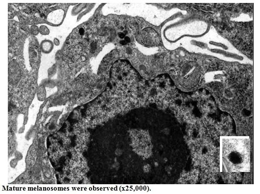
4
MYCN amplification in RMS cell lines

          In September 1985, a tumor on the right abdominal
wall was observed in an 11-year-old female. A cell line desig-
nated SCMC-RM2 was then established from her bone marrow
cells, and transferred to our department for further cellular and
biological characterization.

         Cytoplasmic protein analysis demonstrated a myogenic
origin, with the expression of desmin (Figure 3B), myoglobin,
alpha- and gamma-muscle actin (using the HHF35 monoclonal
antibody) (Figure 3C), MyoD1 mRNA (Figure 1), and alpha-sar-
comeric actin [3,4].

                                                                     Figure. 4 MYCN-amplification (a) and expression (b) in SC-
                                                                     MC-RM2 rhabdomyosarcoma cell line
                                                                     Ewing sarcoma family tumor
                                                                              Classical Ewing sarcoma of the bone, extraskeletal Ew-
                                                                     ing sarcoma (ES), Askin tumor of the thoracic wall, and periph-
                                                                     eral primitive neuroectodermal tumor (PNET), also known as
                                                                     peripheral neuroepithelioma, are small, blue, round tumors that
                                                                     are highly aggressive, and poorly differentiated with unknown
                                                                     histogenesis. Such tumors are classified as part of the Ewing
                                                                     sarcoma family of tumors (ESFT) based on molecular evidence
                                                                     demonstrating shared immunological and genetic traits.
Figure. 3 Cytoskeletal protein of SCMC-RM2 rhabdomyosarco-
ma cell line
                                                                     EWS/FLI1 chimeric gene in established EW sarcoma cell
          According to Barr et al. [8], the PAX3/FKHR chimera
                                                                     lines
product does not only indicate “an embryonal RMS cell line" as
outlined in our previous study [11] but can also refer to alveolar           The t (11;22) (q24;q12) and t (21;22) (q22;q12) chro-
RMS, as highlighted in the example described above (Figure 2).       mosomal translocations are specific to the ESFT, including ES,
                                                                     PNET, and Askin tumors [17].
         Moreover, amplification of the N-Myc proto-oncogene
MYCN has generally been considered to be related to neuro-                    We previously reported two types of EWS/ERG chime-
blastoma [10]; however, MNYC amplification and expression            ric genes in two ES cell lines in addition to six types of EWS/
were observed in the SCMC-RM2 cell line with 8- and 7-fold           FLI-1 chimeric genes in 10 ES cell lines, including KP-EW-YI
elevation as detected by the HL60 cell line in Southern blotting     and KP-EW-MS [17].
(Figure 4A). Northern blots for detection of MYCN mRNA fur-
ther revealed approximately 1/5 the MYCN expression in the                    However, we did not find a clear association between
SCMC-RM2 cell line as compared with the KP-N-RT neuroblas-           the type of chimeric mRNA and clinical features such as sex, age,
toma cell line (Figure 4B) [11]. This SCMC-RM2 cell line repre-      primary site, and histopathology of the patients. All of the chi-
sented one of the first RMS cell lines showing MNYC amplifica-       meric mRNAs were generated from in-frame junctions and are
tion and overexpression. This finding has led to several studies     considered to encode fusion proteins that may be involved in the
examining the relationship between MNYC copy number and              molecular mechanism underlying the progression of ESFT [17].
expression in RMS, along with potential associations with an ad-
verse prognosis in the alveolar subtype [12–16].

 JScholar Publishers                                                                     J Cancer Res Therap Oncol 2019 | Vol 7: 202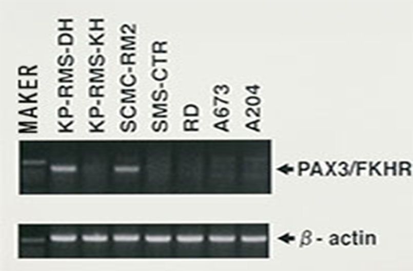
5
C-Myc and focal adhesion kinase (FAK) genes in the                  MRT cell lines
Askin tumor cell line
                                                                    MRT
          Askin tumor is a malignant small, round-cell tumor that
originates from the thoraco pulmonary region and is a member                  MRT was first described as a variant of Wilms' tumor of
of the ESFT. The MP-ASKIN-SA cell line was established from         the kidney in 1978. MRTs are a rare and highly malignant cancer
a tumor identified in the left-posterior pulmonary cavity of        type, which have also been reported outside of the kidney, in-
a 13-year-old boy (Figure 5, Table 1). The MP-ASKIN-SA cell         cluding in the liver, soft tissue, and central nervous system. Sev-
line was identified by the presence of EWS/ERG fusion mRNA          eral cases of primary intracranial MRT have been reported since
with RT-PCR (Figure 6A). Moreover, high expression of C-Myc         its recognition as a separate entity in 1978. The term rhabdoid
(which is associated with enhanced cell growth and proliferation;   was used to describe these cases because if the similarity in ap-
(Figure 6B) and overexpression of the FAK gene (associated with     pearance to RMS under the light microscope. Regardless of the
focal adhesion formation and cell migration) were detected in       location, all rhabdoid tumors are highly aggressive, have a poor
these cells, which appear to play a role in the poor prognosis of   prognosis, and tend to occur in children less than two years of
patients with ESFT [18].                                            age.

                                                                              To date, there are less than 10 MRT cell lines available.
                                                                    Four of these were established in our departments, and thus the
                                                                    cellular and genetic characterization of these cell lines will be
                                                                    useful for new diagnosis methods and novel therapeutic devel-
                                                                    opment.

                                                                    Establishment of four MRT cell lines

                                                                    KP-MRT-NS cell line

                                                                              The KP-MRT-NS cell line was established from a
                                                                    2-month-old infant who presented with abdominal distension
                                                                    and macrohematuria in October 1991. Clinical examination re-
                                                                    vealed a left upper abdominal mass arising from the primary tu-
                                                                    mor from the left kidney (Table l). Morphological observations
Figure. 5 Establishment of the MP-ASKIN-SA cell line from the       of the KP-MRT-NS cell line by electron microscopy showed
left pulmonary thoracopulmonary tumor from a 13 yr-old boy          spindle cells or flat cells (Figure 7A), and a para nuclear whorl
                                                                    of intermediate filaments, which is a common characteristic of
                                                                    MRT cells (Figure 7B). The phenotype of the KP-MRT-NS cell
                                                                    line [19] was similar to that of neuroblastoma cells, indicating a
                                                                    neural crest origin of MRT similar to neuroblastoma [4] (Figure
                                                                    8 and 9).

Figure. 6 EWS/FLl1 chimera gene (a) and c-myc expression (b)
in the MP-ASKIN-SA cell line

 JScholar Publishers                                                                    J Cancer Res Therap Oncol 2019 | Vol 7: 202
6
                                                                  KP-MRT-RY cell line

                                                                           The KP-MRT-RY cell line was established from a
                                                                  1-month-old infant who presented with abdominal distension
                                                                  (Table 1, Figure 10). The left renal tumor was resected, and the
                                                                  KP-MRT-RY cell line was cultured and characterized as de-
                                                                  scribed previously [20].

                                                                  MP-MRT-AN cell line

                                                                           The MP-MRT-AN cell line was cultured from liver bi-
                                                                  opsy specimens (Figure 11). Immunohistochemical assays de-
                                                                  tected the expression of vimentin and cytokeratin. RT-PCR as-
                                                                  says revealed that this cell line did not express smooth muscle
                                                                  myosin heavy chain isoforms or MyoD1 [21].
Figure. 7 Inverted microscopic (a) and electron microscopic (b)
analysis of the KP-MRT-NS cell line

                                                                  Figure. 10 Chest XP and abdominal computed tomographic im-
                                                                  aging of an established KP-MRT-RY cell line
Figure. 8 Indirect immunofluorescence with (a) neurofilament,
(b) alpha-smooth-muscle actin, and (c) SM1 isoforms of the
smooth-muscle-myosin heavy chain in the KP-MRT-NS cell line

Figure 9. Western blot analysis of cytoplasmic proteins of KP-    Figure. 11 Abdominal computed tomographic image showing
MRT-NS cell line with neurofilament, alpha-smooth-muscle, ba-     massive pleural fluid and multiple hepatic tumor lesions in an
sic calponin and SM1 isoforms.                                    established MP-MRT-AN cell line

 JScholar Publishers                                                                 J Cancer Res Therap Oncol 2019 | Vol 7: 202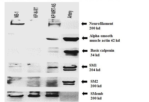
7
KP-MRT-YM cell line                                                   KP-MRT-RY cell line (Figure 13B). Furthermore, INI1 protein
                                                                      expression was not detected in any of the four MRT cell lines by
          The KP-MRT-YM cell line was established from a              western blotting (Figure 13C), confirming that the four cell lines
5-month-old boy who had a thoracic mass without metastasis            were MRT cells.
at the time of diagnosis (Figure 12). The tumor was complete-
ly resected, and histopathologic analysis demonstrated that the
cells were round in shape with vesicular nucleoli and no typical
eosinophilic cytoplasmic inclusions. The cells stained positive for
vimentin and negative for desmin by immunohistochemistry.
Expression of MyoD, PAX3/FKHR, EWS/FLI1, EWS/ERG, and
SSX/SYT chimeric mRNA was absent in the tumor.

          The patient received four cycles of doxorubicin, vin-
cristine, and cyclophosphamide, alternating between ifosfamide
and etoposide. However, after 18 months off-therapy, local re-
currence was detected. He then underwent total resection, addi-
tional chemotherapy, and 30.6-Gy radiation therapy. No further
recurrence has been observed. The patient is alive and well at 4
years post-onset [22, 23].

                                                                      Figure. 13 INI1 gene analysis in four MRT cell lines
                                                                      Preclinical studies of molecular target therapy using ge-
                                                                      finitide in MRT cells

                                                                                The epidermal growth factor receptor (EGFR) was re-
                                                                      cently found to be expressed in MRT cell lines. Gefitinib (mar-
                                                                      keted as Iressa) is an oral and selective EGFR-tyrosine kinase
                                                                      inhibitor with demonstrated efficacy in inhibiting the prolifera-
                                                                      tion of cancer cells in animal models in vivo as well as in clinical
                                                                      trials. These promising results encouraged us to examine the an-
                                                                      titumor effects of gefitinib on our MRT cell lines in vitro and in
                                                                      vivo.

                                                                                The expression of EGFR was confirmed in two MRT
Figure. 12 Chest computed tomographic image showing an en-
                                                                      tumors and their respective established cell lines (MP-MRT-AN
hanced thoracic mass on admission in an established KP-MRT-
YM cell line                                                          and KP-MRT-NS). Immunoblot analysis showed that gefitinib
                                                                      inhibited EGFR-phosphorylation [50% inhibitory concentration
Confirmation of the four cell lines as MRT cells
                                                                      (IC50) < 0.1 µmol/L] (Figure 14). Moreover, gefitinib inhibited
         RT-PCR analysis of the four MRT cell lines did not de-       in vitro cell growth (IC50 = 10–12 µmol/L), and a high concen-
tect INI1CD1 in the KP-MRT-NS, KP-MRT-AN, or KP-MRT-                  tration of gefitinib (20 µmol/L) induced apoptosis in vitro (42.9%
YM cell lines. INI1CD2 was also not detected in the KP-MRT-           MP-MRT-AN and 47.2% KP-MRT-NS) as determined by termi-
AN or KP-MRT-YM cell lines, and only a shortened form of this         nal deoxynucleotidyl transferase-mediated nick end labeling.
gene was detected in the KP-MRT-NS cell line (Figure 13A).            Furthermore, gefitinib at 150 mg/kg had a cytostatic effect on
These results were consistent with previous reports on MRT [19,       established MRT xenografts in athymic mice (MP-MRT-AN, P =
21–24]. However, both INI1CD1 and INI1CD2 were detected               0.039 and 0.0014; and KP-MRT-NS, P = 0.048 and 0.0086; Figure
in our newly established KP-MRT-RY cell line, as well as in the       15).
HL60 cell line, which was used as a positive control. Therefore,
we directly sequenced the amplicon corresponding to INI1 exon
2. A mutation in exon 2 (C157T in codon 53) was observed in the

 JScholar Publishers                                                                       J Cancer Res Therap Oncol 2019 | Vol 7: 202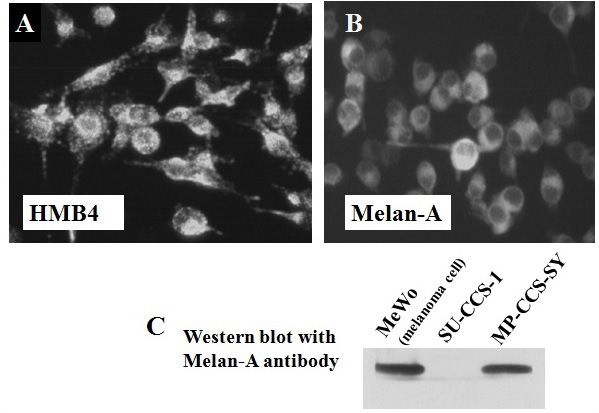
8
                                                                    of the MRT cell lines, whereas the cytotoxicity of trastuzumab
                                                                    against each of the MRT cell lines was significantly increased
                                                                    by the presence of allogeneic and autologous peripheral blood
                                                                    mononuclear cells (P < 0.01). There was a strong correlation
                                                                    coefficient (r = 0.825) between HER-2 expression and the cyto-
                                                                    toxicity enhanced by trastuzumab (Figure 16). Moreover, trastu-
                                                                    zumab in combination with peripheral blood mononuclear cells
                                                                    augmented by interleukin (IL)-2 was significantly more cytotox-
                                                                    ic than trastuzumab alone or IL-2 alone (P < 0.01) (Figure17)
                                                                    [20].

                                                                              These results indicated that (1) trastuzumab can exert
                                                                    antitumor effects on MRT cells using the antibody-dependent
                                                                    cellular cytotoxicity of effector cells and (2) IL-2 can enhance the
Figure. 14 Effect of gefitinib on two MRT cell lines
                                                                    cytotoxicity of trastuzumab against MRT cells [20].

Figure. 15 Inhibition of tumor growth by gefitinib in MRT cell      Figure. 16 Trastuzumab enhances cytotoxicity against MRT cell
lines                                                               lines using allogenic human peripheral blood mononuclear cells
         Overall, these results demonstrated that gefitinib has
antitumor effects in MRT cells in vitro and in vivo, and thus has
promise as a novel and therapeutic strategy for MRT [24].

Preclinical studies on the activation of antibody-depen-
dent cellular cytotoxicity by trastuzumab against MRT
cells

         Trastuzumab, a humanized monoclonal antibody
against human epidermal growth factor receptor-2 (HER-2), has
been shown to be effective against breast cancer and other can-
cers. Therefore, we also examined the expression of HER-2 in
our four MRT cell lines by indirect immunofluorescence, flow
cytometry, and immunohistochemistry [20].

        All four MRT cell lines (KP-MRT-NS, KP-MRT-RY,
                                                                    Figure. 17 Trastuzumab enhances cytotoxicity against KP-MRT-
MP-MRT-AN, KP-MRT-YM) expressed the HER-2 protein.
                                                                    YM cells
Treatment of trastuzumab alone did not reduce the viability

 JScholar Publishers                                                                     J Cancer Res Therap Oncol 2019 | Vol 7: 202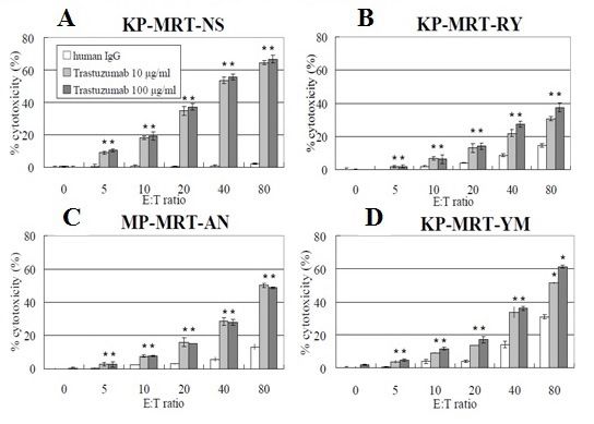
9
CCS cell lines                                                        Establishment of the MP-CCS-SY cell line

Case patient SY in CCS                                                           To date, there are only 10 established cell lines of CCS
                                                                      [26, 27]. We established the MP-CCS-SY cell line from metasta-
          CCS was first described by Enzinger in 1965 [25] as a       sis of the left femoral bone tumor in a 17-year-old SY girl (Figure
rare melanin-producing soft tissue sarcoma. Although CCS is           18).
also referred to as malignant melanoma of soft parts, it is clini-
cally, genetically, and biologically distinct from cutaneous mela-              The tumor cells grew as an adherent monolayer. A small
noma, despite certain histological similarities. CCS of soft parts    number of melanosomes were detected in the cytoplasm by elec-
mainly arises from the tendon or aponeurosis in adults 20–40          tron microscopy (Figure 19), which immunoreacted with two
years of age. Local recurrence is occasionally distinct from cu-      melanoma-associated antibodies, HMB45, and Melan-A (Figure
taneous melanoma, despite certain histological characteristics        20A, B). Western blot analysis further demonstrated the exis-
observed, and this tumor is insensitive to chemotherapy with a        tence of a Melan-A antigen in this cell line (Figure 20C) [26].
poor prognosis.

         A 13-year-old SY girl was referred to us in December
1994 with a left-Achilles tendon tumor that had been growing
for the past eight months (Figure 18). The patient received four
courses of chemotherapy, and wide resection of the primary tu-
mor was performed. Furthermore, mega therapy with autolo-
gous peripheral blood stem cell transplantation was performed
in November 1995. Despite intensive therapy, the disease gradu-
ally progressed, and the tumor metastasized to the left popliteal
lymph nodes, right lung, and left the femoral bone. Although a
transient minor response was observed, the patient passed away
due to progressive disease in December 1999.

                                                                      Figure. 19 Electron microphotograph of MP-CCS-SY cell line

Figure. 18 Enhanced computed tomography scan of left and right
Achilles tendons of a patient with clear cell sarcoma of soft parts

                                                                      Figure. 20 Immunofluorescence and western blot with HMB45
                                                                      and Melan-A antibodies in MP-CCS-SY cell line

 JScholar Publishers                                                                      J Cancer Res Therap Oncol 2019 | Vol 7: 202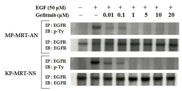
10
          The chimeric EWS/ATF1 transcript was detected in                Acknowledgments
both the SY tumor and MP-CCS-SY cell line (Figure 21A) [26],
as well as in another CCS cell line, SU-CSS-1 (Figure 21A) [27]                     We thank Drs. Jun Minowada, Tadashi Sawada, Yosihi-
by RT-PCR. Overexpression of c-Myc mRNA was detected in                   ro Horii, and Yasuhide Hayashi, and all members of our labora-
both the SY tumor and MP-CCS-SY cell line by northern blot                tory for their valuable guidance and support. We would like to
analysis (Figure 21B) indicating a potential role in the malignant        thank Editage (www.editage.com) for English language editing.
progression of CCS [26]. The availability of this MP-CCS-SY cell
                                                                          Disclaimer: The Japanese review entitled “Contribution of our
line will help to improve understanding of the molecular biology
                                                                          established pediatric malignant cell lines to clinical and basic
of this malignancy and should serve as a useful tool for develop-
                                                                          medical science” was published in Journal of Kyoto Prefectural
ing boron neutron capture therapy [28].
                                                                          University of Medicine 117 (3), 135–165, 2008 in Japanese, which
                                                                          was completely revised, and divided into two English reviews.

Figure. 21 EWS/ATF1 chimeric gene (a) and c-myc mRNA in
MP-CCS-SY cell line and SY tumor cells

Conclusion
           Since August 1984, our frozen vials have been routinely
thawed and cultured for basic and clinical in vitro and in vivo
studies in oncology. In the previous review, we discussed the es-
tablishment of 16 cell lines from 12 neuroblastoma patients and
their characteristics [29]. In the present review, we provided an
overview of the establishment of 13 soft-tissue sarcoma cell lines,
including two alveolar RMS cell lines and one MYCN-amplified
and overexpressed RMS cell line. In addition, we highlight the
establishment of one Askin sarcoma cell line and four rare MRT
cell lines. Two molecular target therapies involving gefinitide
and trastuzumab have been attempted for the refractory MRT
cell lines. The rare CCS cell line of soft parts offers a possible tool
for developing a boron neutron capture therapy. Overall, these
established childhood soft-tissue sarcoma cell lines are expected
to provide novel insights into tumor biology.

 JScholar Publishers                                                                         J Cancer Res Therap Oncol 2019 | Vol 7: 202
11
References sion of the N-myc oncogene. Int. J. Cancer 45: 705–711.
1) Sugimoto T, Gotoh T , Yagyu S, Kuroda H, Iehara T, Hosoi 12) Toffolatti L, Frascella E, Ninfo V, Gambini C, Forni M,Carli
H, et al. (2013) A MYCN-amplified cell line derived from a M, Rosolen A (2002) MYCN expression in human rhabdomyo-
long-term event-free survivor among our sixteen established sarcoma cell lines and tumour samples. J. Pathol 196: 450–458.
neuroblastoma cell lines. Cancer Lett., 331: 115–121.
13) Dias P, Kumar P, Marsden HB, Gattamaneni HR, Heigh-
2) Tsokos M, Webber BL, Parham DM, Wesley RA, Miser JS , way J, Kumar S. (1990) N-myc gene is amplified in alveolar
et al. (1992) Rhabdomyosarcoma. A new classification scheme rhabdomyosarcomas (RMS) but not in embryonal RMS. Int. J.
related to prognosis. Arch. Pathol. Lab. Med., 116: 847–855. Cancer 45: 593–596.
3) Hosoi H, Sugimoto T, Hayashi Y, Inaba J, Horii Y, Morioka 14) Tonelli R, McIntyre A, Camerin C, Walters ZS, Leo K Di,
H, et al. (1992) Differential expression of myogenic regulatory Selfe J, et al. (2012) Antitumor activity of sustained N-myc re-
genes, MyoD1 and myogenin, in human rhabdomyosarcoma duction in rhabdomyosarcomas and transcriptional block by
sublines. Int. J. Cancer 50: 977–983. antigene therapy. Clin. Cancer Res., 18: 796–807.
4) Sugimoto T, Mine H, Horii Y, Takahashi K, Nagai R, et al. 15) Mercado GE, Xia SJ, Zhang C, Ahn EH, Gustafson DM, Laé
(2000) Neuroblastoma cell lines showing smooth muscle cell M, et al. (2008) Identification of PAX3-FKHR-regulated genes
phenotypes. Diagn. Mol. Pathol 9: 221–228. differentially expressed between alveolar and embryonal rhab-
domyosarcoma: focus on MYCN as a biologically relevant target.
5) Davis RL, Weintraub H, Lassar AB (1987) Expression of a sin- Genes Chromosomes Cancer 47: 510–520.
gle transfected cDNA converts fibroblasts to myoblasts. Cell, 51:
987–1000. 16) Williamson D, Lu YJ, Gordon T, Sciot R, Kelsey A, Fisher
C, Poremba C, et al. (2005) Relationship between MYCN copy
6) Tonin PN, Scrable H, Shimada H, Cavenee WK (1991) Mus- number and expression in rhabdomyosarcomas and correlation
cle-specific gene expression in rhabdomyosarcomas and stages with adverse prognosis in the alveolar subtype. J. Clin. Oncol.,
of human fetal skeletal muscle development. Cancer Res 51: 23: 880–888.
5100–5106.
17) Ida K, Kobayashi S, Taki T, Hanada R, Bessho F, et al.(1995)
7) Tapscott SJ, Thayer MJ, Weintraub H (1993) Deficiency in rab- EWS-FLI-1 and EWS-ERG chimeric mRNAs in Ewing’s sarcoma
domyosarcoma of a factor required for MyoD activity and myo- and primitive neuroectodermal tumor. Int. J. Cancer, 63: 500–
genesis. Science 259: 1450–1453. 504.
8) Barr FG, Galili N, Holick J, Biegel JA, Rovera G,Emanuel BS 18) Moritake H, Sugimoto T, Kuroda H, Hidaka F, Takahashi Y,
(1993) Rearrangement of the PAX3 paired box gene in the pe- et al. (2003) Newly established Askin tumor cell line and over-
diatric solid tumor alveolar rhabdomyosarcoma. Nat. Genet 3: expression of focal adhesion kinase in Ewing sarcoma family of
113–117. tumors cell lines. Cancer Genet. Cytogenet 146: 102–109.
9) Sorensen PH, Lynch JC, Qualman SJ, Tirabosco R,Lim JF, et 19) Sugimoto T, Hosoi H, Horii Y, Ishida H, Mine H, Takahashi
al. (2002) PAX3-FKHR and PAX7-FKHR gene fusions are prog- K, et al.et al. (1999) Malignant rhabdoid-tumor cell line showing
nostic indicators in alveolar rhabdomyosarcoma: a report from neural and smooth-muscle-cell phenotypes. Int. J. Cancer 82:
the children’s oncology group. J. Clin. Oncol., 20: 2672–2679. 678–686.
10) Schwab M, Alitalo K, Klempnauer KH, Varmus HE, etal. 20) Katsumi Y, Kuwahara Y, Tamura S, Kikuchi K, Otabe O, et
(1983) Amplified DNA with limited homology to myc cellular al. (2008) Trastuzumab activates allogeneic or autologous anti-
oncogene is shared by human neuroblastoma cell lines and a body-dependent cellular cytotoxicity against malignant rhab-
neuroblastoma tumour. Nature 305: 245–248. doid tumor cells and interleukin-2 augments the cytotoxicity.
Clin. Cancer Res 14: 1192–1199.
11) Hayashi Y, Sugimoto T, Horii Y, Hosoi H, Inazawa J, Kems-
head JT, et al. (1990) Characterization of an embryonal rha- 21) Kuroda H, Moritake H, Sawada K, Kuwahara Y, Imoto I, In-
do-myosarcoma cell line showing amplification and over-expres- azawa J, Sugimoto T (2005) Establishment of a cell line from a
JScholar Publishers J Cancer Res Therap Oncol 2019 | Vol 7: 20212
malignant rhabdoid tumor of the liver lacking the function of
two tumor suppressor genes, hSNF5/INI1 and p16. Cancer Gen-
et. Cytogenet 158:172–179.
22) Hosoi H, Iehara T, Tsuchiya K, Misawa A, Miyaji M, et al.
(2007) Continuous remission in an infant with chest wall malig-
nant rhabdoid tumor after relapse. J. Pediatr. Surg 42: E9–E12.
23) Misawa A, Hosoi H, Imoto I, Iehara T, Sugimoto T, Inazawa
J (2004) Translocation (1; 22) (p36; q11.2) with concurrent del
(22) (q11.2) resulted in the homozygous deletion of SNF5/INI1
in a newly established cell line derived from the extrarenal rhab-
doid tumor. J. Hum. Genet., 49: 586–589.
24) Kuwahara Y, Hosoi H, Osone S, Kita M, Iehara T, Kuroda H,
Sugimoto T (2004) Antitumor activity of gefitinib in malignant
rhabdoid tumor cells in vitro and in vivo. Clin. Cancer Res., 10:
5940–5948.
25) Enzinger FM (1965) Clear-cell sarcoma of tendons and apo-
neuroses. An analysis of 21 cases. Cancer 18: 1163–1174.
26) Moritake H, Sugimoto T, Asada Y, Yoshida M.A, Maehara
Y, et al. (2002) Newly established clear cell sarcoma (malignant
melanoma of soft parts) cell line expressing melanoma-associ-
ated Melan-A antigen and overexpressing C-MYC oncogene.
Cancer Genet. Cytogenet 135: 48–56.
27) Epstein AL, Martin AO, Kempson R (1984) Use of a newly
established human cell line (SU-CCS−1) to demonstrate the re-
lationship of clear cell sarcoma to malignant melanoma. Cancer
Res 44: 1265–1274.
28) Fujimoto T, Andoh T, Sudo T, Fujita I, Moritake H, Sugimoto
T, et al. (2013) Boron neutron capture therapy (BNCT) selective- Submit your manuscript to a JScholar journal
ly destroys human clear cell sarcoma in the mouse model. Appl. and benefit from:
Radiat. Isot 73: 96–100. ¶¶ Convenient online submission
¶¶ Rigorous peer review
29) Sugimoto T, Kuroda H, Yagyu S, Gotoh T, Osone S, Tamura
¶¶ Immediate publication on acceptance
S, Iehara T, Hosoi H (2019) Cellular and molecular characteris-
¶¶ Open access: articles freely available online
tics of established neuroblastoma cell lines. J Cancer Res Therap
¶¶ High visibility within the field
Oncol 7: 1-15.
¶¶ Better discount for your subsequent articles
Submit your manuscript at
http://www.jscholaronline.org/submit-manuscript.php
JScholar Publishers J Cancer Res Therap Oncol 2019 | Vol 7: 202You can also read