CircTLK1 facilitates the proliferation and metastasis of renal cell carcinoma by regulating miR-495-3p/CBL axis
←
→
Page content transcription
If your browser does not render page correctly, please read the page content below
Open Life Sciences 2021; 16: 362–374
Research Article
Xiangli Lei, Meiling Yang, Zhifang Xiao, Heng Zhang, Shuai Tan*
circTLK1 facilitates the proliferation
and metastasis of renal cell carcinoma
by regulating miR-495-3p/CBL axis
https://doi.org/10.1515/biol-2021-0041 Keywords: circTLK1, miR-495-3p, CBL, renal cell carcinoma
received August 29, 2020; accepted January 05, 2021
Abstract: Renal cell carcinoma (RCC) is a common uro-
logical malignancy. Circular RNAs (circRNAs) have been
confirmed to play an important regulatory role in various 1 Introduction
cancers. This study aimed to investigate the role and
potential mechanism of circTLK1 (hsa_circ_0004442) in Renal cell carcinoma (RCC) is a urologic malignancy ori-
RCC. The levels of circTLK1, Cbl proto-oncogene (CBL), ginating from the renal epithelium, which accounts for
and microRNA-495-3p (miR-495-3p) were detected by quan- more than 90% of renal cancers [1]. It is estimated that
titative reverse transcription polymerase chain reaction or there were 403,262 new kidney cancer cases and 175,098
western blot. Cell proliferation, cycle arrest and apoptosis, related deaths worldwide in 2018 [2]. RCC is a genitouri-
migration, and invasion were assessed by colony formation, nary malignant tumor with a mortality rate second only
flow cytometry, scratch, and transwell assays. The levels to bladder cancer [3]. Targeted therapies have become
of E-cadherin and Vimentin were measured by western the main treatment for patients with recurrent or meta-
blot. The targeting relationship between miR-495-3p and static RCC [4]. Because of the radioresistance and chemo-
miR-495-3p or CBL was verified by dual-luciferase reporter resistance of RCC, the 5-year survival rate of metastatic
assay. Tumor growth in vivo was evaluated by xenograft RCC is still as low as about 10% [5]. Therefore, exploring
assay. The results found that circTLK1 and CBL were the potential mechanism of RCC pathogenesis is essential
up-regulated in RCC tissues and cells. Silencing of circTLK1 for the development of effective RCC treatment strategies.
or CBL inhibited proliferation and metastasis and accelerated Circular RNAs (circRNAs) are a special type of tran-
apoptosis in RCC cells. In addition, circTLK1 directly bound scripts characterized by covalent closed loops with no 5′
to miR-495-3p, and CBL was the target of miR-495-3p. to 3′ polarity [6]. Increasing evidence has manifested that
circTLK1 sponged miR-495-3p to increase CBL expression. circRNAs exert crucial effects in various diseases, espe-
Moreover, knockdown of circTLK1 suppressed tumor growth cially cancer, and may be diagnostic or prognostic markers
in vivo. In conclusion, down-regulation of circTLK1 res- as they are more stable than linear RNA [7]. Moreover,
trained proliferation and metastasis and promoted apoptosis substantial studies have corroborated that circRNAs par-
in RCC cells by modulating miR-495-3p/CBL axis. ticipate in the occurrence and development of various
cancers through mediating diverse biological processes
[8]. For example, circ_0000190 hindered cell proliferation
* Corresponding author: Shuai Tan, Department of Oncology, and metastasis in gastric carcinoma by down-regulating
Affiliated Nanhua Hospital, University of South China, 336 Dongfeng microRNA-1252 to up-regulate PAK3 [9]. In addition, hsa_
Road, Zhuhui District, Hengyang, 421000, Hunan, China,
circ_0101145 contributed to epithelial-mesenchymal transi-
tel: +86-073-4835-8008, e-mail: sljxnu@163.com
Xiangli Lei: Department of Nephrology, Affiliated Nanhua Hospital, tion (EMT) in hepatocellular carcinoma through absorbing
University of South China, Hengyang, Hunan, China microRNA-548c-3p to regulate laminin subunit gamma 2
Meiling Yang: Department of Oncology, Affiliated Nanhua Hospital, expression [10]. In non-small cell lung carcinoma, circ_
University of South China, 336 Dongfeng Road, Zhuhui District, 0000376 facilitated tumor progression and elevated
Hengyang, 421000, Hunan, China
chemoresistance via combining with microRNA-384 [11].
Zhifang Xiao: Department of Endocrinology, Affiliated Nanhua
Hospital, University of South China, Hengyang, Hunan, China
Moreover, a recent study suggested that hsa_circ_0004442
Heng Zhang: Department of Hematology, Affiliated Nanhua derived from Tousled-like kinases 1 (TLK1) was remark-
Hospital, University of South China, Hengyang, Hunan, China ably up-regulated in RCC, and circTLK1 expedited RCC
Open Access. © 2021 Xiangli Lei et al., published by De Gruyter. This work is licensed under the Creative Commons Attribution 4.0
International License.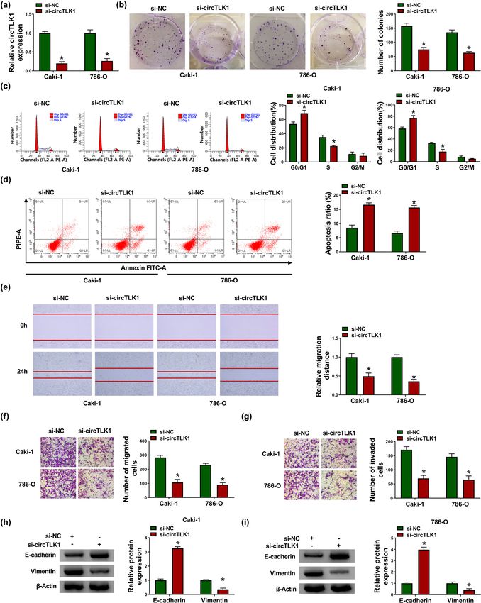
The role of circTLK1 in renal cell carcinoma 363
proliferation and metastasis by modulating microRNA-136- Table 1: Correlation between circTLK1 expression and clinico-
5p/CBX4 pathway [12]. Nevertheless, the exact mechanism of pathological parameters in RCC patients
circTLK1 in RCC development still needs further exploration.
Mounting evidence has verified that microRNAs Clinicopathological Number circTLK1 P-value
factors expression
(miRNAs) suppress mRNA translation or induce mRNA
degradation by directly pairing with mRNA 3′UTR [13]. Low High
Moreover, circRNAs participate in the post-transcrip- (n = 17) (n = 18)
tional regulation of mRNAs by competitively binding to Age
miRNAs [14]. Therefore, we predicted some miRNAs that 0.05
might bind to circTLK1 through bioinformatics analysis. ≥50 years 16 8 8
Gender
Cbl proto-oncogene (CBL) belongs to the E3 ubiquitin
Female 21 10 11 >0.05
ligase family and regulates signal transduction through Male 14 7 7
tyrosine kinase-dependent pathways [15]. Decreasing c-Cbl Tumor size
activity contributes to osteoblast differentiation in mesen- >4 cm 17 5 12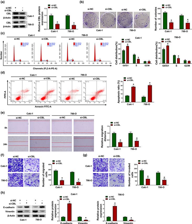
364 Xiangli Lei et al.
Table 2: The primer sequences for qRT-PCR 2.7 Scratch assay
Primer name Sequence (5′–3′) Tm (°C) The transfected Caki-1 and 786-O cells were plated in
circTLK1-F CAGTCAATGGAGCAGAGAA 60.0 6-well plates. Later, a linear wound was created by
circTLK1-R CCATTCTTGTTGCCTTTTTG 59.1 scraping the cells with a sterilized pipette tip. After 24 h
TLK1-F ACGTGGCCACAAAATTAGCG 64.7 of incubation, the migration distance was photographed
TLK1-R GGAGAAGGGCTATTCGGTCG 65.0 using a microscope at 100× magnification and calculated
CBL-F TGACATCTTTACCCGACTC 59.4
using ImageJ 1.8.0 software (National Institutes of Health,
CBL-R CATACCCAATAGCCCAC 57.1
miR-495-3p-F AACACGCAAACAAACATGGTGC 74.5
Bethesda, MD, USA).
miR-495-3p-R CAGTGCAGGGTCCGAGGT 61.1
β-Actin-F GTCACCGGAGTCCATCACGAT 66.8
β-Actin-R TCACCAACTGGGACGACATG 65.1
U6-F CTCGCTTCGGCAGCACA 65.6
2.8 Transwell assay
U6-R AACGCTTCACGAATTTGCGT 64.5
Cell migration and invasion were determined using trans-
well chambers with 8 μm polycarbonate membrane filters
complementary DNA (cDNA) was synthesized using spe- (Corning, Corning, NY, USA). The transfected Caki-1 and
cific reverse transcription kits (Takara, Dalian, China). 786-O cells were injected into the upper chamber.
Then, RNA levels were detected using SYBR Premix Ex Meanwhile, medium with 10% FBS was added as an
Taq (Takara) and calculated using the 2−ΔΔCt method. The attractant in the lower chamber. After 24 h of culture,
PCR amplification procedure included 95°C for 10 min, the cells were fixed with methanol and stained with
followed by 40 cycles of 95°C for 5 s, 60°C for 10 s, and 0.1% crystal violet (Solarbio). The migrated cells were
72°C for 10 s. β-Actin or U6 was considered as an internal counted under a microscope at 100× magnification. For
control. The primers are presented in Table 2. cell invasion test, the difference was that transwell chamber
was pre-coated with Matrigel (Corning).
2.5 Colony formation assay
2.9 Western blot assay
After transfection, Caki-1 and 786-O cells were trypsinized
and then seeded into 6-well plates. The culture medium Total protein was extracted with RIPA buffer (Solarbio). After
was changed every 3 days for 2 weeks. Subsequently, the the protein was quantified using BCA™ Protein Assay Kit
cells were fixed with formaldehyde and stained with crystal (Pierce, Appleton, WI, USA), the equal amounts of protein
violet (Solarbio). Finally, the number of colonies was samples were separated by polyacrylamide gel electrophor-
counted in five randomly selected fields under a microscope. esis and transferred to polyvinylidene fluoride membranes
(Millipore, Billerica, MA, USA). The membranes were blocked
with 5% fat-free milk for 2 h and incubated with primary
antibodies against E-cadherin (1:500, ab15148, Abcam),
2.6 Flow cytometry Vimentin (1:2,000, ab137321, Abcam), CBL (1:5,000, ab32027,
Abcam), or β-actin (1:2,000, ab8227, Abcam). After washing
The transfected Caki-1 and 786-O cells were harvested thrice with tris-buffered saline, the membranes were probed
and trypsinized. Subsequently, the precipitate was washed with horseradish peroxidase-labeled secondary antibody
in phosphate-buffered saline (PBS; Solarbio) and fixed (1:25,000, ab205718, Abcam). Finally, the protein bands
with ethanol for 1 h. After incubation with RNase (Seebio) were measured using the ECL system (Beyotime, Shanghai,
for 30 min, the cells were stained with propidium iodide (PI; China).
Abcam, Cambridge, UK). Finally, cell distribution was
monitored by FACScan Flow Cytometry (BD Biosciences,
San Diego, CA, USA). 2.10 Dual-luciferase reporter assay
Cell apoptosis was assessed using Annexin V-FITC/PI
Apoptosis Detection kit (Vazyme, Nanjing, China) following circTLK1 sequence containing miR-495-3p wild-type or
the manufacturer’s instructions. The apoptosis cells were mutant binding site was cloned into pmirGLO vector
measured by FACScan Flow Cytometry (BD Biosciences). (LMAI Bio, Shanghai, China) to form WT-circTLK1 and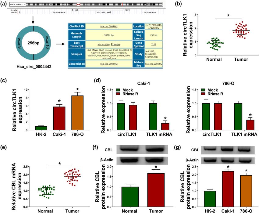
The role of circTLK1 in renal cell carcinoma 365
MUT-circTLK1 vectors. Meanwhile, CBL 3′UTR harboring tissues were analyzed. As shown in Figure 1b, circTLK1
miR-495-3p wild-type or mutant binding site was inserted expression was remarkably higher than that in normal
into pmirGLO vector (LMAI Bio) to form WT-CBL 3′UTR tissues. Meanwhile, circTLK1 level was strikingly increased
and MUT-CBL 3′UTR vectors. Next, the constructed vector in RCC cells (Caki-1 and 786-O) compared with normal
and miR-NC mimic or miR-495-3p mimic was co-transfected human kidney cell line (HK-2) (Figure 1c). Moreover,
into Caki-1 and 786-O cells. Subsequently, the luciferase RNase R digestion assay showed that circTLK1 was not
intensity was detected via Dual-Lucy Assay Kit (Solarbio). affected by RNase R, indicating that circTLK1 was more
stable than TLK1 mRNA in Caki-1 and 786-O cells (Figure 1d).
In addition, CBL mRNA and protein levels in RCC tissues
2.11 Xenograft assay were markedly higher than those in normal tissues
(Figure 1e and f). Simultaneously, CBL protein expression
Five-week-old BALB/c nude mice (n = 10) were randomly in Caki-1 and 786-O cells was significantly increased com-
divided into two groups (n = 5 in each group). Lentivirus pared to HK-2 cells (Figure 1g). As presented in Table 1,
containing circTLK1 short hairpin RNA (sh-circTLK1) or circTLK1 expression was not associated with age and
negative control (sh-NC) was purchased from Genechem. gender, but was associated with tumor size, lymph node
786-O cells (5 × 10⁶) stably expressing sh-circTLK1 or metastasis, and TNM stage. These data hinted that
sh-NC were subcutaneously injected into the back of mice. circTLK1 and CBL might play carcinogenic roles in RCC.
Tumor volume was measured once a week. After 4 weeks,
the mice were killed and the xenograft tumors were weighed.
3.2 Knockdown of circTLK1 inhibits
The levels of circTLK1, miR-495-3p, and CBL in the excised
tumors were measured by qRT-PCR or western blot. proliferation and metastasis and
promotes apoptosis in RCC cells
Ethical approval: The research related to animal use has
been complied with all the relevant national regulations To investigate the function of circTLK1 in RCC develop-
and institutional policies for the care and use of animals ment, we performed loss-of-function experiments by
and has been approved by the Animal Ethics Committee transfecting si-circTLK1 into Caki-1 and 786-O cells.
of Affiliated Nanhua Hospital, University of South China. First, circTLK1 level in the si-circTLK1 group was promi-
nently reduced compared with the si-NC group, sug-
gesting that circTLK1 knockdown efficiency was signifi-
cant (Figure 2a). Colony formation assay showed that
2.12 Statistical analysis circTLK1 silencing inhibited the proliferation of Caki-1 and
786-O cells (Figure 2b). In addition, flow cytometry showed
All data were expressed as mean ± standard deviation that knockdown of circTLK1 induced cell cycle arrest of
using GraphPad Prism 7 software (GraphPad, San Diego, Caki-1 and 786-O cells in G1 phase and accelerated apoptosis
CA, USA). Student’s t-test and one-way analysis of variance (Figure 2c and d). Scratch and transwell assays revealed
were used to analyze the differences. The linear relation- that transfection with si-circTLK1 suppressed the migration
ships among circTLK1, miR-495-3p, and CBL were tested by and invasion of Caki-1 and 786-O cells (Figure 2e–g). More-
Spearman’s correlation coefficient. P < 0.05 was considered over, western blot analysis showed that depletion of
statistically significant. circTLK1 led to a marked increase in E-cadherin level and
a significant decrease in Vimentin level (Figures 2h and i).
Overall, these results indicated that down-regulation of
3 Results circTLK1 suppressed the proliferation and metastasis of
RCC cells and promoted apoptosis.
3.1 circTLK1 and CBL are up-regulated in RCC
tissues and cells 3.3 CBL silencing inhibits proliferation and
metastasis and induces apoptosis in
First, we demonstrated that hsa_circ_0004442 was derived RCC cells
from exons 9 and 10 of TLK1 gene (Figure 1a). To explore the
biological function of circTLK1 in RCC, the expression dif- Next, a series of functional experiments were performed
ferences of circTLK1 in 35 pairs of RCC tissues and normal in Caki-1 and 786-O cells transfected with si-CBL to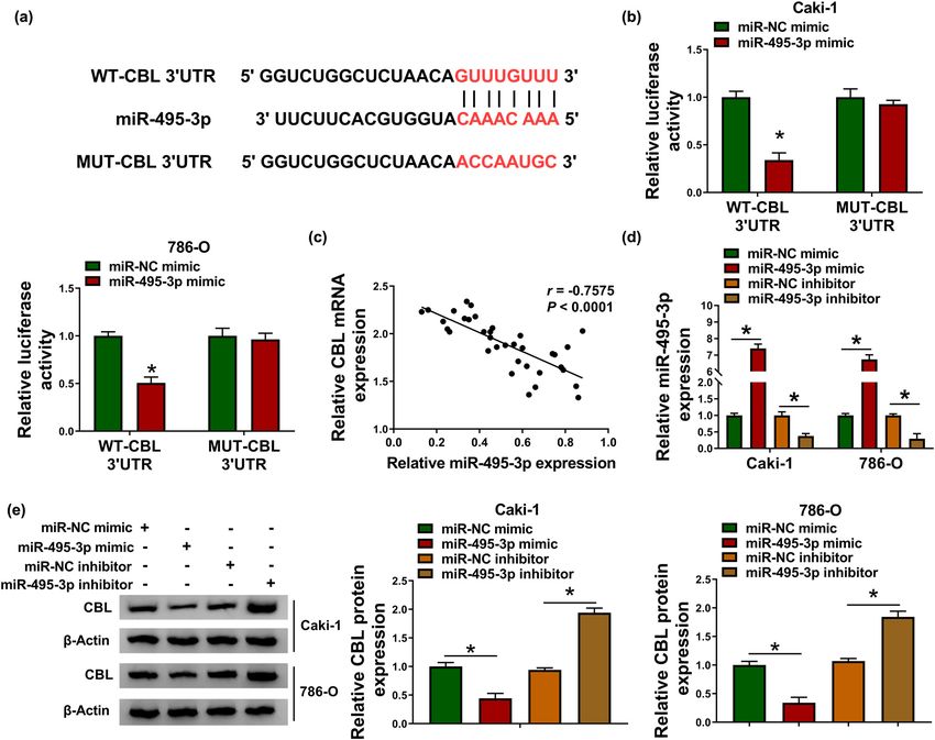
366 Xiangli Lei et al. Figure 1: circTLK1 and CBL are up-regulated in RCC tissues and cells. (a) Schematic diagram showing the origin and formation of circTLK1. (b) circTLK1 expression was detected in RCC tissues (n = 35) and adjacent normal tissues (n = 35) by qRT-PCR. (c) circTLK1 level was measured in HK-2 cells and RCC cells (Caki-1 and 786-O). (d) After RNase R stimulation, the levels of circTLK1 and TLK1 mRNA were examined using qRT-PCR. (e and f) The mRNA and protein levels of CBL in RCC tissues and normal tissues were detected by qRT-PCR and western blot. (g) CBL protein level was measured in HK-2, Caki-1 and 786-O cells. *P < 0.05. explore the role of CBL in RCC progression. First of all, the decreasing Vimentin (Figure 3h). Collectively, these data knockdown efficiency of CBL was determined by western evidenced that knockdown of CBL impeded RCC cell blot assay (Figure 3a). Subsequently, colony formation proliferation and metastasis and induced apoptosis. assay suggested that si-CBL transfection significantly reduced the proliferation ability of Caki-1 and 786-O cells (Figure 3b). Flow cytometry illustrated that silencing of 3.4 CBL overexpression reverses the effect CBL expedited cycle arrest and apoptosis of Caki-1 and of circTLK1 depletion on RCC cell 786-O cells (Figures 3c and d). In addition, scratch assay progression exhibited that knockdown of CBL impeded the migration of Caki-1 and 786-O cells (Figure 3e). Furthermore, transwell In view of the functions of circTLK1 and CBL in RCC cells, we assay showed that the migration and invasion of Caki-1 and further explored whether the influence of circTLK1 on RCC 786-O cells were inhibited after si-CBL transfection (Figure cell progression is related to CBL. As depicted in Figure 4a, 3f and g). In addition, CBL down-regulation blocked EMT CBL protein level in Caki-1 and 786-O cells transfected with in Caki-1 and 786-O cells by increasing E-cadherin and pcDNA-CBL was significantly up-regulated, indicating
The role of circTLK1 in renal cell carcinoma 367 Figure 2: Knockdown of circTLK1 inhibits proliferation and metastasis and promotes apoptosis in RCC cells. (a) The knockdown efficiency of circTLK1 was determined by qRT-PCR. After transfecting Caki-1 and 786-O cells with si-NC or si-circTLK1, colony formation assay and flow cytometry were used to evaluate colony number (b), cell distribution (c), and apoptosis ratio (d). (e–g) Cell migration and invasion were assessed by scratch assay and transwell assay in Caki-1 and 786-O cells transfected with si-NC or si-circTLK1. (h and i) The levels of EMT- related proteins (E-cadherin and Vimentin) were detected using western blot. *P < 0.05.
368 Xiangli Lei et al. Figure 3: CBL silencing inhibits proliferation and metastasis and induces apoptosis in RCC cells. Caki-1 and 786-O cells were introduced with si-NC or si-CBL. (a) CBL protein level was measured by western blot. (b) Cell proliferation was assessed by colony formation assay. (c and d) Cell distribution and apoptosis ratio were determined by flow cytometry. (e–g) Cell migration and invasion were evaluated by scratch assay and transwell assay. (h) The protein levels of E-cadherin and Vimentin were examined by western blot. *P < 0.05. successful transfection. Subsequently, si-circTLK1 and cells to investigate their effects on RCC cell progression. pcDNA-CBL were co-transfected into Caki-1 and 786-O The results showed that knockdown of circTLK1 markedly
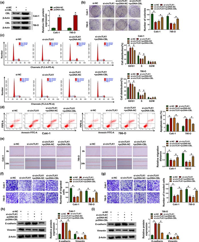
The role of circTLK1 in renal cell carcinoma 369 Figure 4: CBL overexpression reverses the effect of circTLK1 depletion on RCC cell progression. (a) The overexpression efficiency of CBL was tested by western blot. After transducing Caki-1 and 786-O cells with si-NC, si-circTLK1, si-circTLK1 + pcDNA-NC, or si-circTLK1 + pcDNA-CBL, colony number (b), cell distribution (c), apoptosis rate (d), cell migration (e and f), cell invasion (g), and the levels of EMT-related proteins (h and i) were detected via appropriate methods. *P < 0.05.
370 Xiangli Lei et al.
impeded cell proliferation (Figure 4b) and induced used to predict miRNAs with a targeting relationship
cell cycle arrest (Figure 4c) and apoptosis (Figure 4d) with circTLK1. Circular RNA Interactome online software
in Caki-1 and 786-O cells, while these effects were found that circTLK1 and miR-495-3p have a possible
abolished by up-regulating CBL. In addition, inhibition binding site (Figure 5a). Then, dual-luciferase reporter
of circTLK1 remarkably suppressed cell migration assay suggested that miR-495-3p mimic overtly reduced
(Figure 4e and f), invasion (Figure 4g), and EMT the luciferase activity of WT-circTLK1 reporter in Caki-1
(Figure 4h and i) in Caki-1 and 786-O cells, whereas and 786-O cells (Figure 5b and c). In addition, circTLK1
these impacts were abrogated after transfection with pcDNA- expression was strikingly increased after transfection
CBL. These data evidenced that circTLK1 affected RCC cell with oe-circTLK1 (Figure 5d). Moreover, circTLK1 over-
progression by regulating CBL. expression drastically restrained miR-495-3p expression,
while circTLK1 silencing remarkably promoted miR-495-
3p expression (Figure 5e). Compared with normal tissues,
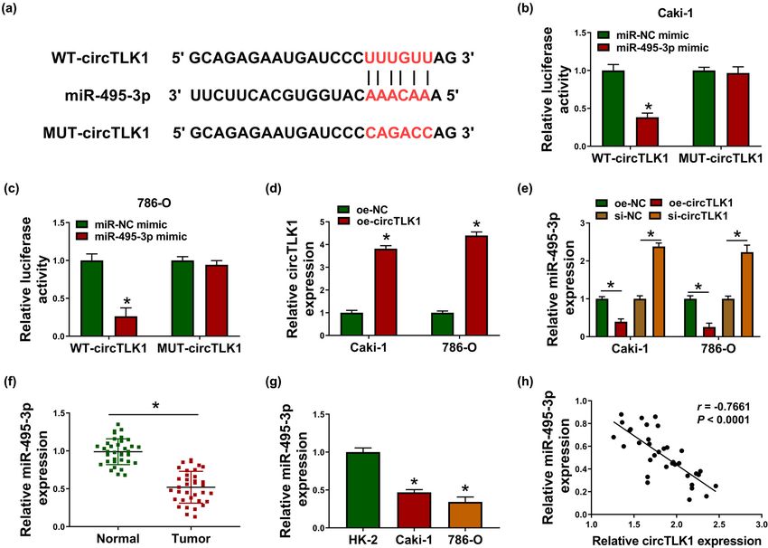
3.5 circTLK1 directly interacts with miR-495-3p level was significantly decreased in RCC tis-
miR-495-3p sues (Figure 5f). Consistently, miR-495-3p level in Caki-1
and 786-O cells was prominently reduced compared to
To discover the molecular mechanism of circTLK1 reg- HK-2 cells (Figure 5g). Spearman’s correlation analysis
ulating RCC progression, bioinformatics analysis was showed that circTLK1 and miR-495-3p were negatively
Figure 5: circTLK1 directly interacts with miR-495-3p. (a) Circular RNA Interactome predicted the putative binding site of circTLK1 and miR-
495-3p. (b and c) The luciferase activity was tested in Caki-1 and 786-O cells co-transfected with WT-circTLK1 or MUT-circTLK1 and miR-NC
mimic or miR-495-3p mimic. (d) circTLK1 expression was detected in Caki-1 and 786-O cells after transfection with oe-NC or oe-circTLK1.
(e) The level of miR-495-3p was measured in Caki-1 and 786-O cells transfected with oe-NC, oe-circTLK1, si-NC, or si-circTLK1. (f and g) miR-
495-3p expression was examined in RCC tissues and cells. (h) The correlation between circTLK1 and miR-495-3p was analyzed by
Spearman’s correlation coefficient. *P < 0.05.The role of circTLK1 in renal cell carcinoma 371
correlated in RCC tissues (Figure 5h). These data indi- introduced with miR-495-3p mimic or miR-495-3p inhibitor,
cated that circTLK1 targeted miR-495-3p in RCC. and qRT-PCR analysis showed significant miR-495-3p over-
expression and knockdown efficiency (Figure 6d). Further-
more, up-regulation of miR-495-3p markedly inhibited CBL
protein expression, whereas down-regulation of miR-495-3p
3.6 miR-495-3p directly targets CBL overtly facilitated CBL protein expression (Figure 6e). These
data evidenced that CBL was a target of miR-495-3p.
Next, bioinformatics software TargetScan predicted that
miR-495-3p might bind to CBL 3′UTR (Figure 6a). Subse-
quently, dual-luciferase reporter analysis showed that
mature miR-495-3p remarkably decreased the luciferase 3.7 circTLK1 regulates CBL by sponging miR-
activity of WT-CBL 3′UTR reporter in Caki-1 and 786-O 495-3p
cells (Figure 6b). Spearman’s correlation coefficient illustrated
that miR-495-3p was negatively correlated with CBL in RCC To elucidate the relationship among circTLK1, miR-495-
tissues (Figure 6c). Besides, Caki-1 and 786-O cells were 3p, and CBL in RCC cells, Caki-1 and 786-O cells were
Figure 6: miR-495-3p directly targets CBL. (a) The predicted binding site of miR-495-3p and CBL 3′UTR was displayed. (b) The luciferase
activity was detected in Caki-1 and 786-O cells after transfection with WT-CBL 3′UTR or MUT-CBL 3′UTR and miR-NC mimic or miR-495-3p
mimic. (c) The correlation between miR-495-3p and CBL in RCC tissues was tested by Spearman’s correlation coefficient. (d and e) The levels of
miR-495-3p and CBL protein were examined in Caki-1 and 786-O cells transfected with miR-NC mimic, miR-495-3p mimic, miR-NC inhibitor, or
miR-495-3p inhibitor. *P < 0.05.372 Xiangli Lei et al.
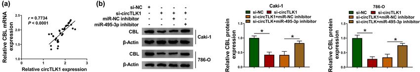
Figure 7: circTLK1 regulates CBL by sponging miR-495-3p. (a) Spearman’s correlation coefficient was used to analyze the correlation
between circTLK1 and CBL mRNA in RCC tissues. (b) CBL protein level was measured by western blot in Caki-1 and 786-O cells transfected
with si-NC, si-circTLK1, si-circTLK1 + miR-NC inhibitor, or si-circTLK1 + miR-495-3p inhibitor. *P < 0.05.
transfected with si-NC, si-circTLK1, si-circTLK1 + miR-NC Therefore, elucidating the molecular mechanism of cir-
inhibitor, or si-circTLK1 + miR-495-3p inhibitor. In RCC cRNAs is essential for RCC treatment. In our research, the
tissues, circTLK1 expression was positively correlated biological function and potential mechanism of circTLK1
with CBL mRNA expression (Figure 7a). As displayed in in RCC were investigated in depth. Previous studies revealed
Figure 7b, co-transfection of si-circTLK1 and miR-495-3p that circTLK1 was conspicuously up-regulated in patients
inhibitor alleviated the reduction in CBL protein level with acute ischemic stroke and RCC, which was consistent
caused by circTLK1 knockdown alone. These results indi- with the results of this research [12,21]. Besides, loss-of-func-
cated that circTLK1 regulated CBL expression by sponging tion experiments indicated that circTLK1 silencing deceler-
miR-495-3p. ated proliferation and metastasis and facilitated apoptosis in
Caki-1 and 786-O cells, which was in agreement with the
previous results [12].
3.8 circTLK1 silencing blocks tumor growth Emerging evidence has suggested that circRNAs act
in vivo as miRNA sponges and regulate biological functions by
mediating gene expression through competing endoge-
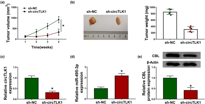
To explore the role of circTLK1 in tumorigenesis in vivo, nous RNA (ceRNA) mechanisms [22]. In this research,
we constructed a xenograft mouse model by introducing we found that circTLK1 might sponge miR-495-3p based
sh-circTLK1 into 786-O cells. As shown in Figure 8a, on bioinformatics analysis. Investigations have shown
tumor volume was remarkably decreased in the sh-circTLK1 that miR-495-3p serves as a tumor-suppressing factor in
group compared to the sh-NC group. Simultaneously, various cancers, including gastric carcinoma [23], mela-
tumor weight in the sh-circTLK1 group was prominently noma [24], and prostate cancer [25]. In clear cell RCC,
lower than that in the sh-NC group (Figure 8b). In addition, LUCAT1 accelerated cell growth and invasion via targeting
the levels of circTLK1 and CBL protein were strikingly miR-495-3p [26]. However, the potential mechanism of
reduced, while miR-495-3p level was significantly increased miR-495-3p in RCC progression needs further study. In the
in the sh-circTLK1 group compared with the sh-NC group present research, circTLK1 directly targeted miR-495-3p to
(Figure 8c–e). These results indicated that circTLK1 knock- regulate CBL expression.
down inhibited tumor growth in vivo. Moreover, increasing evidence has verified that miRNAs
weaken gene expression by binding to 3′UTR of mRNAs [27].
This research confirmed that miR-495-3p was directly
combined with CBL. CBL is a proto-oncogene encoding
4 Discussion E3 ubiquitin ligase [28]. CBL mutations play a crucial role
in many cancers, including acute myeloid leukemia [29].
With the rapid development of high-throughput sequen- In hepatocellular carcinoma, miR-486-5p overexpression
cing technology, multiple circRNAs related to tumor pro- hindered cell proliferation and motility through repres-
gression have been identified [18]. Accumulating evi- sing CBL [30]. In breast cancer, miR-124-3p ameliorated
dence has certified that dysregulation of circRNAs is the malignancy of tumor via down-regulating CBL [31].
extensively implicated in the development of various dis- A previous research unveiled that CBL up-regulation
eases, including cancer [19]. Moreover, circRNAs have alleviated the inhibition of miR-200a-3p overexpression
been confirmed to occupy an important position in the on RCC progression [32]. In the current research, CBL was
pathogenesis of kidney diseases, including RCC [20]. also strikingly up-regulated in RCC. Furthermore, thisThe role of circTLK1 in renal cell carcinoma 373
Figure 8: circTLK1 silencing blocks tumor growth in vivo. 786-O cells transfected with sh-NC or si-circTLK1 were subcutaneously injected into
the nude mice (n = 5 per group). (a) Tumor volume was measured once a week. (b) After 4 weeks, the mice were killed, and the xenograft
tumors were removed and weighed. (c–e) The levels of circTLK1, miR-495-3p, and CBL in xenograft tumors were detected using qRT-PCR or
western blot. *P < 0.05.
research found that silencing of CBL suppressed cell pro- [2] Bray F, Ferlay J, Soerjomataram I, Siegel RL, Torre LA, Jemal A.
liferation and metastasis and triggered apoptosis in RCC. Global cancer statistics 2018: GLOBOCAN estimates of inci-
In conclusion, these findings discovered that circTLK1 dence and mortality worldwide for 36 cancers in 185 countries.
CA Cancer J Clin. 2018;68:394–424.
contributed to the growth and metastasis of RCC cells by
[3] Zarrabi K, Paroya A, Wu S. Emerging therapeutic agents for
sponging miR-495-3p to indirectly modulate CBL. The dis- genitourinary cancers. J Hematol Oncol. 2019;12:89.
covery of the new ceRNA mechanism of circTLK1/miR-495- [4] Posadas EM, Limvorasak S, Figlin RA. Targeted therapies for
3p/CBL might provide a new therapeutic approach for RCC. renal cell carcinoma. Nat Rev Nephrol. 2017;13:496–511.
[5] Strizova Z, Bartunkova J, Smrz D. The challenges of adoptive
cell transfer in the treatment of human renal cell carcinoma.
Funding information: The authors state no funding involved.
Cancer Immunol Immunother. 2019;68:1831–8.
[6] Qian L, Yu S, Chen Z, Meng Z, Huang S, Wang P. The emerging
Conflict of interest: The authors state no conflict of role of circRNAs and their clinical significance in human can-
interest. cers. Biochim Biophys Acta Rev Cancer. 2018;1870:247–60.
[7] Xia L, Song M, Sun M, Wang F, Yang C. Circular RNAs as bio-
markers for cancer. Adv Exp Med Biol. 2018;1087:171–87.
Data availability statement: The datasets generated
[8] Tang Q, Hann SS. Biological roles and mechanisms of circular
during and/or analyzed during the current study are
RNA in human cancers. Onco Targets Ther. 2020;13:2067–92.
available from the corresponding author on reasonable [9] Wang GJ, Yu TY, Li YR, Liu YJ, Deng BB. circ_0000190 sup-
request. presses gastric cancer progression potentially via inhibiting
miR-1252/PAK3 pathway. Cancer Cell Int. 2020;20:351.
[10] Jin J, Liu H, Jin M, Li W, Xu H, Wei F. Silencing of hsa_circ_
0101145 reverses the epithelial-mesenchymal transition in
hepatocellular carcinoma via regulation of the miR-548c-3p/
References LAMC2 axis. Aging (Albany NY). 2020;12:11623–35.
[11] Sun H, Chen Y, Fang YY, Cui TY, Qiao X, Jiang CY, et al.
[1] Hsieh JJ, Purdue MP, Signoretti S, Swanton C, Albiges L, Circ_0000376 enhances the proliferation, metastasis, and
Schmidinger M, et al. Renal cell carcinoma. Nat Rev Dis chemoresistance of NSCLC cells via repressing miR-384.
Primers. 2017;3:17009. Cancer Biomark. 2020;29:463–73.374 Xiangli Lei et al.
[12] Li J, Huang C, Zou Y, Ye J, Yu J, Gui Y. circTLK1 promotes the [24] Xia Y, Zhou Y, Han H, Li P, Wei W, Lin N. lncRNA NEAT1
proliferation and metastasis of renal cell carcinoma by facilitates melanoma cell proliferation, migration, and
sponging miR-136-5p. Mol Cancer. 2020;19:103. invasion via regulating miR-495-3p and E2F3. J Cell Physiol.
[13] Thomas M, Lieberman J, Lal A. Desperately seeking microRNA 2019;234:19592–601.
targets. Nat Struct Mol Biol. 2010;17:1169–74. [25] Chen F, Liu L, Wang S. Long non-coding RNA NORAD exhaus-
[14] Zhao ZJ, Shen J. Circular RNA participates in the carcinogenesis tion represses prostate cancer progression through inhibiting
and the malignant behavior of cancer. RNA Biol. 2017;14:514–21. TRIP13 expression via competitively binding to miR-495-3p.
[15] Rao N, Dodge I, Band H. The Cbl family of ubiquitin ligases: Cancer Cell Int. 2020;20:323.
critical negative regulators of tyrosine kinase signaling in the [26] Wang LN, Zhu XQ, Song XS, Xu Y. Long noncoding RNA lung
immune system. J Leukoc Biol. 2002;71:753–63. cancer associated transcript 1 promotes proliferation and
[16] Severe N, Marie P. Implication of the ubiquitin ligase c-Cbl in bone invasion of clear cell renal cell carcinoma cells by negatively
formation and tumorigenesis. Med Sci (Paris). 2012;28:970–5. regulating miR-495-3p. J Cell Biochem. 2018;119:7599–609.
[17] Tang R, Langdon WY, Zhang J. Regulation of immune responses [27] Zhang Y, Wang Z, Gemeinhart RA. Progress in microRNA
by E3 ubiquitin ligase Cbl-b. Cell Immunol. 2019;340:103878. delivery. J Control Release. 2013;172:962–74.
[18] He J, Xie Q, Xu H, Li J, Li Y. Circular RNAs and cancer. Cancer [28] Liyasova MS, Ma K, Lipkowitz S. Molecular pathways:
Lett. 2017;396:138–44. cbl proteins in tumorigenesis and antitumor immunity-
[19] Bolha L, Ravnik-Glavac M, Glavac D. Circular RNAs: biogenesis, opportunities for cancer treatment. Clin Cancer Res.
function, and a role as possible cancer biomarkers. Int J 2015;21:1789–94.
Genomics. 2017;2017:6218353. [29] Kales SC, Ryan PE, Nau MM, Lipkowitz S. Cbl and human
[20] Jin J, Sun H, Shi C, Yang H, Wu Y, Li W, et al. Circular RNA in myeloid neoplasms: the Cbl oncogene comes of age. Cancer
renal diseases. J Cell Mol Med. 2020;24:6523–33. Res. 2010;70:4789–94.
[21] Wu F, Han B, Wu S, Yang L, Leng S, Li M, et al. Circular RNA TLK1 [30] He J, Xiao B, Li X, He Y, Li L, Sun Z. miR-486-5p suppresses
aggravates neuronal injury and neurological deficits after proliferation and migration of hepatocellular carcinoma cells
ischemic stroke via miR-335-3p/TIPARP. J Neurosci. through downregulation of the E3 ubiquitin ligase CBL.
2019;39:7369–93. Biomed Res Int. 2019;2019:2732057.
[22] Ng WL, Mohd Mohidin TB, Shukla K. Functional role of circular [31] Wang Y, Chen L, Wu Z, Wang M, Jin F, Wang N, et al. miR-124-3p
RNAs in cancer development and progression. RNA Biol. functions as a tumor suppressor in breast cancer by targeting
2018;15:995–1005. CBL. BMC Cancer. 2016;16:826.
[23] Eun JW, Kim HS, Shen Q, Yang HD, Kim SY, Yoon JH, et al. [32] Ding M, Sun X, Zhong J, Zhang C, Tian Y, Ge J, et al. Decreased
MicroRNA-495-3p functions as a tumor suppressor by regu- miR-200a-3p is a key regulator of renal carcinoma growth and
lating multiple epigenetic modifiers in gastric carcinogenesis. migration by directly targeting CBL. J Cell Biochem.
J Pathol. 2018;244:107–19. 2018;119:9974–85.You can also read