OBTENTION AND CHARACTERIZATION OF FERROUS CHLORIDE FECL2 4H2O FROM PICKLING WASTE LIQUORS
←
→
Page content transcription
If your browser does not render page correctly, please read the page content below
Preprints (www.preprints.org) | NOT PEER-REVIEWED | Posted: 20 July 2021                               doi:10.20944/preprints202107.0466.v1

     Article

     Obtention and Characterization of Ferrous Chloride
     FeCl2·4H2O from Pickling Waste Liquors
     Lorena Alcaraz 1,*, Belén Sotillo 2, José F. Marco 3, Francisco J. Alguacil 1, Paloma Fernández 2 and Félix A. López 1

                                       1 Centro Nacional de Investigaciones Metalúrgicas (CENIM), Consejo Superior de Investigaciones Científicas
                                         (CSIC), Avda. Gregorio del Amo, 8, 28040 Madrid, España; alcaraz@cenim.csic.es; fjalgua@cenim.csic.es;
                                         f.lopez@csic.es
                                       2 Departamento de Física de Materiales, Facultad de Ciencias Físicas, Universidad Complutense de Madrid

                                         (UCM), 28040 Madrid, España; bsotillo@fis.ucm.es; arana@fis.ucm.es
                                       3 Instituto de Química Física 'Rocasolano', Consejo Superior de Investigaciones Científicas (CSIC), C/Serrano,

                                         119, 28006 Madrid, España; jfmarco@iqfr.csic.es
                                       * Correspondence: alcaraz@cenim.csic.es

                                       Abstract: As a hazardous waste, pickling waste liquor must be properly treated. An alternative con-
                                       sists in promoting the formation of ferrous salts from this residue, due to its higher ferrous content.
                                       Since FeCl2·4H2O is widely used in several applications, obtaining pure crystals of this material ap-
                                       pears to be an interesting prospect. However, this compound has scarcely been investigated. In the
                                       present work, FeCl2·4H2O crystals have been obtained from pickling waste liquors. Their structural
                                       and morphological characteristics have been investigated by X-ray diffraction, scanning electron
                                       microscopy as well as Mössbauer spectroscopy. In addition, the photoluminescence study of the
                                       obtained samples was also assessed. It was observed that after some aging time, the obtained crys-
                                       tals change in colour from green to more yellowish. So, the aged sample has also been evaluated,
                                       and their structural characteristics are compared with the original crystals. Despite this, the obtained
                                       crystals exhibit a FeCl2·4H2O structure, which is not modified with the aging of the sample.

                                       Keywords: Ferrous chloride; FeCl2·4H2O crystals; pickling waste liquors

                                       1. Introduction
                                             Pickling waste liquor is a common waste generated in the process of steel pickling
                                       and electroplating industry, which usually contains an acid (cleaning reagent) and a large
                                       amount of iron ions in its composition [1,2]. Normally, cleaning agents such as hydrochlo-
                                       ric, sulphuric, hydrofluoric, and nitric acids are widely used in the mentioned process [1].
                                       Due to the acidic nature (30-100 g/L concentration of acid), and the high concentration of
                                       iron ions (at around 60-250 g/L), this type of waste can lead to severe environmental dam-
                                       age and it has been listed as hazardous waste [2]. For this reason, an adequate treatment
                                       for pickling waste liquors is of great importance [3,4].
                                             Several techniques have been used to treat pickling waste liquor including electrodi-
                                       alysis [5], evaporation [6], crystallization [7], selective precipitation [8], neutralization [5],
                                       or ion exchange [9]. In addition, previous investigations have reported the obtention of
                                       functional materials from the pickling waste liquors such as magnetic biochar [10], com-
                                       posites inorganic coagulants [8], or ferrites [12,13].
                                             Another recovery alternative is the direct conversion of the unused acid to its corre-
                                       sponding iron salt [14]. Among the different ferrous salts, there is a great interest in the
                                       FeX2·4H2O systems, especially FeCl2·4H2O, due to their potential applications in chemistry
                                       and composite material areas [15]. FeCl2·4H2O is useful used as reducing [16,17] and elec-
                                       trolytic agent [18], synthesis of pharmaceutical compounds [19,20], or water treatment
                                       [21,22].

               © 2021 by the author(s). Distributed under a Creative Commons CC BY license.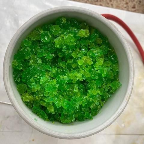
Preprints (www.preprints.org) | NOT PEER-REVIEWED | Posted: 20 July 2021                  doi:10.20944/preprints202107.0466.v1

                                      Despite ferrous chloride has been obtained from pickling waste liquors, to the best of
                                 our knowledge, the structural characterization of FeCl4·4H2O has been scarcely investi-
                                 gated. In the present work, the obtention of FeCl2·4H2O from pickling waste liquor using
                                 an effective process is described. A deep characterization of the structural and optical
                                 properties of the obtained samples was performed. In addition, the effects of aging on the
                                 crystal structure, structural characteristics as well as optical properties have been carried
                                 out.

                                 2. Experimental

                                 2.1. Obtention of the iron chloride from pickling waste liquor
                                      Ferrous chloride was obtained from pickling waste liquor from hydrochloric acid
                                 (HCl) pickling process of carbon steel.
                                      Acid pickling with HCl is a chemical process aimed at dissolving iron oxides from
                                 the metal surface without any significant attack on the steel. The oxidation layer com-
                                 posed of different types of iron oxides (Fe2O3, Fe3O4, and FeO) reacts with HCl to form
                                 ferrous chloride through reactions 1-3 [23]. Meanwhile, hydrochloric acid penetrates the
                                 oxidation layer and attacks the underlying metal. Ferrous chloride and hydrogen gas are
                                 produced by the reaction between hydrochloric acid and base steel (reaction 4) [23]. Due
                                 to these reactions, the oxidized layer is peeled off the surface, which is the main action of
                                 the pickling process.

                                  Fe2 O3 + Fe + 6HCl → 3FeCl2 + 3H2 O              (1)
                                  Fe3 O4 + Fe + 8HCl → 4FeCl2 + 4H2 O              (2)
                                  FeO + 2HCl → FeCl2 + H2 O                        (3)
                                  Fe + 2HCl → FeCl2 + H2                           (4)

                                       When free HCl is exhausted and ferrous chloride accumulates in the pickling liquor
                                 to the point where pickling cannot be carried out effectively, spent pickling solutions are
                                 discharged from the pickling tanks and managed as hazardous waste. It is estimated that
                                 around 380,000 m3 of chlorinated pickling water is produced annually in Europe [24]. One
                                 way to utilize this water is to treat it to recover the ferrous chloride and residual acid.
                                       The pickling water used in this work comes from the company HIASA (Asturias,
                                 Spain). The pickling water (139.9±5.1 g/L Fe 2+, 43.6±8.2 g/L free acid, 237.5±20.5 g/L total
                                 acid) was treated in a pilot evaporation-crystallization plant. The pickling water was
                                 heated to 130 °C in a pre-heater before entering a 500 mm long Vigreux column at the
                                 same temperature. The pickling water inlet flow into the column was 3 L/h. In the column,
                                 the pickling water is split into two streams: a vapour stream that condenses in a condenser
                                 at 65 °C and a liquor enriched in Fe2+ (188 ± 8.6 g/L). The vapour leaving the Vigreux col-
                                 umn is split into two streams in the condenser. A concentrated acid stream (107.7± 21.8
                                 g/L) and a water stream which is treated in a condenser at 6 °C to obtain a water stream
                                 without free acid. The whole system is kept under a vacuum of -640 mbar. The enriched
                                 liquor is crystallized in a crystallizer at 130 °C and filtered to obtain a mass of crystallized
                                 ferrous chloride and a residual solution that was recirculated at the beginning of the pro-
                                 cess. Figure 1 shows the ferrous chloride crystals immediately after crystallization and
                                 filtration. Concentrated acid and water can be reused in the pickling process.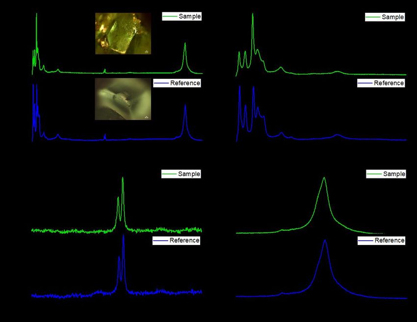
Preprints (www.preprints.org) | NOT PEER-REVIEWED | Posted: 20 July 2021 doi:10.20944/preprints202107.0466.v1
Figure 1. Ferrous chloride crystals obtained from pickling waste liquor by the described method.
2.2. Characterization of the iron chloride obtained from pickling waste liquor
X-ray diffraction measurements (XRD) have been done using a PANalytical Empy-
rean diffractometer using λ Cu Kα radiation with a step size of 0.02° (2θ) and a step time
of 7.9 s. Powder diffraction data were refined by the Rietveld method using the FullProf
software [25].
Micro-Raman measurements were performed at room temperature in a Horiba Jobin
Yvon LabRAM HR800 confocal microscope. Raman spectra were recorded under excita-
tion with the 632.8 nm line of a He-Ne laser. A charge-coupled device (CCD) detector was
used to collect the scattered light dispersed by a 600 lines/mm grating. The spectral reso-
lution of the system used was 1.5 cm-1. Photoluminescence measurements were done in
the same system but using the 325 nm line of a He-Cd laser as excitation source.
Optical micrographs were taken using a Leica DFC295 digital camera mounted on a
Leica MSV266 microscope.
The morphology and chemical composition of the obtained crystals were carried out
by Field Emission Scanning Electronic Microscopy using a Hitachi S-4800 and a FEI In-
spect SEM.
57Fe Mössbauer data were recorded at 298 K in the transmission mode using a con-
ventional constant acceleration spectrometer and a 57Co(Rh) source. An effective absorber
thickness of ca. 5 mg·cm-2 of natural iron was used in all the cases. The velocity scale was
calibrated using a 6 µm thick iron foil. The isomer shifts were referred to the centroid of
the spectrum of -Fe at room temperature. All the spectra were computer-fitted using
Lorentzian lines.
3. Results and discussion
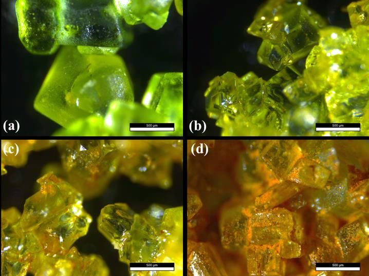
Commercial ferrous chloride (CFC) was analyzed for comparison purposes (Figure
2a). The fresh sample (CFF) was aged for a few days (CFT) at standard conditions (around
298 K, and 100 kPa of pressure). The initial light-green colour of the sample (Figure 2b)
was turned to brown in some areas of the obtained crystals (Figure 2c-d). The change in
colour from green to more yellowish crystals occurring in a few days (figure 2c). If the
samples are left in ambient atmosphere for more than 20 days, the colour progresses to-
wards an orange-brown (Figure 2b-d). No further changes are observed in samples aged
for months, indicating that at some point the degradation of the crystals stops. A close
inspection of the samples shows that orange regions and orange agglomerates appear be-
tween and on the crystals, respectively (Figure 2d). Moreover, although the crystals are
still green in colour, they lost their transparency and the surface is much rougher. These
two effects are behind the macroscopic colour change observed in the aged samples.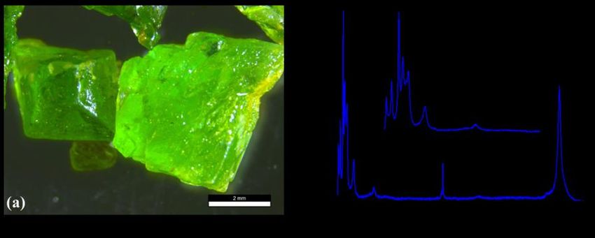
Preprints (www.preprints.org) | NOT PEER-REVIEWED | Posted: 20 July 2021                  doi:10.20944/preprints202107.0466.v1

                                 Figure 2. Optical micrographs of the FeCl2·4H2O crystals: (a) Commercial reference crystals (CFC).
                                 (b) Fresh FeCl2·4H2O sample (CFF). (c)-(d) Images showing different stages of the aging of the
                                 crystals (CFT): (c) few days; (d) months. In all cases, the scale bar is 500 μm.

                                 3.1. X-ray diffraction (XRD)
                                      Figure 3 exhibits comparative X-ray diffraction patterns of the samples investigated.
                                 All diffraction maxima can be indexed to a monoclinic structure with a space group P2 1/c
                                 and Z = 2 (14) (PDF 01-071-0668) compatible with a FeCl2·4H2O phase. No additional peaks
                                 were observed within the sensitivity of the experimental system used indicating the purity
                                 of the obtained samples. Lower intensity diffraction maxima can be observed in the difrac-
                                 togram of the CFC sample, as compared with that of the CFF sample. This result could be
                                 indicating the presence of an amorphous phase/thin layer of degradation products formed
                                 on the surface of the aged sample which goes undetected by XRD. The possible formation
                                 of this thin layer on the surface of ferrous chloride crystals would lead to a decrease of the
                                 diffraction maxima intensity. It should be noted that optical images exhibit a colour
                                 change in some areas of the CFF sample (see figure 2c, and figure 2d).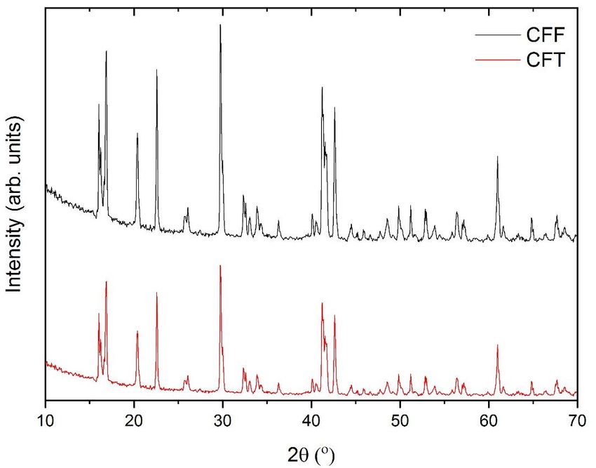
Preprints (www.preprints.org) | NOT PEER-REVIEWED | Posted: 20 July 2021 doi:10.20944/preprints202107.0466.v1
Figure 3. XRD patterns of the obtained ferrous chloride samples.
In order to carry out a more detailed structural characterization, XRD data were re-
fined by the Rietveld method where iron atoms were distributed in the 2a (0, 0, 0) posi-
tions, and chlorine, oxygen, and hydrogen were located in the 4e (x, y, z) sites. FulProf
software was used to calculate the structural parameters of the samples according to the
crystallographic database code ICSD-9198 [26]. Lattice parameters and cell angles of the
obtained samples are shown in Table 1. Slight differences were found in the calculated
cell parameters (i.e. cell parameters, and cell angles) for both, CFF and CFT, ferrous chlo-
ride samples. In addition, these values are similar to previously reported, as well as to the
theoretical values. The obtained results show that the structure of the FeCl2·4H2O samples
obtained from the pickling waste liquors is in good accordance with the theoretical struc-
ture previously reported for this compound [26,27].
Table 1. Lattice parameters, and cell angles calculated from the Rietveld refinements for the ob-
tained samples.
Sample a (Å) b (Å) c (Å) (O) (O) (O)
[26] 5.885(3) 7.174(3) 8.505(4) 90 111.11(5) 90
CFF 5.891(4) 7.117(4) 8.510(1) 90 111.10(2) 90
CFT 5.902(8) 7.121(6) 8.486(3) 90 112.02(8) 90
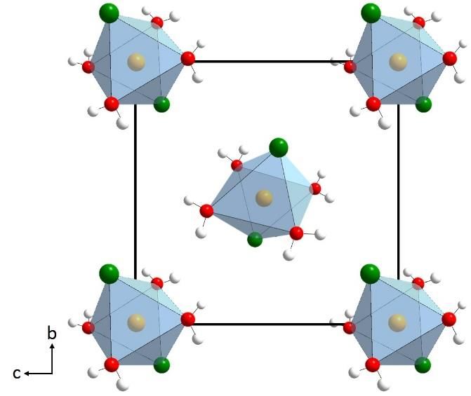
According to the calculated structural parameters, the crystal structure of the ob-
tained FeCl2·4H2O crystals structure is formed by a net of octahedra. Each octahedron has
the metal (Fe) at the center, surrounded by two Cl atoms and 4 water molecules (see Figure
4).
Preprints (www.preprints.org) | NOT PEER-REVIEWED | Posted: 20 July 2021              doi:10.20944/preprints202107.0466.v1

                                 Figure 4. Crystal structure for the FeCl2·4H2O.

                                 3.2. Scanning electron microscopy (SEM)

                                     Figure 5 shows the SEM images of the CFC reference sample and the obtained crystals
                                 of FeCl2·4H2O (CFF fresh sample, and CFT aged sample). In all cases, similar morpholo-
                                 gies were found. SEM micrographs reveal the formation of cubic crystals, in good agree-
                                 ment with those previously reported for samples with the same stoichiometry [27]. How-
                                 ever, a smoother surface was found in the case of the CFF sample (Figure 5b) as compared
                                 with the CFT sample (Figure 5c). In the latter case, some areas of the crystals exhibit a
                                 rougher surface (shown in the inset of Figure 5c), which correspond to the orange areas of
                                 the aged sample, which might be consistent with the formation of small crystallites of a
                                 new phase on the surface of the ferrous chloride crystals. These results again are in agree-
                                 ment with the optical images and XRD results.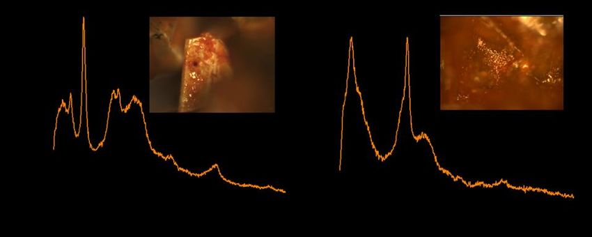
Preprints (www.preprints.org) | NOT PEER-REVIEWED | Posted: 20 July 2021                 doi:10.20944/preprints202107.0466.v1

                                 Figure 5. SEM micrographs of the (a) CFC reference sample, and (b) CFF and (c) CFT obtained crys-
                                 tals.

                                 3.3. µ-Raman spectroscopy and µ-photoluminescence

                                     In order to confirm the crystal structure of the obtained FeCl2·4H2O sample and study
                                 the formation of new phases in the aging process, micro-Raman spectroscopy measure-
                                 ments have been performed. In Figure 6, the Raman spectra of the reference FCF crystals
                                 and the fresh sample CFF are shown. In both cases (Figure 6a), all the modes can be as-
                                 cribed to FeCl2·4H2O with monoclinic C2h5 space group [28], in agreement with the XRD
                                 results. The 30 atoms in the unit cell give rise to 42 Raman active modes. Furthermore, the
                                 observed Raman modes can be divided in different types, also related to different fre-
                                 quency ranges. At frequencies below 100 cm-1, octahedra external modes are detected
                                 (peak at 70 cm-1). The octahedra internal modes appear in the frequency range between
                                 100 cm-1 and 400 cm-1 (Figure 6b, indicated as octahedra internal deformations, OID). The
                                 peaks at 104, 145, and 170 cm-1 (measured on the CFF spectrum) are related to the internal
                                 deformation bending of the octahedra. A shoulder at around 190 cm-1 is associated with
                                 Fe-Cl stretching modes. On the other hand, Fe-O stretching mode peaks are located at 203
                                 and 303 cm-1. The rest of the observed modes come from the vibrations of the water mol-
                                 ecules. The band related to water vibration modes is found at 619 cm-1 (Figure 6a). The
                                 peaks shown in Figure 6b are ascribed to bending vibrations of the water molecules (1632
                                 and 1651 cm-1). Finally, stretching vibrations (symmetric and antisymmetric) of the water
                                 molecules are included in the band centered at 3410 cm-1 (Figure 6d).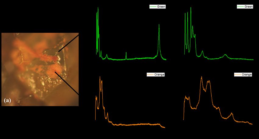
Preprints (www.preprints.org) | NOT PEER-REVIEWED | Posted: 20 July 2021                   doi:10.20944/preprints202107.0466.v1

                                 Figure 6. Micro-Raman spectra recorded on the CFC sample (blue) and the CFF sample (green). The
                                 excitation wavelength is 633 nm. (a) Spectra showing all the Raman peaks obtained. In the inset,
                                 optical images of both reference and obtained crystals are shown. (b) Raman modes appearing in the
                                 low-frequency range (50-1000 cm-1). (c) Intermediate frequency range (1250-2000 cm-1). (d) High-fre-
                                 quency range (3000-3800 cm-1).

                                      As has been already mentioned, as the colour of the samples changes from green to
                                 orange-brown, the aspect of the crystals in the optical microscope images changes (see
                                 figures 2c-d). Two different regions are observed in the CFT crystals: the region that still
                                 has a green colour and the orange region (Figure 7a). We have used micro-Raman meas-
                                 urements to study the differences between the green and the orange regions. The spectra
                                 recorded on the green part of the crystal (Figure 7b-c) reveal that it is still FeCl2·4H2O, as
                                 it exhibits the same modes as those described in figure 6. The main difference is the signal
                                 intensity, which is lower in the aged samples. This can be related to a decrease in the
                                 crystal quality. On the other hand, the spectra recorded on the orange region (Figure 7d-
                                 e) are completely different. The first observation that can be made is the disappearance of
                                 the water molecule vibrations between 1600 and 3800 cm -1 (Figure 7c). Then, the modes
                                 are mainly found in the range between 50 and 1000 cm -1 (Figure 7d).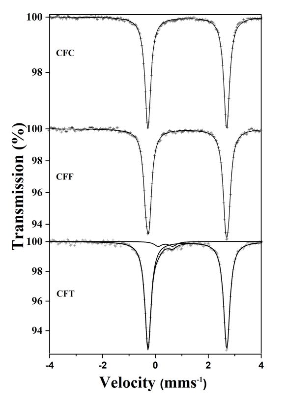
Preprints (www.preprints.org) | NOT PEER-REVIEWED | Posted: 20 July 2021                 doi:10.20944/preprints202107.0466.v1

                                 Figure 7. Micro-Raman spectra recorded on CFT sample. (a) Optical micrograph of a crystal, show-
                                 ing the regions where the spectra were taken. (b)-(c) Raman spectra measured on the green region.
                                 (d)-(e) Raman spectra recorded on the orange region.

                                     Although in some rare cases a peak at 191 cm-1, related to the FeCl3 compound [29]
                                 can be detected (Figure 8a), the most common peaks recorded in the orange region are
                                 those shown in Figure 7e. They are broad bands centered at about 112, 305, 399, 493, 540,
                                 and 717 cm-1. They can be related to different phases of iron oxyhydroxides (FeOOH) [30],
                                 specifically akaganeite (β-FeOOH) [31].

                                      The formation of iron oxyhydroxides has been observed in the corrosion process of
                                 iron in the presence of chloride ions [32]. It has been reported [33] that when iron metals
                                 are exposed to concentrated hydrochloric acid and subsequently to a humid environment,
                                 FeCI2.4H2O crystals are first formed. After some time, a membrane of β-FeOOH covers
                                 the ferrous chloride. β-FeOOH is commonly found on iron exposed to highly chlorinated
                                 water, or on iron immersed in strong solutions of hydrochloric acid or sodium chloride
                                 and then exposed to air [33]. A similar situation could be occurring for our crystals: first,
                                 the formation of the FeCl2·4H2O crystals by the as-described process, an iron compound
                                 exposed to concentrated HCl solution, and then exposed to air for few days (with the cor-
                                 responding air humidity). In addition, iron chloride hydrates are deliquescent. Thus, fer-
                                 rous chloride could absorb air humidity and dissolve in it, leading to a superficial film
                                 solution formed by Fe2+, Cl-, and oxygen, and promoting the oxidation of Fe(II) to Fe(III)
                                 forming the β-FeOOH phase.

                                      For CFT samples aged for a period of months, a shaper peak at 331 cm -1 can also be
                                 detected in some areas (Figure 8b), which can be also related to the akaganeite phase with
                                 Cl [34]. All these results point toward the formation of akaganeite iron oxyhydroxide due
                                 to the presence of HCl on the surface of the crystals that remains after the washing process.
Preprints (www.preprints.org) | NOT PEER-REVIEWED | Posted: 20 July 2021 doi:10.20944/preprints202107.0466.v1
Figure 8. Micro-Raman spectra recorded on CFT sample: (a) showing the peak related to FeCl 3; (b)
showing the peak adscribed to akaganeite phase with Cl. Insets: optical micrographs of the crystals
where the spectra were taken.
Photoluminescence (PL) measurements have been also performed on both green and
orange regions. Again, a clear difference in the signal recorded has been observed (Figure
9). PL spectrum of green crystals is dominated by a sharp peak at 365 nm. No reference in
the literature has been found about the PL emission of FeCl2·4H2O compound, but the same
emission is found in the CFC and CFF samples (Figure 9, blue and green line respectively).
Due to the sharpness and shape of this peak, it can be related to the near band edge emis-
sion of the material. This emission disappears completely in the orange regions of the CFT
sample, in agreement with the material transformation described in the Raman measure-
ments (Figure 9, orange line).
Figure 9. PL emission of CFC sample, CFF sample, and CTF sample, recorded using 325 nm laser
excitation wavelength. In the inset, a zoom of the peak at 365 nm is shown.
3.4. Mössbauer spectroscopy
Figure 10 collects the 298 K 57Fe Mössbauer spectra recorded from a commercial
FeCl2·4H2O sample (CFC), a fresh sample (CFF), and a sample aged for seven days (CFT).
The CFC spectrum consists of a narrow doublet with isomer shift =1.21 mm·s-1 and quad-
rupole splitting =2.98 mm·s-1. These hyperfine parameters are fully characteristic ofPreprints (www.preprints.org) | NOT PEER-REVIEWED | Posted: 20 July 2021 doi:10.20944/preprints202107.0466.v1
FeCl2·4H2O [35]. The spectrum taken from CFF is also a narrow doublet with identical
parameters to those of CFC. Therefore, we can confidently ensure that the fresh sample
CFF is FeCl2·4H2O. Differently to the spectra of these two samples, the spectrum recorded
from the aged sample CFT shows two quadrupole doublets. A major one, accounting for
95% of the spectral area and parameters typical of FeCl2·4H2O, and a minor one, 5%, with
isomer shift =0.38 mms-1 and quadrupole splitting =0.54 mms-1. These Mössbauer pa-
rameters can be assigned to Fe3+ oxyhydroxides such as lepidocrocite (-FeOOH) or ak-
aganeite (-FeOOH) [36]. Since akageneite is usually formed in the presence of chloride
ions it is plausible to think that the Fe3+ doublet can be due to this latter oxyhydroxide.
Also, akaganeite is the compound detected in Raman spectroscopy, as we have described
before. The spectra of samples aged for longer (up to 60 days) did not show higher Fe 3+
concentrations than that shown by CFT.
Figure 10. 298 K 57Fe transmission Mössbauer spectra recorded from the various samples.
Then, the observed colour change can be related to a degradation process of the crys-
tals due to the formation of rust on the surface and intergranular sites (Figure 2d). The rush
formed is composed mainly by akaganeite iron oxyhydroxide (β-FeOOH), as confirmed
by Raman and Mössbauer measurements. In the case of the sample aged for longer, the
formation of a surface film of the akaganeite phase (which is not liquescent) could reduce
the degradation of the ferrous crystals. For this reason, the corrosion stops when this film
is enough to prevent the contact of the FeCl2·4H2O surface with the air humidity.
In addition, it has been possible to obtain bigger FeCl 2·4H2O from the pickling waste
liquors by the ferrous phase slowly concentrated (Figure 11a). The Raman spectrum of
these crystals is the same as those described in Figure 5, confirming that they have thePreprints (www.preprints.org) | NOT PEER-REVIEWED | Posted: 20 July 2021 doi:10.20944/preprints202107.0466.v1
same crystal structure (Figure 11b). More interesting is that these bigger crystals do not
present degradation even if they are left in ambient for a long period of time. Orange ag-
glomerates that may be found on these bigger crystals show the Raman spectrum of FeCl 3
compound, without modes related to the oxyhydroxides.
Figure 11. (a) Optical micrographs of the FeCl2·4H2O crystals of larger size (scale bar of 2 mm). (b)
Micro-Raman spectra recorded on the crystals of part (a).
4. Conclusions
Ferrous chloride crystals with FeCl2·4H2O stoichiometry were obtained from liquors
hydrochloric acid pickling process of carbon steel. The samples were obtained in a pilot
evaporation-crystallization plant using pickling water from the company HIASA (Astu-
rias, Spain). The final obtained crystals were characterized by a wide number of tech-
niques. The effect of the aging of the samples in the structural characterization was also
assessed. XRD shows that in both, fresh and aging samples all diffraction maxima can be
attributed to the FeCl2·4H2O structure. In addition, the results obtained from the Rietveld
refinement exhibit that the structural parameters were similar to the theoretical previously
reported for this type of structure. SEM images reveal the formation of cubic crystals in
all cases. Moreover, the obtained ferrous crystals exhibit a smooth surface, as in the case
of the reference sample, while in the case of the aged sample some rougher surface areas
were found which might be due to the formation of small crystallites of a new phase. µ-
Raman spectra for the reference sample and the obtained ferrous chloride crystals were
similar. All vibration modes can be attributed to FeCl2·4H2O phase, with monoclinic C2h5
space group. Stretching vibrations (symmetric and antisymmetric) of the water molecules
in the structure were also detected. Two different Raman spectra were found in the case
of the aged sample related to the different areas observed. Green regions exhibit the same
Raman spectra attributable to FeCl2·4H2O phase. In the case of orange regions, Raman
peaks that can be related to different phases of iron were found probably due to a corro-
sion process of iron in the presence of chloride ions in the aged sample. Differences in the
intensity PL were observed for the analyzed samples. Despite that, to the best of our
knowledge PL study of this type of compound has not previously been reported, the dom-
inated sharp band could be related to the near band edge emission of the material. Möss-
bauer spectra of both the reference sample and the obtained crystals consists of a narrow
doublet, which are fully characteristic of FeCl2·4H2O. Again, the obtained results reveal
the formation of FeCl2·4H2O crystals. In the case of the aged sample, the spectrum shows
two quadrupole doublets. One of them accounting for 95% of the spectral area, which is
typical of FeCl2·4H2O; while the other one accounting for 5%, and can be assigned to Fe 3+
oxyhydroxides. The spectra of samples aged for longer periods did not show higher Fe3+
concentrations. Thus, all obtained results reveal that it is possible to obtain high-purity
and high-stability FeCl2·4H2O crystals from pickling waste liquors.
Author Contributions: Conceptualization, Félix López; Formal analysis, Lorena Alcaraz, Belén So-
tillo, José F. Marco, Paloma Fernández and Félix López; Investigation, Lorena Alcaraz, Belén Sotillo,Preprints (www.preprints.org) | NOT PEER-REVIEWED | Posted: 20 July 2021 doi:10.20944/preprints202107.0466.v1
José F. Marco, Paloma Fernández and Félix López; Methodology, Lorena Alcaraz, Belén Sotillo,
Francisco Alguacil and Félix López; Writing – original draft, Lorena Alcaraz, Belén Sotillo and José
F. Marco; Writing – review & editing, Lorena Alcaraz, Belén Sotillo, José F. Marco, Francisco Algua-
cil, Paloma Fernández and Félix López.
Funding: Authors from the National Center for Metallurgical Researches, Spanish National Re-
search Council (CENIM-CSIC) would like to thank the financial support thought the project
DECAPVALUE. HIASA Contract ID 20200543.
Acknowledgments: B.S. acknowledges the postdoctoral fellowship from the “Atracción de Talento”
Program funded by the Comunidad de Madrid (2017-T2/IND-5465). P.F. acknowledges Com-
plutense University of Madrid and Banco Santander for support via the project UCM-Santander
2019 (PR87/19-22613). Financial support from the Spanish Agencia Estatal de Investigación through
project RTI2018-095303-B-C51 is gratefully acknowledged.
Conflicts of Interest: The authors declare no conflict of interest.
ReferencesPreprints (www.preprints.org) | NOT PEER-REVIEWED | Posted: 20 July 2021 doi:10.20944/preprints202107.0466.v1
1. Tang, J.; Pei, Y.; Hu, Q.; Pei, D.; Xu, J. The Recycling of Ferric Salt in Steel Pickling Liquors: Preparation of Nano-
sized Iron Oxide. Procedia Environ. Sci. 2016, 31, 778–784, doi:10.1016/j.proenv.2016.02.071.
2. Gao, J.; Du, Z.; Ma, S.; Cheng, F.; Li, P. High-efficiency leaching of valuable metals from saprolite laterite ore
using pickling waste liquor for synthesis of spinel-type ferrites MFe2O4 with excellent magnetic properties. J.
Mater. Res. Technol. 2021, 10, 988–1001, doi:10.1016/j.jmrt.2020.12.063.
3. Negro, C.; Blanco, P.; Dufour, J.; Latorre, R.; Formoso, A.; López, F. The treatment of hydrochloric acid waste
pickle liquors. J. Environ. Sci. Heal. . Part A Environ. Sci. Eng. Toxicol. 1993, 28, 1651–1667,
doi:10.1080/10934529309375969.
4. Schmidt, B.; Wolters, R.; Kaplin, J.; Schneiker, T.; Lobo-Recio, M. de los A.; López, F.; López-Delgado, A.; Alguacil,
F.J. Rinse water regeneration in stainless steel pickling. Desalination 2007, 211, 64–71,
doi:10.1016/j.desal.2006.03.591.
5. Hoak, R.D.; Lewis, C.J.; Hodge, W.W. Treatment of Spent Pickling Liquors with Limestone and Lime. Ind. Eng.
Chem. 1945, 37, 553–559, doi:10.1021/ie50426a017.
6. Rituper, R. High-performance effluent-free pickling plants with fluid bed hydrochloric acid regeneration. Iron
Steel Eng. 1995, 72, 50–54.
7. Marañón, E.; Suárez, F.; Alonso, F.; Fernández, Y.; Sastre, H. Preliminary Study of Iron Removal from
Hydrochloric Pickling Liquor by Ion Exchange. Ind. Eng. Chem. Res. 1999, 38, 2782–2786, doi:10.1021/ie9806895.
8. López-Delgado, A.; Alguacil, F.J.; López, F.A. Recovery of iron from bio-oxidized sulphuric pickling waste water
by precipitation as basic sulphates. Hydrometallurgy 1997, 45, 97–112, doi:10.1016/S0304-386X(96)00066-7.
9. Tomaszewska, M. Recovery of hydrochloric acid from metal pickling solutions by membrane distillation. Sep.
Purif. Technol. 2001, 22–23, 591–600, doi:10.1016/S1383-5866(00)00164-7.
10. Yi, Y.; Tu, G.; Zhao, D.; Tsang, P.E.; Fang, Z. Pyrolysis of different biomass pre-impregnated with steel pickling
waste liquor to prepare magnetic biochars and their use for the degradation of metronidazole. Bioresour. Technol.
2019, 289, 121613, doi:10.1016/j.biortech.2019.121613.
11. Yang, S.; Li, W.; Zhang, H.; Wen, Y.; Ni, Y. Treatment of paper mill wastewater using a composite inorganic
coagulant prepared from steel mill waste pickling liquor. Sep. Purif. Technol. 2019, 209, 238–245,
doi:10.1016/j.seppur.2018.07.049.
12. Ciminelli, V.S.T.; Dias, A.; Braga, H.C. Simultaneous production of impurity-free water and magnetite from steel
pickling liquors by microwave-hydrothermal processing. Hydrometallurgy 2006, 84, 37–42,
doi:10.1016/j.hydromet.2006.03.058.
13. Chen, D.; Hou, J.; Yao, L.; Jin, H.; Qian, G.-R.; Xu, Z.P. Ferrite materials prepared from two industrial wastes:
Electroplating sludge and spent pickle liquor. Sep. Purif. Technol. 2010, 75, 210–217,
doi:10.1016/j.seppur.2010.07.009.
14. Özdemir, T.; Öztin, C.; Kıncal, N.S. Treatment of waste pickling liquors: process synthesis and economicanalysis.Preprints (www.preprints.org) | NOT PEER-REVIEWED | Posted: 20 July 2021 doi:10.20944/preprints202107.0466.v1
Chem. Eng. Commun. 2006, 193, 548–563, doi:10.1080/00986440500192238.
15. Zając, M.; Lipiński, I.E.; Rudowicz, C. Magnetostructural correlations for Fe 2+ ions at orthorhombic sites in
FeCl2·4H2O and FeF2·4H2O crystals modeled by microscopic spin Hamiltonian approach. J. Magn. Magn. Mater.
2016, 401, 1068–1077, doi:10.1016/j.jmmm.2015.11.007.
16. Brisebois, P.P.; Izquierdo, R.; Siaj, M. Room-Temperature Reduction of Graphene Oxide in Water by Metal
Chloride Hydrates: A Cleaner Approach for the Preparation of Graphene@Metal Hybrids. Nanomaterials 2020,
10, 1255, doi:10.3390/nano10071255.
17. Khandanlou, R.; Ahmad, M.; Shameli, K.; Kalantari, K. Synthesis and Characterization of Rice Straw/Fe 3O4
Nanocomposites by a Quick Precipitation Method. Molecules 2013, 18, 6597–6607, doi:10.3390/molecules18066597.
18. Huang, X.; An, D.; Song, J.; Gao, W.; Shen, Y. Persulfate/electrochemical/FeCl 2 system for the degradation of
phenol adsorbed on granular activated carbon and adsorbent regeneration. J. Clean. Prod. 2017, 165, 637–644,
doi:10.1016/j.jclepro.2017.07.171.
19. Kakar, S.; Batra, D.; Singh, R. Preparation and evaluation of magnetic microspheres of mesalamine (5-
aminosalicylic acid) for colon drug delivery. J. Acute Dis. 2013, 2, 226–231, doi:10.1016/S2221-6189(13)60132-8.
20. Shaghaghi, B.; Khoee, S.; Bonakdar, S. Preparation of multifunctional Janus nanoparticles on the basis of SPIONs
as targeted drug delivery system. Int. J. Pharm. 2019, 559, 1–12, doi:10.1016/j.ijpharm.2019.01.020.
21. Ahmad, T.; Ahmad, K.; Alam, M. Characterization of Water Treatment Plant’s Sludge and its Safe Disposal
Options. Procedia Environ. Sci. 2016, 35, 950–955, doi:10.1016/j.proenv.2016.07.088.
22. Kang, S.; Sun, Y.; Deng, S.; Li, S.; Su, Y.; Guo, W.; Li, J. Extraction of Huadian oil shale in subcritical FeCl2 solution.
Fuel Process. Technol. 2021, 211, 106571, doi:10.1016/j.fuproc.2020.106571.
23. Tang, B.; Su, W.; Wang, J.; Fu, F.; Yu, G.; Zhang, J. Minimizing the creation of spent pickling liquors in a pickling
process with high-concentration hydrochloric acid solutions: Mechanism and evaluation method. J. Environ.
Manage. 2012, 98, 147–154, doi:10.1016/j.jenvman.2011.12.027.
24. No Title Available online: CONDORCHEM ENVITECH, S. L. (2017). Life Dime HCl & Zn Recovery.
25. Roisnel, T.; Rodriguez-Carvajal, J. WinPLOTR, a graphic tool for powder diffraction.
26. Verbist, J.J.; Hamilton, W.C.; Koetzle, T.F.; Lehmann, M.S. Neutron Diffraction Study of Iron(II) Chloride
Tetrahydrate, FeCl2· 4H2O. J. Chem. Phys. 1972, 56, 3257–3264, doi:10.1063/1.1677688.
27. Guchhait, S.K.; Sammi, H.; Yadav, K.K.; Rana, S.; Jha, M. New hydrometallurgical approach to obtain uniform
antiferromagnetic ferrous chloride cubes from waste tin cans. J. Mater. Sci. Mater. Electron. 2021, 32, 2965–2972,
doi:10.1007/s10854-020-05048-1.
28. Cariati, F.; Masserano, F.; Martini, M.; Spinolo, G. Raman studies of NiX 2·6H2O and FeCl2·4H2O. J. Raman
Spectrosc. 1989, 20, 773–777, doi:10.1002/jrs.1250201204.
29. Caswell, N.; Solin, S.A. Vibrational excitations of pure FeCl 3 and graphite intercalated with ferric chloride. SolidPreprints (www.preprints.org) | NOT PEER-REVIEWED | Posted: 20 July 2021 doi:10.20944/preprints202107.0466.v1
State Commun. 1978, 27, 961–967, doi:10.1016/0038-1098(78)91015-3.
30. de Faria, D.L.A.; Venâncio Silva, S.; de Oliveira, M.T. Raman microspectroscopy of some iron oxides and
oxyhydroxides. J. Raman Spectrosc. 1997, 28, 873–878, doi:https://doi.org/10.1002/(SICI)1097-
4555(199711)28:113.0.CO;2-B.
31. Nieuwoudt, M.K.; Comins, J.D.; Cukrowski, I. The growth of the passive film on iron in 0.05 M NaOH studied
in situ by Raman micro-spectroscopy and electrochemical polarisation. Part I: Near-resonance enhancement of
the Raman spectra of iron oxide and oxyhydroxide compounds. J. Raman Spectrosc. 2011, 42, 1335–1339,
doi:10.1002/jrs.2837.
32. Criado, M.; Martínez-Ramirez, S.; Bastidas, J.M. A Raman spectroscopy study of steel corrosion products in
activated fly ash mortar containing chlorides. Constr. Build. Mater. 2015, 96, 383–390,
doi:10.1016/j.conbuildmat.2015.08.034.
33. Gilberg, M.R.; Seeley, N.J. The identity of compounds containing chloride ions in marine iron corrosion products:
a critical review. Stud. Conserv. 1981, 26, 50–56, doi:10.1179/sic.1981.26.2.50.
34. Neff, D.; Bellot-Gurlet, L.; Dillmann, P. Deterioration of iron archaeological artefacts: micro-Raman investigation
on Cl-containing corrosion products. 2007, doi:10.1002/jrs.1659.
35. Bull, J.N.; Maclagan, R.G.A.R.; Fitchett, C.M.; Tennant, W.C. A new isomorph of ferrous chloride tetrahydrate:
A 57Fe Mössbauer and X-ray crystallography study. J. Phys. Chem. Solids 2010, 71, 1746–1753,
doi:10.1016/j.jpcs.2010.09.009.
36. Murad, E. Magnetic properties of microcrystalline iron (III) oxides and related materials as reflected in their
Mössbauer spectra. Phys. Chem. Miner. 1996, 23, 248–262, doi:10.1007/bf00207766.You can also read


