Effect of feeding mosquito larvae on the coloration of Siamese fighting fish (Betta splendens) during grow-out
←
→
Page content transcription
If your browser does not render page correctly, please read the page content below
Int Aquat Res (2021) 13:71–79
https://doi.org/10.22034/IAR.2021.1916191.1116

SHORT COMMUNICATION

Effect of feeding mosquito larvae on the coloration of Siamese
fighting fish (Betta splendens) during grow-out

Moises Mejia-Mejia . Elsah Arce                    . Judith García-Rodríguez . Luis M. Burciaga

Received: 26 November 2020 / Accepted: 06 February 2021 / Published online: 28 March 2021
© The Author(s) 2021

Abstract Coloration is one of the most valued features in aquaculture or ornamental fish, and the Siamese
fighting fish (Betta splendens) (Regan 1910) is an excellent model to study coloration. Carotenoids are one
of the pigments that express colour in fish. Microalgae synthesize these pigments, which can be transferred
through feeding first to mosquito larvae, then to fish when they feed on mosquito larvae. We tested the effect
of feeding mosquito larvae on coloration and growth in Siamese fighting fish. Over a 60-day period, 52
individual Siamese fighting fish (32 males and 20 females) were fed with commercial micro pellets (control
diet) or mosquito larvae (experimental diet). We expected that fish fed with mosquito larvae would be more
colourful and larger than fish fed with commercial micro pellets. Consistent with this prediction, Siamese
fighting fish were more colourful when they were fed the mosquito larvae diet than when they were fed a
commercial micro pellet diet. We therefore recommend the use of mosquito larvae for Siamese fighting fish
production. Additionally, since the Siamese fighting fish is an efficient predator of the mosquito larvae, we
suggest the use of this live food as a high quality alternative food and a colour bio-capsule with numerous
carotenoid pigments.

Keywords Carotenoid source . Aquarium fish coloration . Chlorella sp. . Culex quinquefasciatus

Introduction

The Siamese fighting fish, Betta splendens, is a popular fish species for ornamental production due to its
brilliant colours and large fins (Puello-Cruz et al. 2010; Saekhow et al. 2018). The production of Siamese
fighting fish (Betta splendens) (Regan 1910) is an important economic activity in countries like Thailand,
Indonesia, Singapore, China, Malaysia, Japan, USA, and Mexico (Chuan et al. 2003; Thongprajukaew et
al. 2011). Coloration is one of the most valued features in ornamental fish (Barber et al. 2000; Pavlidis
et al. 2006). Since fish cannot produce the pigments that give them their colour endogenously, they must
acquire them through diet (Sefc et al. 2014). One of the classes of pigments that express colour in fish are
carotenoids, and in various studies, the diets of ornamental fish have been enriched with these pigments
in order to improve their coloration and increase their market value (Sommer et al. 1992; Pham et al.
2014). The inclusion of pigments in fish food is also necessary for the nutrition of the fish (Gordon et
al. 1981). Carotenoid pigments have a variety of physiological roles, for example, as immunostimulants
and antioxidants (McGraw and Ardia 2003). Coloration in fish has been evaluated after feeding live food

Moises Mejia-Mejia
Facultad de Ciencias Agropecuarias, Universidad Autónoma del Estado de Morelos, Cuernavaca, Morelos, México

Elsah Arce ( ) . Judith García-Rodríguez
Laboratorio de Acuicultura e Hidrobiología, Departamento de Hidrobiología, Centro de Investigaciones Biológicas, Universidad Autónoma del Estado
de Morelos, Cuernavaca, Morelos, México
e-mail: elsah.arce@uaem.mx

Luis M. Burciaga
Posgrado en Ciencias del Mar y Limnología, Universidad Nacional Autónoma de México; Av. Ciudad Universitaria 3000, C.P. 04510, Coyoacán, Ciudad
de México, México
72                                                                                    Int Aquat Res (2021) 13:71-79

including water fleas, mosquito larvae, and Artemia nauplii as pigment sources (Torrisen and Naevdal 1988;
Thongprajukaew et al. 2014; Arce et al. 2018). Like the fish, these organisms used as food for ornamental
fish (Artemia and mosquito larvae) cannot produce carotenoid pigments themselves, but rather obtain them
from microalgae present in live food cultures (Jones et al. 1993; Nieves et al. 1996; Voltolina et al. 1999).
Microalgae are photosynthetic organisms that synthesize different types of pigments and transport them
to chloroplasts (Siefermann-Harms 1987; Yong and Lee 1991; Bidigareet et al. 1993); the chloroplasts
contain the pigments that transfer the colour to the fish (Siefermann-Harms 1987; Storebakken et al. 1987).
Additionally, it has been observed that live food in general stimulates growth and coloration in fish (Torrisen
1984; Arce et al. 2018; Luna-Figueroa et al. 2019).
    The goal of this study was to evaluate the effect of feeding mosquito larvae on coloration and growth of
Siamese fighting fish. We examined the effect of microalgae present in live food culture on pigment transfer
to mosquito larvae and colour expression in fish. We expected that fish fed with mosquito larvae would be
more colourful and grow more than fish fed with commercial micro pellets. Although coloration and growth
have been assessed before in fish species (Baron et al. 2008; Mat Nawang et al. 2019), this is the first study
to assess coloration using mosquito larvae as food.

Materials and methods

Fish acclimation and control and experimental diets

Four-month-old Siamese fighting fish (Blue veiltail-Betta total length: 42.30 ± 3.48 mm; mass: 1.08 ± 0.23
g) were obtained from multiple parents at fish farms (n= 56). The fish were placed into individual circular
containers with a volume of 500 mL. The walls of these containers were covered with white plastic film
to prevent the fish from seeing each other and to equalize the luminosity because fish use vision to assess
their surroundings (Stevens et al. 2007) and because luminosity and wall colour can affect growth and
body coloration in ornamental fish (Mat Nawang et al. 2019). The physical and chemical conditions of the
water were 24 ± 0.5 °C, pH 7.2 ± 0.1, and the fish were subjected to a 12:12 h light: dark photoperiod using
a tube LED lamp connected to a programmed timer (TORK 40382). Prior to beginning the experimental
diet treatments, all of the fish to be used in the experiment were acclimated for 20 days to a 1:1 mixture
of the control and experimental diets. The control diet, commercial micro pellets, used Chlorella vulgaris
algae as a source of carotenoids (0.52 ppm). The control diet contained 43% protein, 4.5% lipids, and
5.0% carbohydrates. The experimental diet, mosquito larvae (Culex quinquefasciatus, Pham et al. 2014),
contained 43.5% protein, 9.5% lipids and 5.2% carbohydrates. Proximate analysis of the control diet was
performed in dry matter according to the AOAC (1990) methods. Samples were dried in an oven at 105
°C to constant weight, and ash content was estimated by incineration in a muffle furnace at 600 °C for 6
h. Crude protein was determined by acid digestion using the Kjeldahl method, lipids by petroleum ether
extraction, and carbohydrates by subtraction (Yuangsoi et al. 2010). Fish were fed ad libitum twice daily at
9:00 h and 16:00 h (Thongprajukaew et al. 2011; Arce et al. 2018). Faeces and uneaten food were removed
daily (De la Torre et al. 2018).

Experimental diet protocol and growth measurement

Following the 20-day acclimation period, the 56 fish were randomly divided based on the toss of a coin
into two experimental groups of 16 males (n= 32, 1.11 ± 0.04 g, 42.79 ± 0.65 mm) and 12 females (n= 24,
1.03 ± 0.05 g, 42.65 ± 0.69 mm) for the diet experiment (n = 28 per treatment). Each fish was placed in
an individual tank with the same physical and chemical water conditions as during the acclimation period.
At the beginning of the experimental period, the fish were kept without food for 24 h before biometric
analysis to ensure complete gastric evacuation. Fish were weighed with a plate balance (OHAUS; 0.01
g) and measured with callipers (INSIZE; 0.01 mm). For the next 60 days, the control group was fed the
commercial micro pellets and the experimental group received two mosquito larvae (C. quinquefasciatus).
Under both treatments, fish were fed ad libitum, daily at 9:00 h and 16:00 h (Thongprajukaew et al. 2011;
Arce et al. 2018). Growth of each fish was calculated at the end of the experiment as weight gain (mass,
Int Aquat Res (2021) 13:71-79 73 g) and size gain in total length (mm) by subtracting the initial measurement from the final measurement. Live food culture In order to obtain mosquito larvae to use as live food, Chlorella sp. microalgae was cultured in eight 590- L fiberglass tanks fertilized with 1000 mg organic matter (chicken manure; Paniagua-Michel et al. 1987; Voltolina et al. 1999; Ekpo et al. 2016). The temperature of the tanks was 26 ± 1 °C, pH 8.5 ± 1.4, and they were kept in natural light. Once fertilized, the tanks were left for 7 days before live food (mosquito larvae) were collected. The tanks remained under observation, and microalgae were counted and identified daily to estimate abundance. The growth of the microalgae and species identification of microalgae was determined by direct counting in a Neubauer chamber using a LEICA ICC50 HD microscope (Avendaño and Riquelme 1999).When the microalgae abundance reached 20000 cells/mL and/or after seven days, we fertilized another eight tanks. This procedure was repeated as many times as necessary to maintain a consistent supply of nutrients for the microalgae from the acclimation period through the end of the experiment. Because Betta fish do not discriminate between developmental stages of mosquitos as prey (unpublished data), mosquitoes from 1st instar larvae to pupa were considered “larvae” (Griffith and Turner 1996). Mosquito larvae were collected daily using a net with a mesh size of 0.5-mm. To avoid pathogens, mosquito larvae were provided aeration at 0.5 L/min flow and 10 °C and a 5 µg/L disinfectant solution (Wescodyne) was added for 10 minutes prior to using them as fish food (Kent et al. 2009). Carotenoid pigments To determine the concentration of carotenoid pigments in microalgae (mg/m3), 100 mL of water was filtered daily for the 7 days during the culture in each of the eight microalgae culture tanks (n= 56 samples) using a millipore filter (0.45 μm pore aperture). The membrane was centrifuged with 10 mL of methanol for 10 minutes at 1500 rpm. The carotenoid concentration was determined using a spectrophotometer (HACH DR / 2017; Dere et al. 1998). The microalgae collection started 7 days after fertilization, when microalgae were abundant (630,431 ± 23,922 cell/mL; 8 tanks, 7 days; n= 56) and at the end of the experiment, when algal abundance decreased (226,844 ± 10,003 cell/mL; 8 tanks, 7 days; n= 56). To determine the concentration of carotenoid pigments in mosquito larvae, we collected larvae daily for the 7 days that the culture lasted from each microalgae culture tank (100 mL). Mosquito larvae were rinsed with clean water and macerated in 10 ml of acetone, then carotenoid concentration was determined in the same way as for the microalgae (Dere et al. 1998). Fish colour measurements To assess fish coloration, 52 fish (32 males and 20 females) were placed individually into a transparent photographic tank (12 x 12 x 2 cm) at the beginning and end of the experiment with water with the same conditions as those used during maintenance. This photographic tank was placed in a 40 x 40 x 50 cm box with opaque white walls and white light to photograph the fish (Arce et al. 2018). Each fish was photographed in lateral view using a professional digital camera (Panasonic DMC-GH4; Arce et al. 2018). The camera was placed 30 cm from the photographic tank and operated in manual mode to avoid automatic colour modifications. All of the digital images were saved as Tagged Image File Format (TIFF) files (Mat Nawang et al. 2019). A similar area (12.6 mm2) of the peduncle zone of each fish in each picture was analysed for colour using Image J software (Igathinathane et al. 2009; Schneider et al. 2012; Johansson and Nilsson-Örtman 2013). This software detects the colour intensity of an image on an RGB scale, with hue and chroma values on a scale of 0-255. (Touchon and Warkentin 2008; Touchon and Wojdak 2014). Statistics We compared the pigment concentration of the microalgae and mosquito-larvae at the beginning of the microalgae culture and end of the collection period via Student’s t-test. We compared the colour, weight and size of the individuals assigned to each treatment at the beginning and end of the experiments using linear mixed-effects models (LMM; Bates et al. 2007). Sex and diet were included as fixed factors, and fish
74                                                                                                      Int Aquat Res (2021) 13:71-79

                                      140                                                                             f

                                      120         Control
                                                                                               d
                                                 Mosquito larvae                                              e
                                      100

                   Coloration (RGB)
                                      80                                                c

                                      60
                                            a                    b      b
                                                  a
                                      40

                                      20

                                        0
                                            Female                Male          1      Female                  Male
                                                       Initial                                      Final
Fig. 1 Fig. 1 Coloration
       Coloration        in female
                    in female  and and
                                   malemale Siamese
                                        Siamese     fightingfish.
                                                 fighting     fish.Different
                                                                    Different letters
                                                                              letters indicate
                                                                                       indicatesignificant differences
                                                                                                 significant           (p
Int Aquat Res (2021) 13:71-79 75
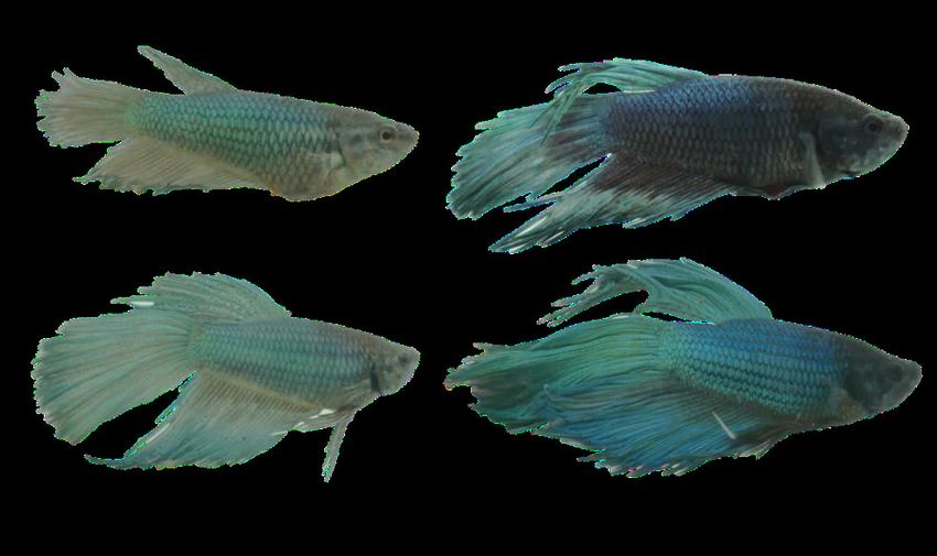
Fig. 2 Final body coloration of Siamese fighting fish. a) female control diet, b) male control diet, c) female mosquito larvae diet, and
d) male mosquito larvae diet.
Fig. 2 Final body coloration of Siamese fighting fish. a) female control diet, b) male control diet, c) female mosquito larvae diet,
and d) male mosquito larvae diet.
2.5 d
Control
c
2.0 Mosquito larvae c
1.5 b
ab
Mass (g)
a a
a
1.0
0.5
0.0
Female Male 1 Female Male
Initial Final
Fig. 3 in
Fig. 3 Mass Mass in female
female and male
and male Siamese
Siamese fighting
fighting fish.
fish. Differentletters
Different letters indicate
indicate significant
significantdifferences (p <
differences (p0.05). Mean
< 0.05). values
Mean and and
values
SE are SE are shown.
shown.
70 c c
Control
60 Mosquito larvae b b
50
Total length (mm)
a a a a
40
30
20
10
0
Female Male 1 Female Male
Initial Final
Fig. 4 Fig.
Total4 Total
lengthlength in female
in female andandmale
maleSiamese
Siamese fighting
fightingfish. Different
fish. letters
Different indicate
letters significant
indicate differences
significant (p < 0.05).(pMean
differences valuesMean
< 0.05).
and SE are shown.
values and SE are shown.
than fish fed with the control diet (Bonferroni; females: p < 0.001; males: p < 0.001; Figure 3). Final total76 Int Aquat Res (2021) 13:71-79
length also differed between diets in both sexes (LMM; χ2= 26.97, d.f.= 1, p < 0.001; Figure 4). Final total
length did not differ between males and females within each treatment (LMM; χ2= 0.42, d.f.= 1, p =0.52;
Figure 4). Fish fed mosquito larvae were longer than those fed the control diet (Bonferroni; females: p <
0.001, males: p < 0.001; Figure 4).
Discussion
Live food culture is an important industry for aquaculture (Le Ruyet et al. 1993; Reitan et al. 1993).
Microalgae are food for zooplankton, which is the most popular live food for fish, and Chlorella sp. is
one the most frequently used microalgae in the culture and collection of live food (Reitan et al. 1993;
Borowitzka 1999; Agwa and Abu 2014). Chlorella was the most abundant microalgae in the culture used in
our research, and although there were other species such as Scenedesmus, and Cosmarium, these accounted
for only 1% of the total microalgae population, so their contribution of carotenoids was not significant.
Chlorella, Scenedesmus, and Cosmarium are Chlorophytes and their essential pigments are carotenoids
(Priyadarshani and Rath 2012). Chlorella is a microorganism with many carotenoid pigments (Herring
1968; Gouveia et al. 1996), so from a consumer’s perspective, microalgae improve carotenoid digestion and
absorption (Becker and Venkataraman 1984; Sun et al. 2012). Due to the abundance of cultured microalgae,
the quantity of carotenoids in water samples at the beginning of the feeding period were greater than at the
end of the experimental period. Microalgae are an essential part of the diet of mosquito larvae (Rey et al.
2009). These microalgae are not only fundamental for the nutrition of the mosquito larvae, but mosquito
larvae also become a nutritious and colourful capsule for accumulating microalgae and delivering them
to fish, even when the microalgae density varies in the water. The carotenoid levels in mosquito larvae
that consumed microalgae remained the same throughout the live food culture period, which indicates
that mosquito larvae are efficient accumulators of carotenoid pigments, regardless of availability; thus,
they offer a high concentration of carotenoid pigments to the next trophic level (fish predator). Carotenoid
pigments are used to produce colour in fish and have a variety of physiological functions, for example, as
immunostimulants or sexual attractants (Evans and Norris 1996; Maan et al. 2006). Colour in males is a
sexually selected trait in many fish (Clotfelter et al. 2007), and it has been demonstrated that carotenoid
supplementation significantly increases the immune response (Alonso-Alvarez et al. 2004; Clotfelter et al.
2007). We found that both females and males were more colourful when fed mosquito larvae. The increased
colour intensity obtained by using mosquito larvae could be attributed to C. quinquefasciatus larvae being
filter feeders (microalgae consumers; Agwa and Abu 2014; Manimegalai and Sukanya 2014). They were
shown to be excellent consumers of these algae, and we observed a lot of microalgae in the digestive tracts
of mosquito larvae, such that mosquito larvae can be considered a colour bio-capsule.
Mosquito larvae have been used as live food, favouring the growth of fish (Barroso et al. 2014;
Thongprajukaew et al. 2014; Luna-Figueroa et al. 2019). In this study, both females and males were longer
and heavier when they were fed mosquito larvae. Fish growth is affected by food quality. An essential
characteristic of mosquito larvae is their high protein content (Luna-Figueroa et al. 2019). The amount
of protein contained in Chlorella sp. is 50%-60% (Spolaore et al. 2006), whereas in larvae used as live
food for fish, it is 43% (Luna-Figueroa et al. 2019). In our research we used living mosquito larvae, since
mosquito larvae may filter microalgae from the culture (Marten 2007). In addition to the nutrient quality of
the diet, our results may be partly due to the rate of consumption of live versus inert food, since fish fed ad
libitum may prefer live prey over inert food (Fernández-Díaz et al. 1993).
Conclusion
We demonstrated a positive effect on colour and growth in Siamese fighting fish fed with mosquito larvae
and we suggest the use of this live food (with appropriate precautions to prevent pathogen transmission)
as a high-quality alternative to pellets and an excellent colour bio-capsule of carotenoid pigments. In
future research we suggest using prepared diets with mosquito larvae fed with Chlorella sp. (e.g. mosquito
larvae prepared in pellet form). Such studies are important, first to test whether mass, size and coloration
improvements with the mosquito-based diet fed here can be replicated without the live food component.Int Aquat Res (2021) 13:71-79 77
Conflict of interest The authors declare that they have no conflict of interest.
Authors’ contribution Conceptualization: M.E.M.M., E.A.; Methodology: M.E.M.M., E.A., J.G.R.; Formal analysis: E.A., L.M.B.;
Investigation: L.M.B., E.A.; Resources: E.A., J.G.R.
Acknowledgements We thank Isela Molina (in memoriam) for helpful comments on the original idea. We thank the “Departamento
de Nutrición Animal y Bioquímica. Facultad de Medicina Veterinaria y Zootecnia, Universidad Nacional Autónoma de México” for
the proximate chemical analysis of mosquito larvae. We thank José Figueroa (in memoriam) and Jorge Luna-Figueroa for helpful
comments. Thanks to Diana Molina, Yuritzi Castillo, Olivia De los Santos, Emmanuel Paniagua, and Marco Franco for technical
assistance. We also thank Lynna Kiere for editing the English text.
References
Agwa OK, Abu GO (2014) Utilization of poultry waste for the cultivation of Chlorella sp. for biomass and lipid production. IJCMAS
3:1036-1047
Alonso-Alvarez C, Bertrand S, Devevey G, Gaillard M, Prost J, Faivre B, Sorci G (2004) An experimental test of the dose-dependent
effect of carotenoids and immune activation on sexual signals and antioxidant activity. Am Nat 164:651-659. https//doi.
org/10.1086/424971
Arce UE, Archundia FMP, Luna-Figueroa J (2018) The effect of live food on the coloration and growth in guppy fish, Poecilia
reticulata. Agric Sci 9:171-179. https//doi.org/10.4236/as.2018.92013
AOAC (1990) Official method of analysis. 13th ed. Washington, DC
Avendaño RE, Riquelme CE (1999) Establishment of mixed‐culture probiotics and microalgae as food for bivalve larvae. Aquacult
Res 30:893-900. https//doi.org/10.1046/j.1365-2109.1999.00420.x
Barber I, Arnott SA, Braithwaite VA, Andrew J, Mullen W, Huntingford, FA (2000) Carotenoid-based sexual coloration and body
condition in nesting male sticklebacks. J Fish Biol 57:777-790. https//doi.org/10.1111/j.1095-8649.2000.tb00274.x
Baron M, Davies S, Alexander L, Snellgrove D, Sloman KA (2008) The effect of dietary pigments on the coloration and behaviour of
flame-red dwarf gourami, Colisa lalia. Anim Behav 75:1041-1051. https://doi.org/10.1016/j.anbehav. 2007. 08.014
Barroso FG, Haro C, Sánchez-Muros MJ, Venegas E, Martínez-Sánchez A, Pérez-Bañón C (2014) The potential of various insect
species for use as food for fish. Aquaculture 422:193-201. https//doi.org/10.1016/j.aquaculture.2013.12.024
Bates D, Sarkar D, Bates MD, Matrix L (2007) The lme4 package. R package version 2:1-29
Becker EW, Venkataraman LV (1984) Production and utilization of blue-green alga Spirulina in India. Biomass 4:105-125. https//doi.
org/10.1016/0144-4565(84)90060-X
Bidigareet RR, Ondrusek ME, Kennicutt MC, Iturriaga R, Harvey HR, Hohan RW, Macko SA (1993) Evidence for a photoprotective
function for secondary carotenoids of snow algae. J Phycol 29:427-434. https//doi.org/10.1111/j.1529-8817.1993.tb00143.x
Borowitzka MA (1999) Commercial production of microalgae: ponds, tanks, and fermenters. Prog Ind Microbiol 35:313-321. https//
doi.org/10.1016/S0079-6352(99)80123-4
Chuan LL, Dhert P, Sorgeloos P (2003) Recent developments in the application of live feeds in the freshwater ornamental fish culture.
Aquaculture 227:319-331. https//doi.org/10.1016/S0044-8486(03)00512-X
Clotfelter ED, Ardia DR, McGraw KJ (2007) Red fish, blue fish: trade-offs between pigmentation and immunity in Betta splendens.
Behav Ecol 18:1139-1145. https//doi.org/10.1093/beheco/arm090
De la Torre ZAM, Arce UE, Luna-Figueroa J, Córdoba-Aguilar A (2018) Native fish, Cichlasoma istlanum, hide for longer, move
and eat less in the presence of a non-native fish, Amatitlania nigrofasciata. Environ Biol Fishes 101:1077-1082. https//doi.
org/10.1007/s10641-018-0761-z
Dere S, Günes T, Sivaci R (1998) Spectrophotometric determination of chlorophyll -A, B and total carotenoid contents of some algae
species using different solvents. Turk J Bot 22:13-17
Ekpo U, Ross AB, Camargo-Valero MA, Williams PT (2016) A comparison of product yields and inorganic content in process streams
following thermal hydrolysis and hydrothermal processing of microalgae, manure and digestate. Bioresour Technol 200:951-960.
https//doi.org/10.1016/j.biortech.2015.11.018
Evans MR, Norris K (1996) The importance of carotenoids in signaling during aggressive interactions between male fire mouth
cichlids (Cichlasoma meeki). Behav Ecol 7:1-6. https//doi.org/10.1093/beheco/7.1.1
Fernández-Díaz A, Pascual E, Yúfera M (1993) Feeding behaviour and prey size selection of gilthead seabream, Sparus aurata, larvae
fed on inert and live food. Mar Biol 118:323-328. https//doi.org/10.1007/BF00349800
Gordon HT, Bauernfeind JC, Furia TE (1981) Carotenoids as food colorants. Crit Rev Food Sci Nutr 18:1-59. https//doi.
org/10.1080/10408398209527357
Gouveia L, Veloso V, Reis A, Fernandes H, Novais J, Empis J (1996) Evolution of pigment composition in Chlorella vulgaris.
Bioresour Technol 57:57-159, 161-163. https//doi.org/10.1016/0960-8524(96)00058-2
Griffith JSR, Turner GD (1996) Culturing Culex quinquefasciatus mosquitoes with a blood substitute diet for the females. Med Vet
Entomol 10:265-268. https//doi.org/10.1111/j.1365-2915.1996.tb00741.x
Herring PJ (1968) The carotenoid pigments of Daphnia magna straus-I. The pigments of animals feed Chlorella pyrenoidosa and pure
carotenoids. Comp Biochem Physiol 24:187-203. https//doi.org/10.1016/0010-406X(68)90967-5
Igathinathane C, Pordesimo LO, Batchelo WD (2009) Major orthogonal dimensions measurement of food grains by machine vision
using ImageJ. Food Res Int 42:76-84. https//doi.org/10.1016/j.foodres.2008.08.013
Johansson F, Nilsson-Örtman V (2013) Predation and the relative importance of larval colour polymorphisms and colour polyphenism
in a damselfly. Evol Ecol 27:579-591. https//doi.org/10.1007/s10682-012-9617-8
Jones D, Kamarudin MS, Le Vay L (1993) The potential for replacement of live feeds in larval culture. J World Aquacult Soc 24:199-
210. https//doi.org/10.1111/j.1749-7345.1993.tb00009.x
Kent ML, Feist SW, Harper C, Hoogstraten-Miller S, Law JM, Sánchez-Morgado JM, Whipps CM (2009) Recommendations
for control of pathogens and infectious diseases in fish research facilities. Comp Biochem Phys C 149:240-248. https//doi.
org/10.1016/j.cbpc.2008.08.00178 Int Aquat Res (2021) 13:71-79
Le Ruyet JP, Alexandre JC, Thebaud L, Mugnier C (1993) Marine fish larvae feeding: formulated diets or live prey? J World Aquacult
Soc 24:211-224. https//doi.org/10.1111/j.1749-7345.1993.tb00010.x
Luna-Figueroa J, Arce UE, Figueroa TJ, Archundia FMP (2019) Pre-adults mosquito in fish species feeding. Int J Aqu Sci 10:55-59
Maan ME, van der Spoel M, Jimenez PQ, van Alphen JJM, Seehausen O (2006) Fitness correlates of male coloration in a Lake
Victoria cichlid fish. Behav Ecol 17:691-699. https//doi.org/10.1093/beheco/ark020
Manimegalai K, Sukanya S (2014) Biology of the filarial vector, Culex quinquefasciatus (Diptera: Culicidae). Int J Curr Microbiol
App Sci 3:781-724
Marten GG (2007) Larvicidal algae. J Am Mosq Control Assoc 23:177-183. https//doi.org/10.2987/8756-971X(2007)23[
177:LA]2.0.CO;2
Mat Nawang SUS, Ching FF, Senoo S (2019) Comparison on growth performance, body coloration changes and stress response of
juvenile river catfish, Pangasius hypophthalmus reared in different tank background colour. Aquac Res 50:2591-2599. https://doi.
org/10.1111/are.14215
McGraw KJ, Ardia DR (2003) Carotenoids, immunocompetence, and the information content of sexual colors: an experimental test.
Am Nat 162:704-712. https//doi.org/10.1086/378904
Nieves M, Voltolina D, Sapien MT, Gerhardus H, Robles AI, Villa M.A (1996) Culturing microalgae with agricultural fertilizers. Riv
Ital Acquacolt 31:81-84
Paniagua-Michel J, Farfan BC, Bückle-Ramirez F (1987) Culture of marine microalgae with natural biodigested resources. Aquaculture
64:249-256. https//doi.org/10.1016/0044-8486(87)90330-9
Pavlidis M, Papandroulakis N, Divanach P (2006) A method for the comparison of chromaticity parameters in fish skin: preliminary
results for coloration pattern of red skin Sparidae. Aquaculture 258:211-219. https//doi.org/1016/j. aquaculture.2006.05.028
Pham MA, Byun HE, Kim KD, Lee SM (2014) Effects of dietary carotenoid source and level on growth, skin pigmentation,
antioxidant activity and chemical composition of juvenile olive flounder Paralichthys olivaceus. Aquaculture 431:65-72. https//
doi.org/10.1016/j.aquaculture.2014.04.019
Priyadarshani I, Rath B (2012) Commercial and industrial applications of micro algae - A review. J Algal Biomass Utln 3:89-100.
Puello-Cruz AC, Velasco-Blanco G, Martínez-Rodríguez IE (2010) Growth and survival of siamese fighting fish, Betta splendens,
larvae at low salinity and with different diets. J World Aquacult Soc 41:823-838. https//doi.org/10.1111/ j.1749-7345.2010.00425.x
R Core Team (2020) A language and environment for statistical computing. R Foundation for Statistical Computing
Reitan KI, Rainuzzo JR, Oie G, Olsen Y (1993) Nutritional effects of algal addition in first-feeding of turbot (Scophthalmus maximus
L.) larvae. Aquaculture 118:257-275. https//doi.org/10.1016/0044-8486(93)90461-7
Rey JR, Hargraves PE, O’Connell SM (2009) Effect of selected marine and freshwater microalgae on development and survival of the
mosquito Aedes aegypti. Aquatic Ecol 43:987-997. https//doi.org/10.1007/s10452-009-9232-0
Saekhow S, Thongprajukaew K, Phromkunthong W, Sae‐khoo H (2018) Minimal water volume for intensively producing male
Siamese fighting fish (Betta splendens Regan, 1910). Fish Physiol Biochem 44:1075-1085. https//doi.org/10.1007/s10695-018-
0495-z
Schneider CA, Rasband WS, Eliceiri KW (2012) NIH Image to ImageJ: 25 Years of Image Analysis. Nat Methods 9:671-675. https//
doi.org/10.1038/nmeth.2089
Sefc KM, Brown AC, Clotfelter ED (2014) Carotenoid-based coloration in cichlid fishes. Comp Biochem Physiol A Mol Integr
Physiol 173:42-51. https//doi.org/10.1016/j.cbpa.2014.03.006
Siefermann-Harms D (1987) The light harvesting and protective functions of carotenoids in photosynthetic membranes. Physiol Plant
69:561-568. https//doi.org/10.1111/j.1399-3054.1987.tb09240.x
Sommer T, Da Souza FML, Morrissy NM (1992) Pigmentation of adult rainbow trout, Oncorhynchus mykiss, using green alga
Haematococcus pluvialis. Aquaculture 106:63-74. https//doi.org/10.1016/0044-8486(92)90250-O
Spolaore P, Joannis-Cassan C, Duran E, Isambert A (2006) Commercial applications of microalgae. J Biosci Bioeng 101:87-96. https//
doi.org/10.1263/jbb.101.87
Stevens M, Párraga C, Cuthill I, Partridge J, Troscianko T (2007) Using digital photography to study animal coloration. Biol J Linn
Soc 90:211-237. https//doi.org/10.1111/j.1095-8312.2007.00725.x
Storebakken T, Foss P, Schiedt K, Austreng E, Liaaen-Jensen S, Manz U (1987) Carotenoids in diets for salmonids. IV. Pigmentation of
Atlantic salmon with astaxanthin, astaxanthin dipalmitate and cantaxanthin. Aquaculture 65:279-292. https//doi.org/10.1016/0044-
8486(87)90241-9
Sun X, Chang Y, Ye Y, Maa Z, Lianga Y, Li T, Jiang N, Xing W, Luo L (2012) The effect of dietary pigments on the coloration of
Japanese ornamental carp (koi, Cyprinus carpio L.). Aquaculture 342:62-68. https//doi.org/10.1016/j. aquaculture.2012.02.019
Thongprajukaew K, Kovitvadhi U, Kovitvadhi S, Somsueb P, Rungruangsak-Torrissen K (2011) Effects of different modified diets
on growth, digestive enzyme activities and muscle compositions in juvenile Siamese fighting fish (Betta splendens Regan, 1910).
Aquaculture 322:1-9. https//doi.org/10.1016/j.aquaculture.2011.10.006
Thongprajukaew K, Kovitvadhi S, Kovitvadhi U, Rungruangsak‐Torrissen K (2014) Pigment deposition and in vitro screening of
natural pigment sources for enhancing pigmentation in male Siamese fighting fish (Betta splendens Regan, 1910). Aquac Res
45:709-719. https//doi.org/10.1111/are.12009
Torrisen OJ (1984) Effects of carotenoids in eggs and start-feeding on survival and growth rate. Aquaculture 43:185-193. https://doi.
org/10.1016/0044-8486(84)90021-8
Torrisen OJ, Naevdal G (1988) Pigmentation of salmonids. Variation in flesh carotenoids of Atlantic salmon. Aquaculture 68:305-310.
https//doi.org/10.1016/0044-8486(88)90244-X
Touchon JC, Warkenti KM (2008) Fish and dragonfly nymph predators induce opposite shifts in color and morphology of tadpoles.
Oikos 117:634-640. https//doi.org/10.1111/j.0030-1299.2008.16354.x
Touchon JC, Wojdak JM (2014) Plastic hatching timing by red-eyed treefrog embryos interacts with larval predator identity and
sublethal predation to affect prey morphology but not performance. PloS ONE, 9(6):e100623. https//doi.org/10.1371/ journal.
pone.0100623
Voltolina D, Nieves M, Piña P (1999) Fertilizers as cheap growth media for microalgae production: a Mexican point of view. Riv Ital
Acquacolt 34:43-45Int Aquat Res (2021) 13:71-79 79 Yong YYR, Lee YK (1991) Do carotenoids play a photoprotective role in the cytoplasm of Haematococcus lacustris. Phycologia 30:257-261. https//doi.org/10.2216/i0031-8884-30-3-257.1 Yuangsoi B, Jintasataporn O, Areechon N, Tabthipwon P (2010) The use of natural carotenoids and growth performance, skin pigmentation, and immune response in fancy carp (Cyprinus carpio). J Appl Aquac 22:267-283. https//doi.org/10.1080/10454 438.2010.500602 Publisher’s Note IAU remains neutral with regard to jurisdictional claims in published maps and institutional affiliations.
You can also read