Identification of Polynucleotide Phosphorylase (PNPase) in Escherichia coli Involved in Persister Formation - bioRxiv
←
→
Page content transcription
If your browser does not render page correctly, please read the page content below
bioRxiv preprint first posted online Apr. 30, 2018; doi: http://dx.doi.org/10.1101/310987. The copyright holder for this preprint
(which was not peer-reviewed) is the author/funder, who has granted bioRxiv a license to display the preprint in perpetuity.
                              It is made available under a CC-BY-NC-ND 4.0 International license.

         Identification of Polynucleotide Phosphorylase (PNPase) in Escherichia coli Involved in
         Persister Formation

         Nan Wu1, Yumeng Zhang1, Shanshan Zhang1, Youhua Yuan1, Shuang Liu1, Peng Cui1,
         Wenhong Zhang1* and Ying Zhang1,2*

         1
             Key Lab of Molecular Virology, Institute of Medical Microbiology, Department of Infectious
         Diseases, Huashan Hospital, Fudan University, Shanghai, China
         2
             Department of Molecular Microbiology and Immunology, Bloomberg School of Public
         Health, Johns Hopkins University, Baltimore, Maryland, USA

         *Correspondence:
         Wenhong Zhang, MD, PhD
         Department of Infectious Diseases, Huashan Hospital,
         Fudan University, Shanghai, China
         Email: zhangwenhong@fudan.edu.cn
         Ying Zhang, MD, PhD
         Department of Molecular Microbiology and Immunology,
         Bloomberg School of Public Health,
         Johns Hopkins University,
         Baltimore, USA
         Email: yzhang@jhsph.edu

         Running title:         PNPase regulates persistence level.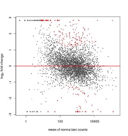
bioRxiv preprint first posted online Apr. 30, 2018; doi: http://dx.doi.org/10.1101/310987. The copyright holder for this preprint
(which was not peer-reviewed) is the author/funder, who has granted bioRxiv a license to display the preprint in perpetuity.
                              It is made available under a CC-BY-NC-ND 4.0 International license.

         ABSTRACT
         Despite the identification of many genes and pathways involved in the persistence
         phenomenon of bacteria, the mechanisms of persistence are not well understood. Here, using
         Escherichia coli as a model, we identified polynucleotide phosphorylase (PNPase) as a key
         regulator in persister formation. We successfully constructed pnp knockout mutant strain and
         its complemented strain, and exposed the pnp knockout mutant and complemented strain to
         antibiotics and stress conditions. The results showed that, compared with the wild-type
         W3110, the pnp knockout strain had defect in persistence to antibiotics and stress conditions,
         and the persistence to antibiotics and stresses was restored upon complementation. RNA-Seq
         was performed to identify the transcriptome profile in the pnp knockout strain compared with
         wild-type strain W3110, and the data revealed that 242 (166 up-regulated, and 76
         down-regulated) genes were differentially expressed in the pnp knockout mutant strain.
         KEGG pathway analysis of the up-regulated genes showed that they were mostly mapped to
         metabolism and virulence pathways, most of which are positively regulated by the global
         regulator cyclic AMP receptor protein (CRP). Similarly, the transcription level of the crp
         gene in the pnp-deletion strain increased 3.22-fold in the early stationary phase. We further
         explored the indicators of cellular metabolism of the pnp-deletion strain, the persistence
         phenotype of the pnp and crp double-deletion mutant, and the transcriptional activity of crp
         gene. Our results indicate that PNPase controls cellular metabolism by negatively regulating
         the crp operon at the post-transcriptional level by targeting the 5’- Untranslated Region (UTR)
         of the crp transcript. This study offers new insight about the persister mechanisms and
         provides new targets for development of new drugs against persisters for more effective
         treatment of persistent bacterial infections.
bioRxiv preprint first posted online Apr. 30, 2018; doi: http://dx.doi.org/10.1101/310987. The copyright holder for this preprint
(which was not peer-reviewed) is the author/funder, who has granted bioRxiv a license to display the preprint in perpetuity.
                              It is made available under a CC-BY-NC-ND 4.0 International license.

         INTRODUCTION
         Persisters are a small fraction of dormant or non-growing bacteria that are tolerant to lethal
         antibiotics or stresses (1). Persisters are distinct from antibiotic-resistant cells in that they are
         genetically identical and remain susceptible to antibiotics when they are growing again (2, 3).
         Persisters pose significant challenges for the treatment of many chronic and persistent
         bacterial infections, such as tuberculosis, urinary tract infections and biofilm infections (4-6) .
         Therefore, it is of great importance to understand the mechanisms of persistence and develop
         new strategies to more effectively cure such persistent infections. Although the phenomenon
         of bacterial persistence was discovered over 70 years ago (7), our understanding of the
         genetic basis of persister formation remains incomplete.

         Because the mechanisms of persistence are highly redundant, new mechanisms of persister
         formation and survival are continually discovered. In bacteria, RNA decay, which is
         necessary for recycling of nucleotides and for rapid changes in the gene expression program
         is mainly carried out by RNA degradosome (8). PNPase encoded by pnp is a major
         component of the RNA degradosome, which is composed of a complex structure with RNase
         E, helicase RhlB, and enolase together with PNPase. In bacteria, mRNA degradation is of
         great significance, as it not only achieves the nucleotide recycling, but also can control gene
         expression in different growth conditions (9, 10). PNPase has been found to be important in
         many aspects of RNA metabolism (8, 11-13). In addition, it also plays important roles in
         post-transcriptional regulation of gene expression (14).

         In this study, to address the role of RNA degradation in bacterial persistence, we constructed
         pnp knockout strain and its complementation strain, and then assessed the survival of the pnp
         gene knockout mutant and the complementation strain upon exposure to antibiotics and stress
         conditions. In addition, RNA-Seq was performed to evaluate the transcriptome of the pnp
         knockout strain compared with the parent strain W3110, and the differential expression was
         analyzed to shed light on the molecular basis of the pnp mediated persistence. We
         demonstrate that PNPase controls cellular metabolism by negatively regulating the global
         regulator cyclic AMP receptor protein CRP        ( )operon at the post-transcriptional level through
         targeting the 5’-Untranslated Region (UTR) of the crp transcript to regulate persister
         formation.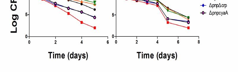
bioRxiv preprint first posted online Apr. 30, 2018; doi: http://dx.doi.org/10.1101/310987. The copyright holder for this preprint
(which was not peer-reviewed) is the author/funder, who has granted bioRxiv a license to display the preprint in perpetuity.
                              It is made available under a CC-BY-NC-ND 4.0 International license.

         MATERIALS AND METHODS
         Bacterial strains and growth media
         The strains used in this work were derived from wild type E. coli K12 W3110
         (F−mcrAmcrBIN(rrnD-rrnE)1 lambda−). Luria-Bertani (LB) broth (0.5% NaCl) and agar
         were used for bacterial cultivation.
         Construction of E. coli W3110 knockout mutants
         The λ Red recombination system was used for construction of pnp knockout in the E. coli
         chromosome (15). The candidate gene was replaced by the chloramphenicol resistance gene,
         which can be removed by pCP20. Primers used for knockout and additional external primers
         used to verify the correct integration of the PCR fragments by homologous recombination are
         shown in Table 1.
         Construction of pBAD202-pnp recombinant plasmid
         The plasmid pBAD202 was used for construction of a recombinant containing a functional
         wild type pnp gene (16). The primers designed based on the pnp gene were F
         (5’-CATGCCATGGACCCACATAGAGCTGGGTTA-3’)                                               and         R        (5’-CCCAAG
         CTTGCAAATGGCAACCTTACT-3’). The PCR products were digested with the restriction
         enzymes NcoI and HindIII (Thermo Fisher, USA) and ligated to the plasmid digested with
         the same enzymes. The recombinant constructs containing the pnp gene and the vector
         control were used to transform the Δpnp deletion strain and wild type W3110 strain by
         electroporation.
         Persister assay for various antibiotics
         Persistence was measured by determining bacterial survival in the form of colony forming
         units (CFUs) upon exposure to three antibiotics, namely, ampicillin at 200 μg/ml, norfloxacin
         at 8 μg/ml, and gentamicin at 40 μg/ml. E. coli cells were grown to stationary phase in LB
         medium, and then were exposed to different antibiotics, where
         undiluted cultures were used for incubation without shaking at 37oC for various times (17).
         The number of CFUs per milliliter was determined by plating dilutions of the bacterial cells
         on LB plates without antibiotics.
         Persister assay for various stresses
         For heat stress, E. coli cells from stationary phase cultures were treated at 52 oC for 30
         minutes. The CFUs were determined after serial dilutions. For acid stress, E. coli cells were
         incubated with acid pH 3 for 4 days at 37oC. For hypertonic saline stress, cultures were
         grown in LB medium containing 3M NaCl at 37oC for 6 days without shaking. E. coli cells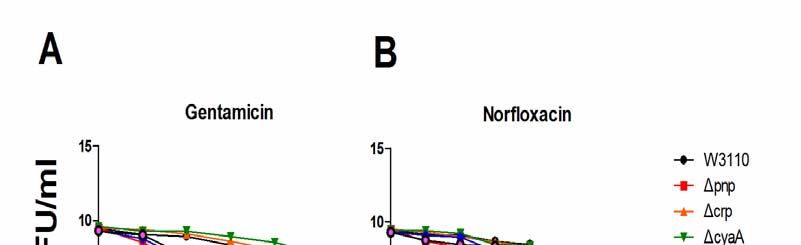
bioRxiv preprint first posted online Apr. 30, 2018; doi: http://dx.doi.org/10.1101/310987. The copyright holder for this preprint
(which was not peer-reviewed) is the author/funder, who has granted bioRxiv a license to display the preprint in perpetuity.
                              It is made available under a CC-BY-NC-ND 4.0 International license.

         were also exposed to 80 mM H2O2 at 37oC for 5 days without shaking. The number of CFUs
         under acid, hypertonic saline, and H2O2 was determined daily.
         RNA extraction and sequencing
         The wild type W3110 cells and the pnp mutants were cultivated at 37oC for 6.5 hours to
         stationary phase. Total RNA was isolated from 1 ml culture, using the RNeasy Mini kit
         (Qiagen, USA), according to the manufacturer’s instructions. All procedures for RNA
         sequencing and alignment of the transcriptome were conducted by Oebiotech (Shanghai,
         China). RNA sequencing was performed using Illumina HiSeqTM2000. Raw reads were
         filtered to remove low quality sequences, adapter sequences, and reads with poly N. The
         clean reads were subjected to BLAST search by tophat/bowtie2. Differential expression
         analysis of two samples was performed using the software DEseq. David Bioinformatics
         Resource 6.7 was used to perform gene ontology and KEGG pathway analysis.

         Detection of bacterial internal redox status
         The concentration of intracellular ATP was detected by BacTiter-Glo Microbial Cell Viability
         Assay Kit (Promega, USA). The intracellular NADH/NAD+ ratio was measured as described
         (18). Carbonyl cyanide m-chlorophenylhydrazone (CCCP), an oxidative phosphorylation
         inhibitor, was purchased from Sigma-Aldrich (St Louis, MO, USA). CCCP (100 μM) and
         antibiotics were added to LB as ATP inhibitors in stationary phase. The number of CFUs per
         milliliter was determined by plating serial dilutions of bacteria on LB plates without
         antibiotics after 24h incubation at 37oC with 210 rpm shaking.
         Detection of transcriptional activity of cyclic adenosine monophosphate (cAMP)
         receptor protein (CRP)
         The β-galactosidase gene lacZ was inserted into the polycloning site of pET-28 vector, and
         the original T7 promoter was replaced with the promoter region of the crp gene or promoter
         region of the crp gene plus 5’-UTR region. The recombinant plasmid constructs
         pET-lacZ-Pcrp/pET-lacZ-Pcrp+5U were transformed into E. coli W3110 and the pnp mutant
         (Δpnp W3110) by electroporation. The activity of β-galactosidase was measured by the β-Gal
         assay kit (Invitrogen, USA) according to the manufacturer’s instructions. Enhanced BCA
         protein assay kit (Beyotime Biotechnology, China) was used to determine the concentration
         of proteins.
bioRxiv preprint first posted online Apr. 30, 2018; doi: http://dx.doi.org/10.1101/310987. The copyright holder for this preprint
(which was not peer-reviewed) is the author/funder, who has granted bioRxiv a license to display the preprint in perpetuity.
It is made available under a CC-BY-NC-ND 4.0 International license.
RESULTS
pnp mutant has defect in persistence to various antibiotics.
The stationary phase cultures of the pnp mutants and the wild-type strain W3110 as a control
were exposed to various antibiotics, including ampicillin (200 μg/ml), norfloxacin (8 μg/ml),
and gentamicin (40 μg/ml). The results showed that the Δpnp mutant was more susceptible
than the parent strain W3110 to all the three antibiotics. Especially upon treatment with
gentamicin, the persister levels of the Δpnp mutant decreased significantly after 24h exposure.
The effect of ampicillin and norfloxacin on sterilization was similar. The Δpnp mutant was
killed by ampicillin and norfloxacin significantly from the second day. Complementation of
the Δpnp mutant with the functional pnp gene conferred increased persistence to the three
antibiotics to the wild-type levels. However, there was no significant change in the persister
level between the pnp gene overexpression strain and wild-type strain W3110 (see Figure 1).
The pnp mutant is more susceptible to various stresses.
Since the persister bacteria are not only tolerant to antibiotics, they can be tolerant to certain
stress conditions (3, 19). Thus, we also tested the survival of the Δpnp mutant under several
stress conditions, including heat, acid pH, hydrogen peroxide, and hypertonic saline stress. As
shown in Figure 2, the stationary phase culture of the Δpnp mutant was more sensitive to the
four stress conditions, than that of wild-type strain W3110. Complementation of the Δpnp
mutant with the functional pnp gene mostly restored the level of persistence of the wild-type
strain. It is worth noting that the persistence level was significantly higher in the pnp
overexpression strains than the wild-type strains under heat stress, but significantly lower in
the pnp overexpression strains than the wild-type strains under hypertonic saline stress.
RNA-seq analysis reveals a higher metabolic status of the pnp deletion mutant strain.
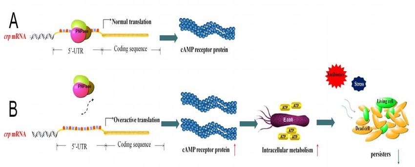
Figure 3 shows the difference in gene expression profiling between the W3110 wild type
strain and the Δpnp mutant. Altogether, 242 genes showed significant differences in the Δpnp
mutant compared with the parent strain W3110, where 166 genes were up-regulated and 76
genes down-regulated.
Pathway analysis of the differentially expressed genes was performed using the KEGG
database, and the significance of the differentially expressed gene enrichment in each
pathway was calculated by the hypergeometric distribution test. The top 20 pathways after
KEGG analysis are shown in Figure 4. Flagellar assembly (PATH: ecj02040), RibosomebioRxiv preprint first posted online Apr. 30, 2018; doi: http://dx.doi.org/10.1101/310987. The copyright holder for this preprint
(which was not peer-reviewed) is the author/funder, who has granted bioRxiv a license to display the preprint in perpetuity.
It is made available under a CC-BY-NC-ND 4.0 International license.
(PATH: ecj03010), ABC transporters (PATH: 02010), Sulfur metabolism (PATH: ecj00920),
Two-component system (PATH: ecj02020), and carbon metabolism (PATH: ecj01200) are the
main enrichment pathways of significant differences in gene transcripts, in which only the
genes in ribosome pathway are down-regulated. We selected 41 genes whose mRNA levels
increased more than 3 fold in the pnp mutant for validation by Real-time quantitative PCR.
The results showed that the expression levels of 32 genes increased more than 4-fold (Table
2).
Detection of internal redox status of bacteria.
Since the RNA-seq results showed that genes involved in metabolic and virulence-related
pathways were expressed at a higher level in the Δpnp mutant than the parent strain, we
measured the intracellular ATP levels and NADH/NAD+ ratios to estimate whether the Δpnp
mutant was in a state of higher metabolism than the parent strain W3110. Since the persister
assay was performed with stationary phase bacteria, we determined the ATP level in Δpnp
mutant and the wild-type strain during the stationary phase. As shown in Figure 5, the ATP
level of Δpnp mutant was higher than that of the wild type strain W3110 in stationary phase,
suggesting that the higher metabolic status in the Δpnp mutant produces excessive ATP and
renders the Δpnp mutant less able to form persisters and thus become more susceptible to
antibiotics and stresses. Consistent with the above observation, the NADH/NAD+ ratio which
reflects the redox status of the microbial cells in the Δpnp mutant was higher than that in the
control parent strain in stationary phase, suggesting that the Δpnp mutant was indeed in a
high metabolic state (Figure 5).
Antibiotic exposure assays of metabolism related gene knockout strains.
Since the RNA-seq analysis indicated that the impact on bacterial metabolism after pnp gene
deletion is extensive, this suggests that maintenance of the normal metabolism of bacteria by
PNPase may be mediated via global regulators. To address this possiblity, we performed
real-time quantitative PCR analysis of several major regulators (ArcA, ArcB, Cra, Crp, CyaA,
Fnr, and RpoS) responsible for global regulation of diverse aspects of metabolism in E. coli.
Significantly, we found that in the early stationary phase, expression ofthe cyaA encoding
adenylate cyclase and crp encoding cAMP receptor protein (CRP) increased 3.13 and 3.22
fold, respectively. therefore, we knocked out the cyaA and crp and assessed their effect on
persister levels in drug exposure experiments. In the killing curve experiments (Figure 6),bioRxiv preprint first posted online Apr. 30, 2018; doi: http://dx.doi.org/10.1101/310987. The copyright holder for this preprint
(which was not peer-reviewed) is the author/funder, who has granted bioRxiv a license to display the preprint in perpetuity.
It is made available under a CC-BY-NC-ND 4.0 International license.
persister levels of the Δcrp deletion strain and ΔcyaA deletion strain were significantly higher
than the wild-type W3110 under gentamicin treatment. The persister levels of the ΔpnpΔcrp
double deletion strain and ΔpnpΔcyaA deletion strain under gentamicin and norfloxacin
treatment were much higher than that of the Δpnp mutant.
Effect of energy inhibitor CCCP on persister level.
In order to further verify the increased sensitivity to antibiotics was caused by high level of
metabolism in the bacteria, we performed ATP inhibition experiments. The results showed
that addition of CCCP (100 μM) caused significantly higher tolerance to antibiotics. The
results are shown in Figure 7.
Transcriptional activity of crp gene in W3110 and pnp deletion strains.
Previous results suggested that PNPase is likely to maintain the downstream metabolic
enzymes at the normal level by inhibiting CRP. The 5’-UTR region of prokaryotes is one of
the important components of post-transcriptional regulation and can affect the initiation of
mRNA translation. It has been found that C-1a PNPase in E. coli can bind to 5’-UTR region
of the operon pgaABCD and inhibit the formation of biofilm by inhibiting the expression of
acetylglucosamine (14). Therefore, we hypothesized that PNPase may also bind to the
5’-UTR region of crp mRNA to inhibit CRP protein translation. To address this, we made
crp-lacZ reporter construct and transformed into the Δpnp mutant and the parent strain
W3110. As shown in Figure 8, the results of β-galactosidase activity assay showed that the
β-galactosidase activity was 8.3-fold higher in the Δpnp/PET-lacZ-Pcrp+5u than that in the
parent strain W3110/PET-lacZ-Pcrp+5u at early stationary phase, and 3.6-fold higher at the
end of stationary phase. The activity of β-galactosidase in W3110/PET-lacZ-Pcrp was
5.1-fold that in W3110/PET-lacZ-Pcrp+5u at early stationary phase, and 4.1-fold at the end of
stationary phase. At any stage, the activity of β-galactosidase in the Δpnp/ PET-lacZ-Pcrp was
the highest. These findings suggest that PNPase may inhibit the expression of crp mRNA or
CRP protein translation, leading to higher metabolic status of the Δpnp mutant.
DISCUSSION
In this study, we identified a new mechanism of persistence mediated by PNPase, part of the
RNA degradasome. PNPase catalyzes the polymerization of nucleoside diphosphate and the
phosphorylation of polynucleotides in vitro. But in vivo, PNPase is not only the mainbioRxiv preprint first posted online Apr. 30, 2018; doi: http://dx.doi.org/10.1101/310987. The copyright holder for this preprint
(which was not peer-reviewed) is the author/funder, who has granted bioRxiv a license to display the preprint in perpetuity.
It is made available under a CC-BY-NC-ND 4.0 International license.
component of RNA degradasome, which catalyzes decomposition of RNA and promote
metabolism and stability of mRNA, but also is independently involved in regulation of
bacterial pathogenicity. For example, in Salmonella enterica, PNPase acts as a global
regulator of virulence gene expression (20).
We found that, compared to wild-type, the Δpnp deletion strain was highly susceptible to
three different antibiotics and stresses, indicating that pnp plays an important role in the
formation or maintenance of persisters. In order to confirm that PNPase was the direct cause
of persister formation, we constructed pnp complementation strain and found that the
persistence phenotype was restored in the pnp complementation strain. These in vitro
experiments have shown that PNPase participates in persister formation or survival. It is
worth noting that phenotype of the Δpnp deletion strain mainly appeared in early stationary
phase and mid- stationary phase. When the cultures were incubated to late stationary phase,
these differences became less obvious, which indicates that the role of PNPase in maintaining
the persistence state likely occurs in the early stage.
To shed new insight on the mechanism of by which PNPase mediates persistence, we
performed RNA-seq analysis of the Δpnp deletion strain compared with the wild type W3110
strain, and found a number of genes belonging to metabolism or virulence pathway are
up-regulated in the Δpnp deletion strain. These finding suggest that elevated metabolism in
the Δpnp deletion strain is likely present. This is confirmed by measurement of the ATP levels
and NADH/NAD+, two indicators of redox state in bacteria, and indicate that the Δpnp
deletion strain had significantly higher metabolism than that of the wild type W3110 strain.
The Escherichia coli CRP is an important transcription factor that its DNA-binding can result
in positive or negative regulation of more than 100 genes mainly involved in catabolism of
carbon sources other than glucose. In our study, the results of validation of differentially
expressed genes showed that the mRNA levels of genes encoding carbohydrate transport
systems (mglBAC, lamB, malK), TCA cycle (sdhA, sdhD, fumA, mdh, and acnB),
Glycerolipid metabolism (glpD, glpK, glpT), etc., were significantly up-regulated.
Interestingly, the metabolic master regulator CRP has a positive regulatory effect on these
genes (21, 22). In addition, the cAMP-CRP complex is known to be involved in regulation of
biofilm formation, quorum sensing systems, and the transcription of the nitrogen regulatory
system (23-26). When the culture environment lacks glucose, CRP binds to the effectorbioRxiv preprint first posted online Apr. 30, 2018; doi: http://dx.doi.org/10.1101/310987. The copyright holder for this preprint
(which was not peer-reviewed) is the author/funder, who has granted bioRxiv a license to display the preprint in perpetuity.
It is made available under a CC-BY-NC-ND 4.0 International license.
cAMP, and the activated CRP binds to the TGTGAnnnnnnTCACA sequence near the
promoter of the regulatory gene, thereby recruiting RNA polymerase to initiate the
transcription of the downstream gene (27). CRP was found to play an important role in the
metabolic process in E. coli, and the perturbation of cAMP-CRP can significantly diminish
the metabolic capabilities of persisters, and can prevent drugs like aminoglycosides from
getting into the bacterial cells, thus weakening the ability of such drugs to kill persisters (28).
We knocked out crp and cyaA gene in Δpnp deletion strain, respectively, and found that the
ability of persister formation was significantly higher in the double ΔcrpΔpnp deletion strain
and ΔcyaAΔpnp deletion strain than that in the Δpnp deletion strain. PNPase can bind to its
own 5’-UTR region to realize the autologous expression regulation by promoting degradation
of pnp mRNA in the presence of RNase III (29-31). We inferred that PNPase in E. coli can
inhibit the translation of the crp mRNA, which controls the CRP level in the normal range, to
maintain the balance of intracellular metabolism. To confirm this inference, we measured the
activity of β-galactosidase to reflect the transcriptional activity of crp in the wild type W3110
strain and the Δpnp deletion strain. We found that the transcriptional activity of crp in the
Δpnp deletion strain had 8.3-fold increase in the early stationary phase, indicating that
PNPase can inhibit the expression of CRP protein at the transcriptional level.
The metabolic status is the critical basis in the formation of persisters in bacteria (32-34).
Although some researchers believe that dormancy is not a necessary condition for the
formation of persisters (35), the idea that “persistence can be considered a termination of a
metabolic procedure, that is bacteria enters a dormant state” remains the mainstream thinking.
Based on our findings, we propose that PNPase in E. coli can bind to the 5’-UTR of the crp
mRNA to cause a negative regulation and maintain a constant level of CRP protein, and
thatdeletion of PNPase could cause overexpression of CRP, the downstream regulation of a
series of metabolic-related gene expression levels increased abnormally, leading to failure to
enter the dormant state, causing decreased persistence capacity in the Δpnp deletion strain
and its inability to tolerate antibiotics and stress conditions.
In summary, our study established a new mechanism of persister formation mediated by
PNPase regulation in E. coli (Figure 9). We found that PNPase controls cellular metabolism
by negatively regulating the global regulator cyclic AMP receptor protein(CRP) operon at
post-transcriptional level through targeting the 5’- UTR region of the crp transcript tobioRxiv preprint first posted online Apr. 30, 2018; doi: http://dx.doi.org/10.1101/310987. The copyright holder for this preprint
(which was not peer-reviewed) is the author/funder, who has granted bioRxiv a license to display the preprint in perpetuity.
It is made available under a CC-BY-NC-ND 4.0 International license.
regulate persister formation. The results of the in vitro studies need to be further tested in
animal models to determine if PNPase has any role in persistence and virulence in vivo in the
future. PNPase, as a regulator of persistence and virulence, is a promising new drug target for
developing improved treatment of persistent bacterial infections.
ACKNOWLEDGEMENTS
This work was supported in part by the National Natural Science Foundation of China
(81572046, 81772231).bioRxiv preprint first posted online Apr. 30, 2018; doi: http://dx.doi.org/10.1101/310987. The copyright holder for this preprint
(which was not peer-reviewed) is the author/funder, who has granted bioRxiv a license to display the preprint in perpetuity.
It is made available under a CC-BY-NC-ND 4.0 International license.
REFERENCES:
1. Lewis K. Persister cells. Annu Rev Microbiol. 2010;64:357-72.
2. Balaban NQ, Merrin J, Chait R, Kowalik L, Leibler S. Bacterial persistence as a
phenotypic switch. Science. 2004;305(5690):1622-5.
3. Zhang Y. Persisters, persistent infections and the Yin-Yang model. Emerging
microbes & infections. 2014;3(1):e3.
4. Ojha AK, Baughn AD, Sambandan D, Hsu T, Trivelli X, Guerardel Y, et al. Growth
of Mycobacterium tuberculosis biofilms containing free mycolic acids and harbouring
drug-tolerant bacteria. Mol Microbiol. 2008;69(1):164-74.
5. Connolly LE, Edelstein PH, Ramakrishnan L. Why is long-term therapy required
to cure tuberculosis? PLoS Med. 2007;4(3):e120.
6. Fox W, Ellard GA, Mitchison DA. Studies on the treatment of tuberculosis
undertaken by the British Medical Research Council tuberculosis units, 1946-1986,
with relevant subsequent publications. Int J Tuberc Lung Dis. 1999;3(10 Suppl
2):S231-79.
7. Hobby GL, K. Meyer, Chaffee E. Observations on the mechanism of action of
penicillin. Proc Soc Exp Biol NY 1942;50:281-5.
8. Briani F, Carzaniga T, Deho G. Regulation and functions of bacterial PNPase.
Wiley interdisciplinary reviews RNA. 2016;7(2):241-58.
9. Hui MP, Foley PL, Belasco JG. Messenger RNA degradation in bacterial cells.
Annual review of genetics. 2014;48:537-59.
10. Laalami S, Zig L, Putzer H. Initiation of mRNA decay in bacteria. Cellular and
molecular life sciences : CMLS. 2014;71(10):1799-828.
11. Phadtare S. Escherichia coli cold-shock gene profiles in response to
over-expression/deletion of CsdA, RNase R and PNPase and relevance to
low-temperature RNA metabolism. Genes to cells : devoted to molecular & cellular
mechanisms. 2012;17(10):850-74.
12. Wong AG, McBurney KL, Thompson KJ, Stickney LM, Mackie GA. S1 and KH
domains of polynucleotide phosphorylase determine the efficiency of RNA binding
and autoregulation. Journal of bacteriology. 2013;195(9):2021-31.
13. Dominguez-Malfavon L, Islas LD, Luisi BF, Garcia-Villegas R, Garcia-Mena J.
The assembly and distribution in vivo of the Escherichia coli RNA degradosome.
Biochimie. 2013;95(11):2034-41.bioRxiv preprint first posted online Apr. 30, 2018; doi: http://dx.doi.org/10.1101/310987. The copyright holder for this preprint
(which was not peer-reviewed) is the author/funder, who has granted bioRxiv a license to display the preprint in perpetuity.
It is made available under a CC-BY-NC-ND 4.0 International license.
14. Carzaniga T, Antoniani D, Deho G, Briani F, Landini P. The RNA processing
enzyme polynucleotide phosphorylase negatively controls biofilm formation by
repressing poly-N-acetylglucosamine (PNAG) production in Escherichia coli C. BMC
microbiology. 2012;12:270.
15. Datsenko KA, Wanner BL. One-step inactivation of chromosomal genes in
Escherichia coli K-12 using PCR products. P Natl Acad Sci USA.
2000;97(12):6640-5.
16. Guzman LM, Belin D, Carson MJ, Beckwith J. Tight regulation, modulation, and
high-level expression by vectors containing the arabinose PBAD promoter. Journal of
bacteriology. 1995;177(14):4121-30.
17. Li J, Ji L, Shi W, Xie J, Zhang Y. Trans-translation mediates tolerance to multiple
antibiotics and stresses in Escherichia coli. The Journal of antimicrobial
chemotherapy. 2013;68(11):2477-81.
18. Vilcheze C, Weisbrod TR, Chen B, Kremer L, Hazbon MH, Wang F, et al. Altered
NADH/NAD+ ratio mediates coresistance to isoniazid and ethionamide in
mycobacteria. Antimicrob Agents Chemother. 2005;49(2):708-20.
19. Li Y, Zhang Y. PhoU is a persistence switch involved in persister formation and
tolerance to multiple antibiotics and stresses in Escherichia coli. Antimicrobial agents
and chemotherapy. 2007;51(6):2092-9.
20. Clements MO, Eriksson S, Thompson A, Lucchini S, Hinton JCD, Normark S, et
al. Polynucleotide phosphorylase is a global regulator of virulence and persistency in
Salmonella enterica. P Natl Acad Sci USA. 2002;99(13):8784-9.
21. Zheng D, Constantinidou C, Hobman JL, Minchin SD. Identification of the CRP
regulon using in vitro and in vivo transcriptional profiling. Nucleic acids research.
2004;32(19):5874-93.
22. Gosset G, Zhang Z, Nayyar S, Cuevas WA, Saier MH, Jr. Transcriptome analysis
of Crp-dependent catabolite control of gene expression in Escherichia coli. Journal of
bacteriology. 2004;186(11):3516-24.
23. Gutierrez-Rios RM, Freyre-Gonzalez JA, Resendis O, Collado-Vides J, Saier M,
Gosset G. Identification of regulatory network topological units coordinating the
genome-wide transcriptional response to glucose in Escherichia coli. BMC Microbiol.
2007;7:53.
24. De Lay N, Gottesman S. The Crp-activated small noncoding regulatory RNAbioRxiv preprint first posted online Apr. 30, 2018; doi: http://dx.doi.org/10.1101/310987. The copyright holder for this preprint
(which was not peer-reviewed) is the author/funder, who has granted bioRxiv a license to display the preprint in perpetuity.
It is made available under a CC-BY-NC-ND 4.0 International license.
CyaR (RyeE) links nutritional status to group behavior. J Bacteriol.
2009;191(2):461-76.
25. Muller CM, Aberg A, Straseviciene J, Emody L, Uhlin BE, Balsalobre C. Type 1
fimbriae, a colonization factor of uropathogenic Escherichia coli, are controlled by the
metabolic sensor CRP-cAMP. PLoS Pathog. 2009;5(2):e1000303.
26. Zheng DL, Constantinidou C, Hobman JL, Minchin SD. Identification of the CRP
regulon using in vitro and in vivo transcriptional profiling. Nucleic Acids Res.
2004;32(19):5874-93.
27. Fic E, Bonarek P, Gorecki A, Kedracka-Krok S, Mikolajczak J, Polit A, et al. cAMP
Receptor Protein from Escherichia coli as a Model of Signal Transduction in Proteins
- A Review. J Mol Microb Biotech. 2009;17(1):1-11.
28. Mok WW, Orman MA, Brynildsen MP. Impacts of global transcriptional regulators
on persister metabolism. Antimicrob Agents Chemother. 2015;59(5):2713-9.
29. Robert-Le Meur M, Portier C. E.coli polynucleotide phosphorylase expression is
autoregulated through an RNase III-dependent mechanism. EMBO J.
1992;11(7):2633-41.
30. Robert-Le Meur M, Portier C. Polynucleotide phosphorylase of Escherichia coli
induces the degradation of its RNase III processed messenger by preventing its
translation. Nucleic Acids Res. 1994;22(3):397-403.
31. Jarrige AC, Mathy N, Portier C. PNPase autocontrols its expression by degrading
a double-stranded structure in the pnp mRNA leader. EMBO J. 2001;20(23):6845-55.
32. Bernier SP, Lebeaux D, DeFrancesco AS, Valomon A, Soubigou G, Coppee JY,
et al. Starvation, together with the SOS response, mediates high biofilm-specific
tolerance to the fluoroquinolone ofloxacin. PLoS Genet. 2013;9(1):e1003144.
33. Amato SM, Brynildsen MP. Nutrient transitions are a source of persisters in
Escherichia coli biofilms. PLoS One. 2014;9(3):e93110.
34. Grant SS, Kaufmann BB, Chand NS, Haseley N, Hung DT. Eradication of
bacterial persisters with antibiotic-generated hydroxyl radicals. Proc Natl Acad Sci U
S A. 2012;109(30):12147-52.
35. Orman MA, Brynildsen MP. Dormancy is not necessary or sufficient for bacterial
persistence. Antimicrob Agents Chemother. 2013;57(7):3230-9.bioRxiv preprint first posted online Apr. 30, 2018; doi: http://dx.doi.org/10.1101/310987. The copyright holder for this preprint
(which was not peer-reviewed) is the author/funder, who has granted bioRxiv a license to display the preprint in perpetuity.
It is made available under a CC-BY-NC-ND 4.0 International license.
Table 1 Primers used in construction of E. coli W3110 knockout mutants
Gene Primer (5’-3’)
pnp KO S CTGCCCGGTTAAAAGCCCCCCGCCGCAGCGGAGGGCAAA
TGGCAACCTTAATGGGAATTAGCCATGGTCC
pnp KO A AGAGGCTTTACCCACATAGAGCTGGGTTAGGGTTGTCATT
AGTCGCGAGGGTGTAGGCTGGAGCTGCTTC
pnp ex S CGCAAAAAAGGCTTCATTTCCCACT
pnp ex A TGGAAACTAATGTATTGTTGCTATG
crp KO S AGCGGCGTTATCTGGCTCTGGAGAAAGCTTATAACAGAG
GATAACCGCGCATGGGAATTAGCCATGGTCC
crp KO A GGGAAACAAAATGGCGCGCTACCAGGTAACGCGCCACTC
CGACGGGATTAGTGTAGGCTGGAGCTGCTTC
cyaA KO S GATGTTGGCGGAATCACAGTCATGACGGGTAGCAAATCA
GGCGATACGTCATGGGAATTAGCCATGGTCC
cyaA KO A TCCGCTAAGATTGCATGCCGGATAAGCCTCGCTTTCCGGC
ACGTTCATCAGTGTAGGCTGGAGCTGCTTC
crp ex S AGCGGCGTTATCTGGCTCTGG
crp ex A GGGAAACAAAATGGCGCGCTAC
cyaA ex S GATGTTGGCGGAATCACAGTCATGA
cyaA ex A TCCGCTAAGATTGCATGCCGGATAAbioRxiv preprint first posted online Apr. 30, 2018; doi: http://dx.doi.org/10.1101/310987. The copyright holder for this preprint
(which was not peer-reviewed) is the author/funder, who has granted bioRxiv a license to display the preprint in perpetuity.
It is made available under a CC-BY-NC-ND 4.0 International license.
Table 2 32 genes whose expression ratios were more than 4-fold in pnp mutant verified by
qPCR
Fold
Gene pval Change Description Pathway
(pnp/WT)
acnB 0.0001 7.71 aconitate hydratase 2 Citrate cycle
aldA 0.0402 4.01 aldehyde dehydrogenase A Pyruvate metabolism
cyoA 0.0368 6.88 cytochrome bo(3) ubiquinol oxidase subunit II Oxidative phosphorylation
cysD 0.0001 6.07 sulfate adenylyltransferase, subunit 2 Sulfur metabolism
cysU 0.0001 5.93 sulfate/thiosulfate ABC transporter permease Sulfur metabolism/ ABC transporter
fimC 0.0001 14.49 periplasmic chaperone
fimD 0.0001 6.18 fimbrial usher outer membrane porin protein
fimbrial protein involved in type 1 pilus
fimI 0.0001 9.06
biosynthesis
fliE 0.0001 7.58 flagellar basal-body component Flagellar assembly
flagellar motor switching and energizing
fliN 0.0004 4.72 Bacterial chemotaxis/Flagellar assembly
component
fliO 0.0005 4.22 flagellar biosynthesis protein Flagellar assembly
fliQ 0.0001 5.68 flagellar biosynthesis protein Flagellar assembly
fumA 0.0170 4.71 fumarate hydratase Citrate cycle
glpD 0.0141 5.15 glycerol-3-phosphatedehydrogenase Glycerophospholipid metabolism
glpK 0.0046 4.59 glycerol kinase Glycerolipid metabolism
glpT 0.0003 5.16 Glycerol-3-phosphate transporter
lamB 0.0350 5.36 maltose outer membrane porin
malK 0.0251 4.25 maltose ABC transportor ATPase ABC transporter
Cysteine and methionine metabolism
mdh 0.0237 4.68 malate dehydrogenase
/Citrate cyclebioRxiv preprint first posted online Apr. 30, 2018; doi: http://dx.doi.org/10.1101/310987. The copyright holder for this preprint
(which was not peer-reviewed) is the author/funder, who has granted bioRxiv a license to display the preprint in perpetuity.
It is made available under a CC-BY-NC-ND 4.0 International license.
ATP-binding cassette protein for galactose
mglA 0.0001 5.54 ABC transporter
uptake
mglB 0.0051 6.77 methyl-galactoside transporter subunit Bacterial chemotaxis/ABC transporter
mglC 0.0020 4.64 Galactose permease protein ABC transporter
nmpC 0.0055 4.58 Outer membrane porin protein
ompF 0.0002 9.32 Outer membrane protein F Two-component system
putP 0.0023 4.22 Sodium/proline symporter
sdhA 0.0404 6.53 succinate dehydrogenase Citrate cycle
sdhD 0.0304 6.39 succinate dehydrogenase, membrane subunit Citrate cycle
glucitol/sorbitol-specific enzyme IIB
srlE 0.0135 4.08 Phosphotransferase system(PTS)
component of PTS
srlD 0.0359 4.41 sorbitol-6-phosphate dehydrogenase Fructose and mannose metabolism
xapA 0.0232 5.67 purine nucleoside phosphorylase 2 Purine metabolism
ydcA 0.0001 10.74 Uncharacterized protein
yfdF 0.0312 5.77 Uncharacterized proteinbioRxiv preprint first posted online Apr. 30, 2018; doi: http://dx.doi.org/10.1101/310987. The copyright holder for this preprint
(which was not peer-reviewed) is the author/funder, who has granted bioRxiv a license to display the preprint in perpetuity.
It is made available under a CC-BY-NC-ND 4.0 International license.
Figure1 PNPase mutant has defective persister levels in antibiotic exposure assays.
Stationary phase culture of the Δpnp mutant, its complemented strain, the parent strain
W3110, and overexpression strain were exposed to no antibiotic (A), ampicillin (200 μg/ml)
(B), norfloxacin (8 μg/ml) (C), gentamicin (40 μg/ml) (D), for various times. Aliquots of
cultures were taken at different timepoints and plated for CFU determination on LB plates.
The vertical axis represents CFU values on a log scale and the horizontal axis represents time
of antibiotic exposure in days.bioRxiv preprint first posted online Apr. 30, 2018; doi: http://dx.doi.org/10.1101/310987. The copyright holder for this preprint
(which was not peer-reviewed) is the author/funder, who has granted bioRxiv a license to display the preprint in perpetuity.
It is made available under a CC-BY-NC-ND 4.0 International license.
Figure 2 Comparison of susceptibility of four mutants to stresses. The Δpnp mutant and
its complemented strain, the wild type control W3110, and the overexpression strain to a
variety of stress conditions: heat at 52 oC (A); acid pH 3.0 (B); hydrogen peroxide (80 mM)
(C); NaCl 3M (D). All strains were cultured to stationary phase and exposed to different
stresses followed by CFU determination. The vertical axis represents CFU values on a log
scale and the horizontal axis represents time of stress exposure in hours or days.bioRxiv preprint first posted online Apr. 30, 2018; doi: http://dx.doi.org/10.1101/310987. The copyright holder for this preprint
(which was not peer-reviewed) is the author/funder, who has granted bioRxiv a license to display the preprint in perpetuity.
It is made available under a CC-BY-NC-ND 4.0 International license.
Figure 3 MA map of differential expression of W3110 and the Δpnp mutant. Vertical axis
represents fold changes in log scale, and horizontal axis represents the mean value of all
sample expression for comparison after normalization. Red dots represent the differential
genes of p-valuebioRxiv preprint first posted online Apr. 30, 2018; doi: http://dx.doi.org/10.1101/310987. The copyright holder for this preprint
(which was not peer-reviewed) is the author/funder, who has granted bioRxiv a license to display the preprint in perpetuity.
It is made available under a CC-BY-NC-ND 4.0 International license.
Figure 4 KEGG of differential expression of W3110 and Δpnp W3110.bioRxiv preprint first posted online Apr. 30, 2018; doi: http://dx.doi.org/10.1101/310987. The copyright holder for this preprint
(which was not peer-reviewed) is the author/funder, who has granted bioRxiv a license to display the preprint in perpetuity.
It is made available under a CC-BY-NC-ND 4.0 International license.
Figure 5 Detection of ATP and NADH/NAD+ in W3110 and the Δpnp mutant. (A) The
ATP at each stage of the W3110 and the Δpnp mutant was detected and the absolute
concentration of ATP is calculated by the ATP standard curve. The time points 6.5 h, 12 h and
18 h represent the early stage, the middle stage of stability and the end of stability,
respectively. The ATP concentration of the Δpnp mutant at all stages is higher than that of the
wild type W3110. (B) NADH/NAD+ at different stages of W3110 and the Δpnp mutant were
measured. The Δpnp mutant had higher NADH/NAD+ levels than the wild type W3110.bioRxiv preprint first posted online Apr. 30, 2018; doi: http://dx.doi.org/10.1101/310987. The copyright holder for this preprint
(which was not peer-reviewed) is the author/funder, who has granted bioRxiv a license to display the preprint in perpetuity.
It is made available under a CC-BY-NC-ND 4.0 International license.
Figure 6 Double KO mutants, ΔpnpΔcrp and ΔpnpΔcyaA show severe defect in
persistence in antibiotic exposure assays. Stationary phase cultures of the control strain
W3110 and the mutants Δpnp, Δcrp, ΔcyaA, ΔpnpΔcrp and ΔpnpΔcyaA were exposed to
antibiotics, (A), gentamicin (40 μg/ml) and (B), norfloxacin (40 μg/ml), for various times.
Aliquots of cultures were taken at different time points and plated for CFU determination on
LB plates. The vertical axis represents CFU values on a log scale and the horizontal axis
represents time of antibiotic exposure in days.bioRxiv preprint first posted online Apr. 30, 2018; doi: http://dx.doi.org/10.1101/310987. The copyright holder for this preprint
(which was not peer-reviewed) is the author/funder, who has granted bioRxiv a license to display the preprint in perpetuity.
It is made available under a CC-BY-NC-ND 4.0 International license.
Figure 7 CCCP addition increases the tolerance of W3110 and Δpnp mutant to
antibiotics. W3110 and the Δpnp mutant were exposed to gentamicin (A) or norfloxacin
(B)with or without CCCP. CCCP addition significantly increased the persister level. Gen,
Gentamicin; Norf, Norfloxacin; CCCP, Carbonyl cyanide m-chlorophenylhydrazone. t tests
were performed to compare the effect of CCCP addition. *: P < 0.05, ***: P < 0.0001.bioRxiv preprint first posted online Apr. 30, 2018; doi: http://dx.doi.org/10.1101/310987. The copyright holder for this preprint
(which was not peer-reviewed) is the author/funder, who has granted bioRxiv a license to display the preprint in perpetuity.
It is made available under a CC-BY-NC-ND 4.0 International license.
Figure 8 PNPase regulates CRP activity through binding to the 5’-UTR region of crp
mRNA. (A) The color reaction of β-Gal assay of four strains in 96 well plate. (B) Elevated
expression of CRP transcriptional activity (β-Gal specific activity) in the PNPase mutant.bioRxiv preprint first posted online Apr. 30, 2018; doi: http://dx.doi.org/10.1101/310987. The copyright holder for this preprint
(which was not peer-reviewed) is the author/funder, who has granted bioRxiv a license to display the preprint in perpetuity.
It is made available under a CC-BY-NC-ND 4.0 International license.
Figure 9 PNPase regulates formation of bacterial persisters through CRP.
(A) PNPase binds to the 5’UTR of crp mRNA. (B) PNPase deletion leads to higher level of
metabolism as seen by elevated redox (NADH/NAD+ ratio) and ATP levels and decreased
number of persisters.You can also read