Long term surgical outcomes of primary retropupillary iris claw intraocular lens implantation for the treatment of intraocular lens dislocation ...
←
→
Page content transcription
If your browser does not render page correctly, please read the page content below
www.nature.com/scientificreports
OPEN Long‑term surgical outcomes
of primary retropupillary iris claw
intraocular lens implantation
for the treatment of intraocular
lens dislocation
Eun Young Choi1,3, Chul Hee Lee2,3, Hyun Goo Kang2, Jae Yong Han2, Suk Ho Byeon1,
Sung Soo Kim1, Hyoung Jun Koh1 & Min Kim2*
We aimed to investigate the efficacy and safety of primary retropupillary iris claw intraocular lens
(R-IOL) implantation in patients with complete intraocular lens (IOL) dislocation. In this single-
center retrospective case series, we reviewed the medical records of patients who underwent R-IOL
implantation surgery with pars plana vitrectomy for the treatment of IOL dislocation between
September 2014 and July 2019. The primary outcome was change in visual acuity (VA) up to 24 months
postoperatively. The secondary outcomes included changes in intraocular pressure (IOP), refractive
errors, and endothelial cell count (ECC) over the same period. Data of 103 eyes (98 patients) were
analyzed. The mean uncorrected VA was significantly improved at one month postoperatively (− 0.69
logMAR, P < 0.001), compared to the preoperative value. IOP (− 2.3 mmHg, P = 0.008) and ECC
(− 333.4 cells/mm2, P = 0.027) significantly decreased one month post-surgery and remained stable
thereafter. Postoperative mean spherical equivalents were similar to the prediction error throughout
the follow-up period. IOP elevation (n = 8, 7.8%), cystoid macular edema (n = 4, 3.9%), and dislocation
of the R-IOL (n = 10, 9.7%) were managed successfully. Overall, primary R-IOL implantation with pars
plana vitrectomy is effective and safe for correcting IOL dislocation due to various causes.
The widespread use of phacoemulsification, which strongly increases the proficiency of cataract operations, has
steadily increased over the past 32 y ears1. Consequently, the incidence of intraocular lens (IOL) dislocation has
increased with the increasing number of cataract surgeries that were performed. Therefore, the treatment for
IOL dislocation is an emerging surgical dilemma and may impose a relatively large public health care burden in
the future. In particular, complete IOL dislocation may predispose patients to serious retinal complications, such
as vitreous hemorrhage and retinal d etachment2. In general, surgical management of complete IOL dislocation
involves either pars plana vitrectomy and removal of the dislocated IOL, followed by secondary IOL implantation
at the surgeon’s discretion, or re-fixation of the dislocated IOL without IOL removal.
Many clinicians have tried to develop an effective secondary IOL implantation technique that would yield
the best visual outcome with minimal complications. Conventional secondary IOL implantation via scleral
sutures has been the treatment of choice, as the position of the scleral-fixated IOL (SF-IOL) is ideal with respect
to the anatomy of the eye. However, this technique requires the use of sutures and sufficient operator experience,
and has a relatively long operation t ime3. Secondary IOL scleral fixation may lead to other severe intraocu-
lar complications, such as vitreous hemorrhage, retinal detachment, choroidal detachment, and suture-related
complications4. An alternative procedure, secondary IOL implantation of anterior chamber IOLs, was popular-
ized in the 1980s due to its simplicity and efficacy. However, a high risk of developing postoperative secondary
glaucoma, uveitis, and endothelial cell decompensation has been reported in cases of anterior chamber IOL5
with a particularly inflexible closed-loop d esign6.
1
Department of Ophthalmology, Institute of Vision Research, Severance Eye Hospital, Yonsei University College
of Medicine, 50‑1, Yonseiro, Seodaemun‑gu, Seoul 03722, Korea. 2Department of Ophthalmology, Institute of
Vision Research, Gangnam Severance Hospital, Yonsei University College of Medicine, 211, Eonjuro, Gangnam‑gu,
Seoul 06273, Korea. 3These authors contributed equally: Eun Young Choi and Chul Hee Lee. *email: minkim76@
gmail.com
Scientific Reports | (2021) 11:726 | https://doi.org/10.1038/s41598-020-80292-3 1
Vol.:(0123456789)www.nature.com/scientificreports/
Recently, the retropupillary iris claw intraocular lens (R-IOL) has emerged as a viable option for secondary
IOL implantation. In the 1980s, Amar first proposed the fixation of an iris claw lens at the posterior surface of
the iris7. This R-IOL implantation technique gained popularity after Mohr et al. reported the 1-year clinical
outcomes for this technique in 2 0028. Structurally, R-IOL provides better stability and lower risks of IOL tilt or
dislocation than iris- or scleral-sutured lenses. In addition, the absence of sutures for IOL scleral fixation lowers
the risk for suture-related complications, such as conjunctival erosion, scleromalacia, and e ndophthalmitis9,10.
Many previous studies have shown that secondary R-IOL is a safe and efficacious technique for correcting vari-
ous aphakic conditions with11–17 or without IOL d islocation3,18. However, the long-term safety and efficacy of
simultaneous primary R-IOL implantation and vitrectomy in patients with complete IOL dislocation have not
been previously assessed.
Herein, we present our surgical method for correcting IOL dislocation using R-IOL and investigate the long-
term efficacy and safety of primary R-IOL implantation in patients with complete IOL dislocation.
Materials and methods
This was a single-center, retrospective case series study. We reviewed the records and operative reports of all
patients who underwent R-IOL implantation (performed by a single surgeon—M. Kim) at the Department of
Ophthalmology, Gangnam Severance Hospital, between September 2014 and July 2019. The study was conducted
in accordance with the Declaration of Helsinki, and the protocol was approved by the Institutional Review Board
at Gangnam Severance Hospital (IRB approval number: 3-2020-0213). The requirement for informed patient
consent was waived because the data were anonymized before the analysis.
We have selectively included cases of IOL dislocation involving total dislocation of the in-the-bag IOL into
the posterior segment or cases of recurrent complete dislocation of the SF-IOL, which were treated by the
removal of the dislocated IOL followed by R-IOL implantation. Cases in which dislocated IOLs were rescued
and repositioned were not included in this study. We included all patients in the analysis, including those with
various complicated retinal pathologies.
Preoperative examination. All past medical history and preoperative ophthalmologic data for each
patient were reviewed. Preoperative uncorrected visual acuity (VA) and best-corrected VA (BCVA) obtained
using the Snellen vision chart, which were converted to logMAR values for statistical analysis, were obtained.
Preoperative intraocular pressure (IOP) was measured using a non-contact tonometer. Preoperative refrac-
tive data were obtained using an auto kerato-refractometer (KR-1, Topcon Medical Systems, Inc.; Oakland, NJ,
USA); spherical equivalent (SE) and corneal astigmatism were also calculated. The auto KR values were used as
the starting point to perform manifest refraction, and the best subjective correction for a distance of 40 cm was
found after adjustment of spherical power, as well as the cylinder axis and power, by ± 0.25 D in 1° increments.
Biometry measurements obtained using a ZEISS IOLMaster 500 (Carl Zeiss AG; Heidenheim, Germany) were
reviewed and the ‘prediction error’, which is the predicted target refraction before surgery, was recorded. IOL
calculations were always performed with the SRK/T formula using an A-constant of 116.9, which is the manu-
facturer’s recommendation for retropupillary fixation. This constant was used in the laser interferometry biom-
etry mode. Finally, endothelial cell count (ECC) performed automatically using specular microscopy (CellChek
XL, Konan Medical USA Inc.; Irvine, CA, USA) was analyzed.
Surgical technique. A 25-gauge pars plana vitrectomy was performed (CONSTELLATION Vision System,
Alcon; Fort Worth, TX, USA) for each patient. After the core vitrectomy and peripheral shaving to free the dis-
located IOL from the vitreous, the anterior chamber was filled with dispersive viscoelastics (VISCOAT, Alcon;
Fort Worth, TX, USA), and the dislocated IOL complex was lifted up into the anterior chamber. Then, a con-
junctival incision was made from the 11 o’clock position to the 1 o’clock position using spring scissors. A 5.5 mm
scleral flap was created using a crescent blade and a sclerocorneal incision was made using a steel keratome. The
dislocated IOL was gently removed through the scleral tunnel. Two paracentesis incisions were made at the 2
o’clock and 10 o’clock positions using a sharp blade, and the R-IOL (ARTISAN Aphakia model 205, OPHTEC
BV; Groningen, Netherlands) was prepared for implantation. After filling the anterior chamber with viscoelastic
material, the R-IOL was inserted through the previously formed scleral tunnel using ARTISAN forceps (OPH-
TEC BV; Groningen, Netherlands). The claws of the R-IOL were fixated at the 3 o’clock and 9 o’clock positions of
the posterior iris using an enclavation needle (OPHTEC BV; Groningen, Netherlands). After closing the scleral
tunnel incision using 10-0 nylon sutures, the remaining viscoelastic material was removed by automatic irriga-
tion and aspiration. The superior conjunctival incision was closed using 7-0 vicryl sutures. Figure 1 presents a
detailed description of the surgical technique.
Postoperative examination. All postoperative data (uncorrected VA, BCVA, IOP, refractive errors, and
ECC) were acquired at 1, 6, 12, and 24 months postoperatively. The SE outcome after surgery was described
as the ‘postoperative refraction’. If there were any missing values in the postoperative data, the subjects were
excluded from the analysis. Postoperative complications and corresponding treatments were documented for
each case.
Statistical analyses. All statistical analyses were performed using Statistical package for the social sciences
(SPSS/IBM Corporation; Chicago, IL, USA) version 23.0 software for Windows. The Shapiro–Wilk test was used
to examine normality. A repeated measures analysis of variance (RM-ANOVA) with a post-hoc Fisher’s least
significant difference (LSD) test was performed to analyze the differences in variables between multiple time-
Scientific Reports | (2021) 11:726 | https://doi.org/10.1038/s41598-020-80292-3 2
Vol:.(1234567890)www.nature.com/scientificreports/
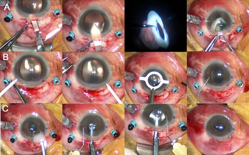
Figure 1. Intraoperative photographs showing the surgical procedures for retropupillary iris claw intraocular
lens (R-IOL) implantation. An 80-year-old male patient underwent R-IOL implantation in the left eye for the
treatment of intraocular lens (IOL) dislocation under local anesthesia. A 25-gauge trans pars plana vitrectomy
was performed. After performing the core vitrectomy and peripheral shaving, (A) a 5.5 mm scleral flap was
made using a round blade under the conjunctival flap. A scleral tunnel incision was made using a steel keratome.
After grasping the posteriorly dislocated capsular bag-IOL complex using a 25-gauge vitrectomy probe, the
dislocated capsular bag-IOL complex was translocated into the anterior chamber and gently removed through
the scleral tunnel using IOL forceps. (B) Two paracentesis incisions were made at the 2 o’clock and 10 o’clock
positions using a sharp blade. The horizontal plane indicating the IOL haptic enclavation sites was marked using
a marking pen. Viscoelastic material was injected into the anterior chamber through the scleral incision site. (C)
The R-IOL was prepared for implantation and inserted into the anterior chamber through the previously formed
scleral tunnel using ARTISAN forceps and was rotated. The haptics of the R-IOL were fixated at the 3 o’clock and
9 o’clock positions of the iris using an enclavation needle. The remaining viscoelastic material was removed, and
the scleral tunnel incision was closed using 10-0 nylon sutures. The superior conjunctival flap was closed using
7-0 vicryl sutures.
points. A paired t-test was used to compare two time-points. P values < 0.05 were considered to be statistically
significant. Data are presented as the mean ± standard deviation, unless otherwise noted.
Results
Preoperative baseline characteristics. Among the 134 patients who underwent R-IOL implantation
surgery, 29 patients who underwent R-IOL implantation for other causes (aphakia correction, n = 12; posterior
capsular rupture or zonular dialysis during cataract surgery, n = 17) were excluded. Additionally, seven patients
were excluded due to loss of follow-up within 24 months postoperatively. Therefore, a total of 103 eyes of 98
patients that met the inclusion criteria were included in the present study.
Table 1 summarizes the preoperative baseline characteristics of patients enrolled in the study. The mean
age of the study population was 60.9 ± 12.3 years (range, 18–88 years). The average follow-up duration was
38.2 ± 6.2 months (range, 24–61 months) and the study population was predominantly comprised of men (90.8%).
Preoperatively, the mean uncorrected VA was 0.97 ± 0.75 and the mean BCVA was 0.34 ± 0.58 on the logarithm of
the minimum angle of resolution (logMAR) scale. The preoperative SE was + 4.96 ± 5.99 diopters (D) (range, − 19
to + 17.75 D), and corneal astigmatism was + 1.12 ± 0.82 D (range, 0 to + 4.88 D). The mean axial length was
25.1 ± 2.3 mm (range, 21.69–34.85 mm). The average IOP was 17.0 ± 7.7 mmHg and the endothelial cell density
was 2164.1 ± 686.2 cells/mm2. We speculated that IOL dislocation was caused by zonular weakness in 94 eyes
(91.3%), which included six eyes with a history of pseudoexfoliation, four eyes had damage to trauma, 24 eyes
(23.3%) exhibited high myopia (axial length ≥ 26.5 mm), eight eyes (7.8%) had previously undergone vitreoretinal
surgery (e.g., retinal detachment, phakic subluxation, and endophthalmitis), and two eyes (1.9%) had previously
Scientific Reports | (2021) 11:726 | https://doi.org/10.1038/s41598-020-80292-3 3
Vol.:(0123456789)www.nature.com/scientificreports/
Total number of patients/ eyes 98/ 103
Age (years) 60.9 ± 12.3
Men/Women 89 (90.8)/9 (9.2)
Mean follow-up duration (months) 38.2 ± 6.2
Preoperative measurements
Uncorrected visual acuity (logMAR) 0.97 ± 0.75
Best-corrected visual acuity (logMAR) 0.34 ± 0.58
Spherical equivalent (diopter) 4.96 ± 5.99
Corneal astigmatism (diopter) 1.12 ± 0.82
Axial length (mm) 25.1 ± 2.3
Intraocular pressure (mmHg) 17.0 ± 7.7
Endothelial cell count (/mm2) 2164.1 ± 686.2
Speculated cause of IOL dislocation
Zonular weakness 94 (91.3)
High myopia (axial length ≥ 26.5 mm) 24 (23.3)
Previous vitreoretinal surgery 8 (7.8)
A history of pseudoexfoliation 6 (6.1)
Post-traumatic 4 (4.0)
Previous intravitreal injections 2 (1.9)
Unknown cause 50 (48.5)
Dislocation of scleral-fixated IOLs 9 (8.7)
Table 1. Baseline characteristics of the patients who underwent retropupillary iris claw intraocular lens (IOL)
implantation surgery with pars plana vitrectomy for the treatment of an IOL dislocation. logMAR, logarithm of
the minimum angle of resolution. Values are expressed as means ± standard deviations, except where indicated
as number (percentage) of subjects.
received intravitreal injections (e.g., for diabetic retinopathy and retinal vein occlusion). In-the-bag IOL disloca-
tions were noted in 88 eyes (85.4%), whereas out-of-the-bag IOL dislocations were observed in 15 eyes (14.6%).
Among the latter 15 eyes, dislocation of previous SF-IOLs was noted in nine cases (8.7%).
Visual acuity. The changes in vision before and after the R-IOL implantation surgery are shown in Fig. 2A.
The mean uncorrected VA values 1, 6, 12, and 24 months postoperatively were 0.28 ± 0.44, 0.22 ± 0.46, 0.19 ± 0.45,
and 0.22 ± 0.46 logMAR, respectively. The uncorrected VA one month postoperatively showed significant
improvement (P < 0.001; RM-ANOVA with the Fisher’s LSD method) when compared with the preoperative
uncorrected VA (0.97 ± 0.75 logMAR); it was similar to the preoperative BCVA (0.34 ± 0.58 logMAR, P = 0.99).
The improvement in VA at one month postoperatively did not change significantly during the follow-up period
(all P values < 0.05). The mean postoperative BCVA at one month after surgery (0.20 ± 0.29 logMAR) was simi-
lar to the uncorrected VA measured at the same time point (P = 0.91; paired t-test); this indicated a significant
improvement when compared with the preoperative BCVA (P = 0.013; paired t-test).
Intraocular pressure. Figure 2B shows the change in IOP before and after R-IOL surgery. The mean IOP
values 1, 6, 12, and 24 months after the operation were 14.7 ± 4.9, 14.3 ± 4.2, 14.0 ± 3.5, and 14.1 ± 3.3 mmHg,
respectively. The IOP was significantly decreased one month after the surgery (mean difference, − 2.3 mmHg,
P = 0.008) when compared with the preoperative IOP (17.0 ± 7.7 mmHg); there were no additional significant
changes during the follow-up period (P values > 0.05; RM-ANOVA with Fisher’s LSD method).
Refractive error. The changes in SE before and after R-IOL surgery are presented in Fig. 2C. The mean PR
values at 1, 6, 12, and 24 months were − 0.41 ± 1.26, − 0.18 ± 2.00, − 0.40 ± 1.29, and − 0.68 ± 1.37 D, respectively.
When compared to the preoperative SE (+ 4.96 ± 5.99 D), a significant change was noted only at one month post-
operatively (P < 0.001; RM-ANOVA with Fisher’s LSD method). There was no significant difference between the
mean prediction error (− 0.56 ± 0.98 D) and the 1-month postoperative refraction (P = 0.21; paired t-test). The
1-month postoperative refraction was within ± 0.5 D, with prediction error observed in 74 eyes (71.8%). There
were no additional significant changes in postoperative refraction between one month and 24 months after
surgery (all P values > 0.05; RM-ANOVA with Fisher’s LSD method). A slight increase in the mean cornea astig-
matism was observed at the end of the follow-up period (1.12 ± 0.83 to 1.65 ± 1.23 D, P < 0.001; paired t-test).
According to the slit lamp examination results, there was no apparent IOL tilt or decentration in any patients
from our case series.
Endothelial cell count. The changes in endothelial cell density are shown in Fig. 2D. The mean ECC values
1, 6, 12, and 24 months after the operation were 1676.8 ± 861.7, 1703.7 ± 915.3, 1740.3 ± 907.5, and 1637.8 ± 870.0
cells/mm2, respectively. A RM-ANOVA showed an overall decline in ECC over time (P = 0.021). A significant
Scientific Reports | (2021) 11:726 | https://doi.org/10.1038/s41598-020-80292-3 4
Vol:.(1234567890)www.nature.com/scientificreports/
Figure 2. Changes in (A) visual acuity, (B) intraocular pressure, (C) spherical equivalent, and (D) endothelial
cell count before and after retropupillary iris claw intraocular lens implantation for the treatment of intraocular
lens dislocation. LogMAR, logarithm of the minimum angle of resolution; Pre, preoperative; POD, postoperative
days; M, month; BC, best-corrected; UC, uncorrected; D, diopter; PE, prediction error. Repeated measures
analysis of variance with post-hoc Fisher’s LSD test for statistical analysis was performed (*P < 0.05, †P < 0.01,
‡
P < 0.001). The small black dots represent mean data points and error bars represent standard deviations.
Postoperative complications N (%)
None 71 (68.9)
Iris atrophy/ focal defect 12 (11.7)
Transient elevation of intraocular pressure 8 (7.8)
Disenclavation 10 (9.7)
Cystoid macular edema 4 (3.9)
Table 2. Postoperative complications of eyes that underwent retropupillary iris claw intraocular lens (IOL)
implantation surgery combined with pars plana vitrectomy for the treatment of an IOL dislocation.
reduction in ECC was observed, but only for the period from before the surgery to one month after the surgery
(− 16.6%, P = 0.027; Fisher’s LSD method).
Postoperative complications. Complications that developed after the R-IOL implantation surgery are
summarized in Table 2. A mild degree of iris atrophy or a focal defect at the enclavation site was noted in 12
eyes (11.7%). Eight eyes (7.8%) had transient IOP elevations. Of these eight eyes, IOP was normalized in six
eyes after instillation of anti-glaucoma eye drops for one week. In one patient, IOP elevation was normalized six
months after instillation of dorzolamide-timolol and latanoprost. Delayed IOP elevation three months after the
operation was noted in another patient who had a history of chronic angle closure and required treatment with
dorzolamide-timolol, latanoprost, and oral acetazolamide for one month.
Scientific Reports | (2021) 11:726 | https://doi.org/10.1038/s41598-020-80292-3 5
Vol.:(0123456789)www.nature.com/scientificreports/
R-IOL disenclavations were encountered in 10 eyes (9.7%). Disenclavation of the R-IOL occurred significantly
more frequently in eyes with iris atrophy (five eyes [41.7%]) than in those without it (five eyes [5.5%]) (P = 0.002;
Fisher’s exact test). The R-IOL disenclavations occurred shortly after the surgery in three cases; in two cases, the
R-IOL disenclavation occurred three months postoperatively, and in one case, it occurred one week postopera-
tively. Patients with a R-IOL disenclavation required simple re-fixation using enclavation needles. The R-IOLs
were stable after the re-fixation and did not require additional manipulation thereafter. There were no significant
changes in BCVA, refractive errors, IOP, or ECC before and after R-IOL re-enclavation.
In addition, four patients (3.9%) had cystoid macular edema (CME). The incidence of CME was significantly
higher in eyes that had previous re-enclavation (two eyes [20%]) than in those that did not (two eyes [2.2%])
(P = 0.046; Fisher’s exact test). CME occurred within six months after the operation in three cases. These cases
of early-onset CME were treated with an intravitreal injection of bevacizumab (1.25 mg) in addition to topical
nonsteroidal anti-inflammatory drugs. One month after the injection, the CMEs were completely resolved in
all cases. At 1-year follow-up, the three patients with early-onset CME had a BCVA that was maintained at 0.1
logMAR, without any further recurrence of CME. Chronic CME recurrence was observed 12 months postop-
eratively in only one patient who had underlying diabetic retinopathy.
Discussion
Herein, we investigated the safety and efficacy of primary R-IOL implantation for correcting an IOL dislocation.
R-IOL implantation combined with IOL removal and pars plana vitrectomy provided significant improvements
in uncorrected VA (average, − 0.69 logMAR) at one month postoperatively as compared to the preoperative
values. The VA improvement after R-IOL implantation for an IOL dislocation was well-sustained during the
24 months after the surgery.
Despite concerns of IOP elevation, IOP was not elevated in most cases in our study. In fact, the postopera-
tive mean IOP was significantly lower than the preoperative IOP at the 1-month follow-up postoperatively and
remained stable thereafter. In agreement with our results, previous studies demonstrated that the mean IOP at
the time of last follow-up after R-IOL implantation was significantly lower t han19 or similar to the preoperative
IOP16,17. Furthermore, six of the eight eyes that experienced IOP elevation required just a week of treatment with
IOP lowering agents, indicating that the IOP elevation may be due to post-vitrectomy inflammation rather than
the R-IOL itself. Baykara et al. showed that the anatomic characteristics of the anterior segment are preserved
after R-IOL implantation20. This indicates that R-IOL does not induce angle closure or pupillary block under
normal circumstances. In addition, there were no cases of peripheral anterior synechiae even 24 months after
the surgery, which also indicates that there is a low risk of chronic angle closure after R-IOL implantation.
There was no significant difference between the prediction error and postoperative refraction of the implanted
IOL. Subsequently, the patients had very satisfactory visual quality as none of them who underwent this surgery
had any postoperative discomfort affecting visual quality. Considerable SE error and, more commonly, IOL tilt
that affect visual quality have been observed with SF-IOLs in previous s tudies14,21–23. These complications result
in higher-order aberrations and a deterioration of visual q uality24. In contrast, R-IOL implantation may result in
small SE variations (− 0.50 to − 0.94 D) but, it has a low risk of inducing IOL tilt12,17, as new needle sclerotomies
would not be required for R-IOL. The mean surgically induced astigmatism after a 5.5 mm scleral incision for
R-IOL implantation has been shown to be as subtle as + 0.65 D at 171°25. However, refraction changes have been
observed due to R-IOL shift, depending on the positioning of the head26. Further studies comparing the IOL tilt
and SE error between eyes that underwent R-IOL implantation and eyes that underwent scleral IOL fixation are
required to make definite conclusions.
R-IOL fixation resulted in a 16.6% decline in ECCs at the 1-month follow-up postoperatively. However,
there was no further ECC reduction until 24 months after the surgery. Furthermore, no patient developed bul-
lous keratopathy postoperatively. In a recent randomized clinical trial that studied cases with IOL dislocations
for six months, an average ECC loss of 10% was observed in patients who underwent IOL replacement with
R-IOL, whereas a 3% ECC decompensation was observed in the IOL repositioning g roup13. However, in other
studies, R-IOL implantation for correcting aphakia did not significantly decrease ECC, even in patients who
had undergone penetrating keratoplasty for bullous keratopathy27–29 Recently, the simultaneous performance
of R-IOL implantation and Descemet endothelial keratoplasty has been proposed as a safe technique to treat
aphakia with corneal decompensation30,31. No significant difference in ECC between IC-IOL (anteropupillary)
and in-the-bag IOL has been r eported32. In addition, no significant changes in ECC have been observed in either
anterior or posterior iris-claw implantation procedures within five years of follow-up14. Stürmer argues that the
ECC decline might be what one would expect after surgical IOL exchange and not necessarily from the use of
R-IOL33. We believe that IOL repositioning and aphakia correction using the R-IOL would result in a smaller risk
of endothelial injury because nothing is removed from the intraocular space. Additionally, IOL replacement itself
presents a risk for endothelial decompensation since the maneuvers related to the removal of the dislocated IOL
through the scleral tunnel may cause some trauma to the corneal endothelium. Furthermore, our study shows
that the ECC decline did not significantly progress after one month postoperatively, which may signify that the
ECC decline was primarily due to surgical factors related to IOL removal rather than problems of R-IOL itself.
The accuracy of ECC could be influenced by variations in cell area and image clarity, as we used an automated
measurement technique.
Previous studies have reported high postoperative complication rates related with scleral-fixation of IOLs.
Vote et al. reported that 30 of the 61 (49%) SF-IOL eyes required two or more procedures to reverse significant
peri- or postoperative complications. The main indication, which accounted for 57% cases requiring reoperations,
was caused by breakage of the polypropylene s uture34. In a study performed in an Asian population, a postopera-
tive complication rate of 24.0% (25 of 104 eyes), mostly suture-related, was reported for SF-IOLs35. Although
Scientific Reports | (2021) 11:726 | https://doi.org/10.1038/s41598-020-80292-3 6
Vol:.(1234567890)www.nature.com/scientificreports/
our study group has previously shown that suture-related complication rates decrease when simultaneous IOL
rescue with suture-less intrascleral tunnel fixation is performed, this technique is limited in that it can only be
used in cases with 3-piece IOL dislocation36.
In our study, 9.7% of the eyes required additional procedures for the correction of partially disenclavated
R-IOLs during the 2-year follow-up period. Previously reported disenclavation rates of R-IOL have varied
between studies. Similar to our results, a 6.6% disenclavation rate of 122 eyes was reported over 7.5 m onths17,
whereas Gonnermann et al. reported a higher disenclavation rate (14% of 137 R-IOL eyes) over 6.7 m onths19.
In other studies of R-IOL eyes, disenclavation has been observed at a lower rate; for example, one reported
disenclavation in 3/320 (0.9%) eyes12 and another reported 2/93 (2.1%) eyes being affected14 during a 5-year
follow-up period. Although there are limitations that prevent direct comparisons between previous reports,
the disenclavation rates of R-IOL (0.9–14%) seem to be relatively lower than the dislocation rates of SF-IOL
(7.9–25.9%)3,34,37. Therefore, a long-term enclavation state of R-IOL might be more stable or sustainable than a
conventional scleral fixation state. In our study, the three cases of early disenclavation may indicate insufficient
tissue enclavation during the first operation. The difficulty associated with visualization of the posterior surface
of the iris during surgery hampers the assessment of the amount of enclaved iris tissue. Simple R-IOL re-fixation
using an enclavation needle was sufficient to correct all cases of R-IOL disenclavation. Since complete vitrectomy
was performed previously to remove the dislocated IOL, there was no need for additional vitrectomies.
Since a previous study on iris-sutured IOLs reported severe inflammation immediately after the o peration38,
questions have been raised regarding the risk of postoperative inflammation of R-IOL, because it is a surgery
that requires iris manipulation. Madhivanan et al. reported that postoperative iritis was more common in R-IOL
than in SF-IOL15. However, there was no significant difference between the two in other comparative s tudies3,14.
In our study, no patient developed significant anterior or posterior inflammation after R-IOL implantation. The
reason for the low incidence of inflammation after R-IOL implantation may be due to the difference in the IOL
material and the method used to fixate the IOL on the iris. While iris-sutured IOLs are primarily acrylic and
require sutures that can injure the blood-aqueous barrier at the endothelium of the iris vessels, R-IOLs (Artisan®
Aphakia model 205, Ophtec, Groningen, Netherlands) are suture-less and are composed of polymethyl meth-
acrylate, which induces lesser leukocytic chemotaxis and inflammation than acrylic IOLs39. Therefore, R-IOLs
may be safer in terms of inflammation than iris-sutured IOLs.
Although the incidence of postoperative CME was low (3.9%) in this study, Gonnermann et al. reported a
CME incidence rate of 8.7% (average follow-up duration, 6.7 months). The inclusion of many cases with pos-
sible uveal pathologies, such as previous ocular trauma (37.5%), intracapsular cataract extraction (28.1%), and
uveitis (6.3%), may have affected the study results of Gonnermann et al.19. In addition, they did not perform
total vitrectomy for all patients, which also may increase the risk of CME due to vitreomacular traction19,40.
Recently, a 4.9% incidence of CME was reported for 122 eyes over a 7.5-month follow-up period, including cases
in which CME was present p reoperatively17. In contrast, patients with uveitis were not included in our study
population, and all included patients underwent total pars plana vitrectomy. This may explain our low incidence
rate of postoperative CME despite a long follow-up duration. Three patients with delayed CME, at an average
of 8 months (two cases at 6 months and one case at 12 months), did not have preoperative risk factors, such as
diabetes, preoperative prostaglandin use, epiretinal membrane, or retinal vein occlusion41. Additionally, they had
no pre-existing vitreoretinal pathologies preoperatively. Surgical risk factors, such as vitreous loss, vitreomacular
traction, and vitreous traction at incision sites40, were precluded by total pars plana vitrectomy. Fluorescein angi-
ography showed a typical petalloid pattern in late-phase frames without signs of secondary causes of CME. We
speculated that the three cases of CME were of delayed Irvine-Gass syndrome, which were successfully treated
with an intravitreal injection of bevacizumab (1.25 mg)42–44 and topical nonsteroidal anti-inflammatory drugs.
Mild iris atrophy or a focal defect at the enclavation site was observed in 12 eyes (11.7%). These patients did
not experience related uncomfortable symptoms, such as photophobia, glare, or multiplopia, during the mean
follow-up period of 28 months. However, iris atrophy appears to be associated with a higher risk of disenclava-
tion. The mechanism for iris atrophy may be ischemia or mechanical stress on the iris sphincter muscles at the
enclavation site. Further studies are required to clarify why iris atrophy occurs after R-IOL implantation, and a
longer follow-up duration may be needed to confirm whether cases of iris atrophy are negligible in clinical prac-
tice. Pupil ovalization occurs frequently after R-IOL implantation. Gonnermann et al. reported that it occurred in
13.9% of the patients examined, with a mean follow-up duration of five m onths19. Baykara et al. found persistent
pupil ovalization after posterior iris claw IOL implantation in 12.7% of eyes examined, with a follow-up duration
of nine m onths20. In our study, iris ovalization was observed in 10.7% of the included eyes.
There was a higher percentage of male than female patients who underwent R-IOL implantation surgery for
the treatment of IOL dislocation, which is consistent with previous s tudies45–47. Men are more prone to ocular
trauma that might have occurred decades ago, which can be ignored and left untreated until the time of cataract
surgery. In addition, several causes of zonule weakness are known to be predominant in men, including intraop-
erative floppy iris syndrome associated with the use of alpha-adrenergic receptor antagonists for treating benign
prostate hyperplasia48 and connective tissue disorders, such as Marfan syndrome49.
The limitations of this study include its retrospective design, the heterogeneous ophthalmologic history,
and the absence of control groups. It was not possible to conduct a comparative analysis for this study because
of lack of sufficient data based on other methods of IOL scleral fixation. There have, however, been a few ran-
domized trials that have reported comparable outcomes for SF-IOL and R-IOL techniques; for example, one
study assessed 30 cases of pediatric aphakia following the removal of congenital cataracts3 and another assessed
85 cases with partially dislocated in-the-bag I OL13. Further studies are therefore required to compare the results
of primary R-IOL implantation with those of other IOL implantation methods in patients with IOL dislocation,
with a focus on the stability of IOLs and the associated complications. In addition, more long-term outcomes
Scientific Reports | (2021) 11:726 | https://doi.org/10.1038/s41598-020-80292-3 7
Vol.:(0123456789)www.nature.com/scientificreports/
should be investigated in terms of postoperative chronic inflammation and ECC reduction especially in cases of
re-enclavation after primary R-IOL implantation.
To the best of our knowledge, this is the largest study on the efficacy and safety of primary R-IOL implantation
solely for the treatment of IOL dislocation. We report visual outcomes and complications that are comparable
to or better than those reported for scleral-fixated IOLs. Although there is no consensus on the best surgical
technique for the treatment of IOL dislocation, we believe that R-IOL implantation combined with pars plana
vitrectomy and the removal of the dislocated IOL is an effective and safe surgical option for the correction of
IOL dislocation due to various causes.
Received: 5 August 2020; Accepted: 9 November 2020
References
1. Gollogly, H. E., Hodge, D. O., St Sauver, J. L. & Erie, J. C. Increasing incidence of cataract surgery: Population-based study. J Cataract
Refract Surg 39, 1383–1389. https://doi.org/10.1016/j.jcrs.2013.03.027 (2013).
2. Smiddy, W. E., Flynn, H. W. Jr. & Kim, J. E. Retinal detachment in patients with retained lens fragments or dislocated posterior
chamber intraocular lenses. Ophthalmic Surg. Lasers 27, 856–861 (1996).
3. Shuaib, A. M., El Sayed, Y., Kamal, A., El Sanabary, Z. & Elhilali, H. Transscleral sutureless intraocular lens versus retropupillary
iris-claw lens fixation for paediatric aphakia without capsular support: A randomized study. Acta Ophthalmol. 97, e850–e859.
https://doi.org/10.1111/aos.14090 (2019).
4. Yilmaz, A., Baser, Z., Yurdakul, N. S. & Maden, A. Posterior chamber lens implantation techniques in posterior capsular rupture.
Eur. J. Ophthalmol. 14, 7–13 (2004).
5. Sawada, T. et al. Long-term follow-up of primary anterior chamber intraocular lens implantation. J. Cataract. Refract. Surg. 24,
1515–1520 (1998).
6. Donaldson, K. E. et al. Anterior chamber and sutured posterior chamber intraocular lenses in eyes with poor capsular support. J.
Cataract. Refract. Surg. 31, 903–909. https://doi.org/10.1016/j.jcrs.2004.10.061 (2005).
7. Amar, L. Posterior chamber implant (Amar’s type) using forceps after intracapsular surgery of cataract. Bull. Soc. Ophtalmol. Fr.
82, 847–850 (1982).
8. Mohr, A., Hengerer, F. & Eckardt, C. Retropupillary fixation of the iris claw lens in aphakia. 1 year outcome of a new implantation
techniques. Ophthalmologe 99, 580–583 (2002).
9. Kwong, Y. Y. et al. Comparison of outcomes of primary scleral-fixated versus primary anterior chamber intraocular lens implanta-
tion in complicated cataract surgeries. Ophthalmology 114, 80–85. https://doi.org/10.1016/j.ophtha.2005.11.024 (2007).
10. Sen, P. et al. Surgical outcomes and complications of sutured scleral fixated intraocular lenses in pediatric eyes. Can. J. Ophthalmol.
53, 49–55. https://doi.org/10.1016/j.jcjo.2017.07.015 (2018).
11. Brandner, M. et al. Retropupillary fixation of iris-claw intraocular lens for aphakic eyes in children. PLoS ONE 10, e0126614. https
://doi.org/10.1371/journal.pone.0126614 (2015).
12. Forlini, M. et al. Long-term follow-up of retropupillary iris-claw intraocular lens implantation: A retrospective analysis. BMC
Ophthalmol. 15, 143. https://doi.org/10.1186/s12886-015-0146-4 (2015).
13. Kristianslund, O., Raen, M., Ostern, A. E. & Drolsum, L. Late in-the-bag intraocular lens dislocation: A randomized clinical
trial comparing lens repositioning and lens exchange. Ophthalmology 124, 151–159. https://doi.org/10.1016/j.ophtha.2016.10.024
(2017).
14. Toro, M. D. et al. Five-year follow-up of secondary iris-claw intraocular lens implantation for the treatment of aphakia: Anterior
chamber versus retropupillary implantation. PLoS ONE 14, e0214140. https://doi.org/10.1371/journal.pone.0214140 (2019).
15. Madhivanan, N. et al. Comparative analysis of retropupillary iris claw versus scleral-fixated intraocular lens in the management
of post-cataract aphakia. Indian J. Ophthalmol. 67, 59–63. https://doi.org/10.4103/ijo.IJO_326_18 (2019).
16. Sumitha, C. V., Pai, V. & Thulasidas, M. Retropupillary iris-claw intraocular lens implantation in aphakic patients. Indian J. Oph-
thalmol. 68, 597–602. https://doi.org/10.4103/ijo.IJO_1043_19 (2020).
17. Mansoori, T., Agraharam, S. G., Sannapuri, S., Manwani, S. & Balakrishna, N. Surgical outcomes of retropupillary-fixated iris-claw
intraocular lens. J. Curr. Ophthalmol. 32, 149–153. https://doi.org/10.4103/JOCO.JOCO_92_20 (2020).
18. Rao, R. & Sasidharan, A. Iris claw intraocular lens: A viable option in monocular surgical aphakia. Indian J. Ophthalmol. 61, 74–75.
https://doi.org/10.4103/0301-4738.107198 (2013).
19. Gonnermann, J. et al. Visual outcome and complications after posterior iris-claw aphakic intraocular lens implantation. J. Cataract.
Refract. Surg. 38, 2139–2143. https://doi.org/10.1016/j.jcrs.2012.07.035 (2012).
20. Baykara, M., Ozcetin, H., Yilmaz, S. & Timucin, O. B. Posterior iris fixation of the iris-claw intraocular lens implantation through
a scleral tunnel incision. Am. J. Ophthalmol. 144, 586–591. https://doi.org/10.1016/j.ajo.2007.06.009 (2007).
21. Hayashi, K., Hayashi, H., Nakao, F. & Hayashi, F. Intraocular lens tilt and decentration, anterior chamber depth, and refractive
error after trans-scleral suture fixation surgery. Ophthalmology 106, 878–882. https://doi.org/10.1016/S0161-6420(99)00504-7
(1999).
22. Durak, A., Oner, H. F., Kocak, N. & Kaynak, S. Tilt and decentration after primary and secondary transsclerally sutured posterior
chamber intraocular lens implantation. J. Cataract. Refract. Surg. 27, 227–232 (2001).
23. Hyun, D. W., Lee, T. G. & Cho, S. W. Unilateral scleral fixation of posterior chamber intraocular lenses in pediatric complicated
traumatic cataracts. Korean J. Ophthalmol. 23, 148–152. https://doi.org/10.3341/kjo.2009.23.3.148 (2009).
24. Matsuki, N., Inoue, M., Itoh, Y., Nagamoto, T. & Hirakata, A. Changes in higher-order aberrations of intraocular lenses with
intrascleral fixation. Br. J. Ophthalmol. 99, 1732–1738. https://doi.org/10.1136/bjophthalmol-2015-307106 (2015).
25. Kristianslund, O., Ostern, A. E. & Drolsum, L. Astigmatism and refractive outcome after late in-the-bag intraocular lens dislocation
surgery: A randomized clinical trial. Invest. Ophthalmol. Vis. Sci. 58, 4747–4753. https://doi.org/10.1167/iovs.17-22723 (2017).
26. Schopfer, K. et al. Position-dependent accommodative shift of retropupillary fixated iris-claw lenses. Graefes Arch. Clin. Exp.
Ophthalmol. 250, 1827–1834. https://doi.org/10.1007/s00417-012-2020-x (2012).
27. Acar, N. et al. Secondary iris claw intraocular lens implantation for the correction of aphakia after pars plana vitrectomy. Retina
30, 131–139. https://doi.org/10.1097/IAE.0b013e3181b32eef (2010).
28. Guell, J. L. et al. Secondary Artisan-Verysise aphakic lens implantation. J. Cataract. Refract. Surg. 31, 2266–2271. https://doi.
org/10.1016/j.jcrs.2005.06.047 (2005).
29. Gicquel, J. J. et al. Ultrasound biomicroscopy study of the Verisyse aphakic intraocular lens combined with penetrating keratoplasty
in pseudophakic bullous keratopathy. J. Cataract. Refract. Surg. 33, 455–464. https://doi.org/10.1016/j.jcrs.2006.11.017 (2007).
30. Mikropoulos, D. G. et al. Combined pupilloplasty and retropupillary iris-claw intraocular lens implantation with DSAEK in a
patient with Traumatic iridoplegia aphakia and corneal decompensation. Ophthalmol. Ther. 8, 497–500. https://doi.org/10.1007/
s40123-019-0198-2 (2019).
Scientific Reports | (2021) 11:726 | https://doi.org/10.1038/s41598-020-80292-3 8
Vol:.(1234567890)www.nature.com/scientificreports/
31. Kymionis, G. D., Voulgari, N., Hashemi, K., Grentzelos, M. A. & Mikropoulos, D. Combined DSAEK and intraocular lens flipping
with retropupillary fixation in a patient with anterior chamber iris-claw intraocular lens and corneal edema. J. Cataract. Refract.
Surg. 45, 1346–1348. https://doi.org/10.1016/j.jcrs.2019.04.014 (2019).
32. Guell, J. L. et al. Unilateral iris-claw intraocular lens implantation for aphakia: A paired-eye comparison. Cornea 35, 1326–1332.
https://doi.org/10.1097/ICO.0000000000001000 (2016).
33. Sturmer, J. Lens exchange for subluxation of posterior chamber lenses implanted in the capsular bag or in the ciliary sulcus. Klin.
Monbl. Augenheilkd. 230, 317–322. https://doi.org/10.1055/s-0032-1328336 (2013).
34. Vote, B. J., Tranos, P., Bunce, C., Charteris, D. G. & Da Cruz, L. Long-term outcome of combined pars plana vitrectomy and scle-
ral fixated sutured posterior chamber intraocular lens implantation. Am. J. Ophthalmol. 141, 308–312. https://doi.org/10.1016/j.
ajo.2005.09.012 (2006).
35. Luk, A. S., Young, A. L. & Cheng, L. L. Long-term outcome of scleral-fixated intraocular lens implantation. Br. J. Ophthalmol. 97,
1308–1311. https://doi.org/10.1136/bjophthalmol-2013-303625 (2013).
36. Kim, M., Lee, D. H., Koh, H. J., Lee, S. C. & Kim, S. S. Surgical outcome of simultaneous intraocular lens rescue and sutureless
intrascleral tunnel fixation of dislocated intraocular lenses. Retina 35, 1450–1457. https://doi.org/10.1097/IAE.000000000000048
4 (2015).
37. Kandemir Besek, N. et al. Comparative evaluation of re-use or replacement of dislocated 3-piece intraocular lenses with a scleral
fixation technique. J. Fr. Ophtalmol. 43, 139–144. https://doi.org/10.1016/j.jfo.2019.06.013 (2020).
38. Kim, K. H. & Kim, W. S. Comparison of clinical outcomes of iris fixation and scleral fixation as treatment for intraocular lens
dislocation. Am. J. Ophthalmol. 160, 463-469 e461. https://doi.org/10.1016/j.ajo.2015.06.010 (2015).
39. Ozdal, P. C. et al. Chemoattraction of inflammatory cells by various intraocular lens materials. Ocul. Immunol. Inflamm. 13,
435–438. https://doi.org/10.1080/09273940591004124 (2005).
40. Flach, A. J. The incidence, pathogenesis and treatment of cystoid macular edema following cataract surgery. Trans. Am. Ophthalmol.
Soc. 96, 557–634 (1998).
41. Henderson, B. A. et al. Clinical pseudophakic cystoid macular edema. Risk factors for development and duration after treatment.
J. Cataract. Refract. Surg. 33, 1550–1558. https://doi.org/10.1016/j.jcrs.2007.05.013 (2007).
42. Barone, A., Russo, V., Prascina, F. & Delle Noci, N. Short-term safety and efficacy of intravitreal bevacizumab for pseudophakic
cystoid macular edema. Retina 29, 33–37. https://doi.org/10.1097/IAE.0b013e31818a1fbc (2009).
43. Spitzer, M. S. et al. Efficacy of intravitreal bevacizumab in treating postoperative pseudophakic cystoid macular edema. J. Cataract.
Refract. Surg. 34, 70–75. https://doi.org/10.1016/j.jcrs.2007.08.021 (2008).
44. Daruich, A. et al. Mechanisms of macular edema: Beyond the surface. Prog. Retin. Eye Res. 63, 20–68. https://doi.org/10.1016/j.
preteyeres.2017.10.006 (2018).
45. Hayashi, K., Hirata, A. & Hayashi, H. Possible predisposing factors for in-the-bag and out-of-the-bag intraocular lens dislocation
and outcomes of intraocular lens exchange surgery. Ophthalmology 114, 969–975. https://doi.org/10.1016/j.ophtha.2006.09.017
(2007).
46. Szigiato, A. A., Schlenker, M. B. & Ahmed, I. I. K. Reply: Population-based analysis of intraocular lens exchange and repositioning.
J. Cataract. Refract. Surg. 43, 1484–1485. https://doi.org/10.1016/j.jcrs.2017.09.019 (2017).
47. Monestam, E. Frequency of intraocular lens dislocation and pseudophacodonesis, 20 years after cataract surgery—A prospective
study. Am. J. Ophthalmol. 198, 215–222. https://doi.org/10.1016/j.ajo.2018.10.020 (2019).
48. Jan Teper, S., Dobrowolski, D. & Wylegala, E. Complications of cataract surgery in patients with BPH treated with alpha 1A-block-
ers. Cent. Eur. J. Urol. 64, 62–66. https://doi.org/10.5173/ceju.2011.02.art2 (2011).
49. Ladewig, M. S., Robinson, P. N., Neumann, L. M., Holz, F. G. & Foerster, M. H. Ocular manifestations and surgical results in
patients with Marfan syndrome. Ophthalmologe 103, 777–782. https://doi.org/10.1007/s00347-006-1386-8 (2006).
Author contributions
(1) Conceptualization, Methodology, and Validation: E.Y.C., C.H.L., and M.K. (2) Investigation, Formal Analy-
sis, and Visualization: E.Y.C. and C.H.L. (3) Resources and Data Curation: S.H.B., S.S.K., H.J.K., and M.K. (4)
Writing—Original Draft Preparation: C.H.L. and M.K. (5) Writing—Review & Editing: E.Y.C. and M.K. (6)
Supervision: H.G.K., J.Y.H., and M.K. (7) Funding Acquisition: M.K.
Funding
This study was supported by the National Research Foundation of Korea (NRF) grant funded by the Korea
government (MSIT) (No. NRF-2019R1G1A1008122) and by the Korean Association of Retinal Degeneration.
Competing interests
The authors declare no competing interests.
Additional information
Correspondence and requests for materials should be addressed to M.K.
Reprints and permissions information is available at www.nature.com/reprints.
Publisher’s note Springer Nature remains neutral with regard to jurisdictional claims in published maps and
institutional affiliations.
Open Access This article is licensed under a Creative Commons Attribution 4.0 International
License, which permits use, sharing, adaptation, distribution and reproduction in any medium or
format, as long as you give appropriate credit to the original author(s) and the source, provide a link to the
Creative Commons licence, and indicate if changes were made. The images or other third party material in this
article are included in the article’s Creative Commons licence, unless indicated otherwise in a credit line to the
material. If material is not included in the article’s Creative Commons licence and your intended use is not
permitted by statutory regulation or exceeds the permitted use, you will need to obtain permission directly from
the copyright holder. To view a copy of this licence, visit http://creativecommons.org/licenses/by/4.0/.
© The Author(s) 2021
Scientific Reports | (2021) 11:726 | https://doi.org/10.1038/s41598-020-80292-3 9
Vol.:(0123456789)You can also read