The FCGR3A 158 V/V genotype is associated with decreased survival of renal allografts with chronic active antibody mediated rejection - Nature
←
→
Page content transcription
If your browser does not render page correctly, please read the page content below
www.nature.com/scientificreports
OPEN The FCGR3A 158 V/V‑genotype
is associated with decreased
survival of renal allografts
with chronic active
antibody‑mediated rejection
Nicolle Litjens*, Annemiek Peeters, Judith Kal‑van Gestel, Mariska Klepper & Michiel Betjes
Natural killer (NK) cells express the Fc-gamma receptor CD16 (FCGR3A) and could therefore mediate
renal endothelial cell damage in cases of chronic-active antibody mediated rejection (c-aABMR). The
V/V-genotype of the FCGR3A 158 F/V polymorphism is associated with increased CD16 expression
and cytotoxicity by NK cells. This study evaluated whether this genotype is associated with the
diagnosis of c-aABMR and renal allograft loss. The distribution of the FGCR3A 158 F/V-genotypes was
not different for c-aABMR cases (N = 133) compared to control kidney transplant recipients (N = 116,
P = 0.65). The V-allele was associated with increased median fluorescence intensity (MFI) of CD16 by
NK cells (MFI 3.5 × 10⁴ versus 1.3 × 10⁴ for V/V and F/F-genotype, P < 0.001). Increased expression of
CD16 correlated with CD16-dependent degranulation of NK cells (R = 0.4; P = 0.02). Moreover, the V/V-
genotype was significantly associated with a higher glomerulitis score and an independent risk factor
(HR 1.98; P = 0.04) for decreased allograft survival. Death-censored graft survival in c-aABMR cases
at 3 years follow-up was 33% for the FCGR3A 158 V/V-genotype versus 62% for the F/F-genotype. In
conclusion, the FCGR3A V/V-genotype increases CD16-mediated NK cell cytotoxicity and is associated
with a higher glomerulitis score and decreased graft survival in cases with c-aABMR.
Abbreviations
ABO Blood group ABO
ADCC Antibody-dependent cellular cytotoxicity
APC(-H7) Allophycocyanin(-H7)
BV Brilliant violet
c-aABMR(h) Chronic active antibody-mediated rejection (with histopathologic features only)
CAV Cardiac allograft vasculopathy
CD Cluster of differentiation
CFSE Carboxyfluorescein succinimidyl ester
DRI Downregulation index
DSAs Donor-specific antibodies
eGFR Estimated glomerular filtration rate
FCGR Fc gamma receptor
FVS780 Fixeable viability stain-780
HLA Human leukocyte antigen
IgG Immunoglobulin G
IQR Interquartile range
MFI Median fluorescence intensity
NK Natural killer
NK-CHAT Natural killer-cellular humoral activation test
PE Phycoerythrin
PerCPCy5.5 Peridinin-chlorophyll protein-Cy5.5
Department of Internal Medicine, Section Nephrology and Transplantation, Na516, Erasmus MC, University Medical
Center, P.O. Box 2040, 3000 CA Rotterdam, The Netherlands. *email: n.litjens@erasmusmc.nl
Scientific Reports | (2021) 11:7903 | https://doi.org/10.1038/s41598-021-86943-3 1
Vol.:(0123456789)www.nature.com/scientificreports/
PMN Polymorphonuclear neutrophils
PRA Panel reactive antibodies
SNP Single nucleotide polymorphism
URI Upregulation index
Long-term allograft survival has shown little improvement over the last decades. An important factor com-
promising long-term allograft survival in kidney transplantation is chronic-active antibody mediated rejec-
tion (c-aABMR)1,2. The histomorphological lesions of c-aABMR develop over time and are associated with
recurrent and episodic endothelial cell activation caused by immunoglobulin G (IgG)-antibodies recognizing
donor-specific human leukocyte antigens (HLA) (DSAs) expressed on renal endothelial cells. Antibodies can
also be formed against non-HLA antigens3. Both complement-dependent and -independent mechanisms have
been proposed to contribute to the DSA-mediated graft i njury4. Complement-independent mechanisms involve
antibody-mediated cellular cytotoxicity (ADCC) exerted by γδ T cells, polymorphonuclear neutrophils (PMN)
or natural killer (NK) cells through interaction of Fc gamma receptors (FCGRs) with DSAs bound to endothe-
lial cells5–7. These FCGRs differ in IgG affinity and cellular distribution and signaling mechanisms. There are
two types of FCGRs, inhibiting and activating. Amongst activating FCGRs, 4 have low (FCGR2A, FCGR2C,
FCGR3A and FCGR3B) and 1 high IgG-binding affinity (FCGR1)5. Evaluation of transcriptomic signatures in
renal allograft biopsies have revealed FCGR3A transcripts to be enriched which correlated with DSAs and ABMR.
Together with enrichment of other NK-associated transcripts this supported the role of antibody-dependent
cellular cytotoxicity (ADCC) in chronic rejecting renal allografts8–10. In addition, increased expression of the
FCGR3A (known as CD16) was noted on circulating NK cells of kidney transplant recipients with a diagnosis
of c-aABMR11 and markers that reflect CD16-dependent activation of circulating NK cells may identify heart
transplant recipients at risk for developing cardiac allograft v asculopathy12.
Genetic variation in FCGRgenes may affect susceptibility to antibody-mediated rejection. A single nucleotide
substitution within FCGR3A gene results in allelic variation in amino acid 158 (phenylalanine-F or valine-V) in
the IgG binding domain of the CD16 receptor, which impacts the expression of CD16 and antibody-dependent
cellular cytotoxicity (ADCC) function of NK cells.
After diagnosis of c-aABMR progression to renal allograft failure and response to therapy is highly variable
and not readily explained by clinical characteristics and Banff s cores13–15. In addition, some patients have only
subclinical c-aABMR, which is only detected by a protocol biopsy. A recent study by Arnold et al.16, showed the
V-allele of this FCGR3A 158 F/V single nucleotide polymorphism (SNP) to be associated with a higher level of
microvascular inflammation in a cohort of DSA-positive kidney transplant recipients diagnosed with ABMR.
The aim of our study was to evaluate whether the V/V-genotype of this FCGR3A 158 F/V SNP might identify
kidney transplant recipients at higher risk to have a clinical relevant c-aABMR and may influence the rate of of
renal allograft loss.
Results
Characteristics of the study population. This study included 133 kidney transplant recipients, diag-
nosed with c-aABMR. The baseline characteristics (prior to kidney transplantation) as well as those at time of
biopsy of the study population is given in Table 1. The median age at time of transplantation was 46 (IQ range
23) years. The majority of patients received a kidney from a living donor and 37 patients were retransplanted.
Eleven transplantations were ABO-incompatible (ABOi). ABOi kidney transplantations are not associated with
inferior long-term graft survival or increased incidence of c-aABMR17 and were therefore not excluded from the
study cohort. A for-cause biopsy was performed at a median of 5.3 years after transplantation and most kidney
transplant recipients were on dual immunosuppression (72%), receiving tacrolimus-based immunosuppression.
All c-aABMRh cases received the combination of IVIG and methylprednisolone, which is the standard treat-
ment for c-aABMR in our c enter15. The median (interquartile range, IQR) eGFR at time of diagnosis was 34 (18)
mL/min/1.73 m2.
The FCGR3A 158 V/V‑genotype is not a risk factor for developing c‑aABMR after kidney trans‑
plantation. To evaluate whether the FCGR3A 158 FV/V-genotype could stratify kidney transplant recipi-
ents at risk for developing c-aABMR, we included a cohort of kidney transplant recipients, transplanted within
the same period, but without a diagnosis of c-aABMR. As ethnicity was found to influence the distribution of
many FCGR polymorphisms18,19, we compared the distribution of ethnicity (P = 0.85), which was similar for
both groups. The proportion of male kidney transplant recipients was not different for both groups (P = 0.31).
The c-aABMRh group was younger than control population (P = 0.04), i.e. the median (IQR) age amounted to
46 (23) years and 50 (20) years, respectively.
Genotyping for FCGR3A 158 F/V polymorphism was performed using gene-specific primers and PCR
amplification20 as briefly described in Materials and Methods section. The genotype frequencies of FCGR3A
158 F/V are shown in Table 2. The genotype distribution did not deviate from the expected genotype frequencies
at the Hardy–Weinberg equilibrium, calculated from allele frequencies in both cohorts (Table 2). Moreover, no
significant difference (P = 0.65) was observed for the distribution of the FCGR3A 158 F/V genotypes between
c-aABMRh and control kidney transplant recipients. Frequencies amounted to 16% versus 15%, 44% versus
40% and 40% versus 46% with respect to the V/V, F/V and F/F-genotypes for c-aABMRh and control kidney
transplant recipients, respectively. In summary, the FCGR3A V/V-genotype does not identify kidney transplant
recipients at risk for developing c-aABMR following kidney transplantation.
Scientific Reports | (2021) 11:7903 | https://doi.org/10.1038/s41598-021-86943-3 2
Vol:.(1234567890)www.nature.com/scientificreports/
Kidney transplant recipients diagnosed with c-aABMRh
Parameters Total (N = 133) V/V genotype (N = 21) F/V genotype (N = 59) F/F genotype (N = 53) P value
At time of transplantation
Recipient age (years) 46 (23) 47 (21) 46 (21) 46 (23) 1.00
Donor age (years) 50 (16) 50 (11) 49 (18) 52 (19) 0.85
Recipient gender (male) 58% 57% 53% 64% 0.46
Deceased donor 30% 38% 24% 34% 0.34
ABO-incompatible 8% 5% 10% 6% 0.58
Re-transplantation 28% 33% 20% 34% 0.23
HLA mismatch in A, B, and DR 3 (3) 4 (3) 3 (2) 3 (3) 0.24
Current PRA 0 (5) 0 (8) 0 (7) 0 (4) 0.78
Historic PRA 4 (48) 5 (28) 5 (43) 4 (61) 0.96
Induction therapy, N (%) 0.001
Basiliximab 43 (33%) 5 (21%) 18 (31%) 20 (38%)
Anti-thymocyte globulin 5 (4%) 0 (0%) 0 (0%) 5 (9%)
Pre-transplant immunoadsorp-
10 (8%) 1 (5%) 5 (8%) 4 (8%)
tion
At time of biopsy
Maintenance immunosuppression, N (%) 0.01
Triple immunosuppressio n 29 (22%) 3 (14%) 9 (15%) 17 (32%)
Dual immunosuppressio n 96 (72%) 17 (81%) 45 (76%) 34 (64%)
Single immunosuppression 8 (6%) 1 (5%) 5 (9%) 2 (4%)
Immunosuppressive agents, N (%)
Tacrolimus 112 (84%) 18 (90%) 48 (81%) 46 (87%)
Cyclosporin A 2 (2%) 0 (0%) 2 (3%) 0 (0%)
mTOR inhibitor 9 (7%) 2 (10%) 3 (5%) 4 (8%)
MPA or azathioprine 120 (90%) 20 (95%) 52 (88%) 48 (91%)
Steroid 45 (34%) 4 (19%) 17 (29%) 24 (45%)
DSAs 70 13 28 29
Positive 28 (40%) 7 (54%) 9 (32%) 12 (41%) 0.41
Negative 42 6 19 17
C4d in PTC 113 18 49 46
Positive 24 (21%) 3 (17%) 9 (18%) 12 (26%) 0.57
Negative 89 15 40 34
2)a
eGFR (mL/min/1.73 m 34 (18) 29 (18) 35 (15) 35 (18) 0.11
Time to biopsy (years) 5.3 (5.6) 2.7 (5.1) 5.4 (4.8) 5 (6.2) 0.37
Recipient age (years) 53 (19) 52 (17) 52 (20) 54 (21) 0.96
Table 1. Demographic and patient characteristics at baseline and time of diagnosis of c-aABMRh. Data
represent median (IQR) and number (proportion of total), respectively. a Estimated glomerular filtration rate
(eGFR): calculated using the CKD-EPI formula, multiplied by 1.159 when a patient has the African/Caribbean
ethnicity.
Kidney transplant recipients
c-aABMRh (N = 133) Controls (N = 116) P value
Genotype Observed (N, %) Expected (N, %)a Observed (N, %) Expected (N, %)a c-aABMRh+ vs. control
V/V 21 (15.8) 23 (17.7) 17 (14.7) 20 (16.8) 0.65
F/V 59 (44.4) 65 (48.7) 46 (39.7) 56 (48.4)
F/F 53 (39.8) 45 (33.6) 53 (45.7) 40 (34.8)
P value (observed vs.
0.60 0.22
expected)
Allelic frequency
V158 80 (42) 63 (41)
F158 112 (58) 99 (59)
Table 2. FCGR3A 158 F/V-genotype frequencies. a Expected genotype frequencies at Hardy Weinberg
equilibrium were calculated from allele frequencies in c-aABMRh and control subjects.
Scientific Reports | (2021) 11:7903 | https://doi.org/10.1038/s41598-021-86943-3 3
Vol.:(0123456789)www.nature.com/scientificreports/
Figure 1. FCGR3A 158 F/V -genotype and flowcytometric analysis of CD16 expression and CD16-dependent
NK cell function. A typical example of the flowcytometric analysis of CD16 expression and CD16-mediated NK
cell function is depicted for PBMCs co-cultured with CFSE-labeled Raji (target cells). Briefly, viable CD56 + NK
cells were identified within CFSE-negative PBMCs (A, left plot) by excluding dead cells, cells expressing either
CD14 (A, middle plot) and CD3 (A, right plot). Median fluorescence intensity of CD16 as well as percentages
of CD107a-expressing NK cells were depicted for a kidney transplant recipient having the V/V- (B) and F/F-
genotype (C). The histograms illustrate CD16 downregulation following co-culture in presence of 10 μg/mL
anti-hCD20-hIgG1, with the open and grey histograms reflecting the expression in absence and presence of
anti-hCD20-hIgG1, respectively. The middle dotplots (B,C) depict the expression of CD107a (percentage) in
absence of and those on the right (B,C) reflect that in presence of anti-hCD20-hIgG1, respectively.
The V‑allele of FCGR3A is associated with increased CD16 expression and CD16‑mediated NK
cell function. Next, we assessed whether the FCGR3A 158 F/V-genotype is related to the phenotype and
function of circulating NK cells by assessing the median fluorescence intensity (MFI) of CD16 on circulating NK
cells and CD16-mediated NK cell function expressed as CD16 downregulation index (CD16 DRI) and CD107a
upregulation index (CD107a URI). The gating strategy (Fig. 1A) as well as a typical flowcytometric example for a
kidney transplant recipient having the V/V-genotype (Fig. 1B) and F/F- genotype (Fig. 1C) are depicted in Fig. 1.
The MFI of CD16 on NK cells was significantly higher (P < 0.001) for the V/V-genotype and F/V-genotype when
compared to the F/F-genotype (Fig. 2A). Mean MFI for CD16 were 3.5 × 10⁴ and 3.2 × 10⁴ and 1.3 × 10⁴ for the
V/V-, F/V- and F/F-genotype, respectively. Presence of anti-human CD20-human-IgG1, resulted in downregu-
lation of CD16 on circulating NK cells and FCGR3A 158 F/V-genotypes tended to be associated with the CD16
Scientific Reports | (2021) 11:7903 | https://doi.org/10.1038/s41598-021-86943-3 4
Vol:.(1234567890)www.nature.com/scientificreports/
80000 Pwww.nature.com/scientificreports/
c-aABMRh+ kidney transplant recipients
Overall (N = 133) V/V(N = 21) F/V (N = 59) F/F (N = 53) P value
Kaplan Meier survival 3 years after c-aABMRh diagnosis
% patient survival 95 95 91 100 0.01
% death-censored graft survival 61 33 69 62 < 0.01
% overall graft survival 56 29 61 62 < 0.01
Table 3. FCGR3A 158 F/V genotypes of c-aABMRh kidney transplant recipients and survival.
DRI (R = − 0.31; P = 0.06). Mean (95% CI) CD16 DRI amounted to 40.0 (22.9–57.1), 35.1 (26.3–43.8) and 22.3
(13.5–31.0) for the V/V-, F/V- and the F/F-genotype, respectively (Fig. 2B). In addition, the CD16 MFI of NK
cells was positively associated (Pearson’s correlation coefficient; R = 0.40; P = 0.02) with the CD16-dependent
degranulation potential of NK cells, expressed as CD107a URI (Fig. 2C). Summarizing, our results revealed the
V-allele to be associated with an increased expression of CD16 on circulating NK cells and CD16-dependent
NK cell function.
The V‑allele of FCGR3A is associated with renal microvascular inflammation. The FCGR3A 158
F/V-genotypes were related to degree of microvascular inflammation [MVI] as measured by the Banff scores
for glomerulitis (g) and peritubular capillaritis (ptc). Cases having the V-allele had significantly higher g-scores
at time of renal allograft biopsy. The median g-score amounted to 2, 2 versus 1 for the V/V-, F/V- versus F/F-
genotype (P = 0.01), respectively. In early ABMR but not c aABMR14, presence of DSAs were found to be associ-
ated with extent of microvascular inflammation21. We evaluated whether the presence of DSAs was associated
with the degree of renal microvascular inflammation in cases with c-aABMRh in the this cohort. Although, the
degree of MVI tended to be associated with presence of DSAs (P = 0.09), no significance was reached for the
association with g (P = 0.27) or ptc (P = 0.68) scores.
Only 28 out of 70 cases tested for DSAs were DSA-positive (Table 1), which limits the evaluation of associa-
tion with FCGR3A 158 F/V-genotypes with level of inflammation for this subgroup. For DSA-negative c-aABMR
cases (N = 42), the level of glomerulitis tended to be associated with the different genotypes (P = 0.05), i.e. similar
to that observed for the total cohort.
In addition, ptc expressed as ptc score (P = 1.00) or as extent of ptc, i.e. being focal (< 50%) versus diffuse
(≥ 50%) (P = 0.96) was not associated with FCGR3A 158 F/V-genotypes and this was also not observed for DSA-
negative cases.
Positivity of C4d within PTC is associated with complement activation, which represent another mechanism
by which DSAs can inflict damage to the renal a llograft1,7. In our study, positivity for C4d was only detected in
21% of the cases diagnosed with c-aABMR (Table 1) and although associated with presence of DSAs (P = 0.001),
no association with extent of microvascular inflammation was observed (P = 0.28). Moreover, FCGR3A 158 F/V-
genotypes were not associated to C4d-positivity (P = 0.57), which might relate to the fact that they represent
different mechanisms of mediating damage to the graft.
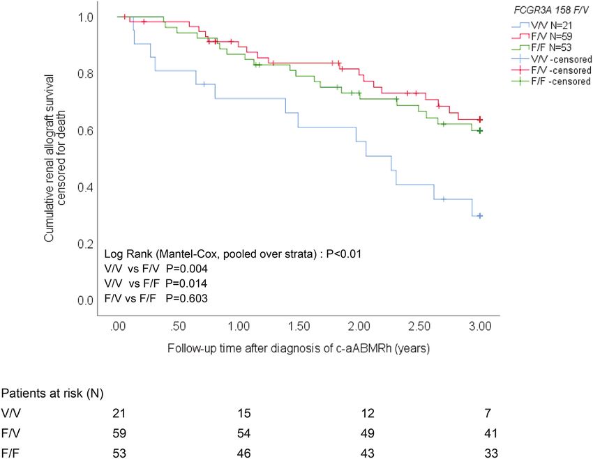
The FCGR3A 158 V/V‑genotype is associated with decreased renal allograft survival. In order
to evaluate whether this single nucleotide polymorphism within FCGR3A (158 F/V) is associated with renal
allograft survival, Kaplan–Meier curves for the different FCGR3A 158 F/V-genotypes were generated for the
c-aABMRh cohort. We evaluated survival of renal allografts at 3 years following diagnosis of c-aABMRh. Fifty-
two out of 133 (39%) c-aABMRh kidney transplant recipients lost their renal allograft due to graft failure and
6 out of 133 (5%) died with a functioning renal allograft (Table 3). The presence or absence of DSAs was not
associated with graft survival (P = 0.70) as was reported before by our g roup14 and others22–25. Overall renal
allograft survival was associated with the FCGR3A 158 F/V genotype (P < 0.01; Fig. 3) and similar results were
obtained for DSA-negative cases (P = 0.04). The V/V-genotype showed a significant lower renal allograft survival
compared to the F/V- as well as the F/F-genotype, using the pairwise between strata comparison (Mantel–Cox
log rank statistical analysis) for the total cohort (P < 0.01 and P = 0.01, respectively) as well as DSA-negative cases
(P = 0.03). Median (min–max) renal allograft survival times amounted to 2.1 (0.1–3), 2.7 (0.1–3) and 3.0 (0.4–3)
years after diagnosis of c-aABMRh for the FCGR3A 158 V/V-, F/V- and F/F-genotype, respectively. Graft sur-
vival was not affected by the FCGR3A 158 F/V-genotype for the control cohort of kidney transplant recipients
without rejection (P = 0.90; data not shown).
To determine whether the V/V-genotype was an independent factor contributing to the risk of renal allograft
loss, relevant clinical characteristics from Table 1, like age of the kidney transplant recipient at transplantation,
gender of kidney transplant recipient, age of the kidney donor, type of kidney donor (living or deceased and ABO-
compatible or ABO-incompatible), re-transplantation and eGFR at time of the for-cause biopsy were included
in a multivariate Cox regression model. Out of these variables only the FCGR3A 158 V/V-genotype (P = 0.04)
together with eGFR at time of diagnosis (HR 0.94, 95% CI 0.91–0.96, P < 0.01) were independently associated
with renal allograft survival). Chronic-aABMRh kidney transplant recipients having the FCGR3A 158 V/V-
genotype had a 1.98-fold (95% CI 1.01–3.82) increased risk (P = 0.04) for losing their allograft within 3 years.
Scientific Reports | (2021) 11:7903 | https://doi.org/10.1038/s41598-021-86943-3 6
Vol:.(1234567890)www.nature.com/scientificreports/
Figure 3. FCGR3A 158 F/V-genotype and renal allograft survival in c-aABMRh kidney transplant recipients.
The c-aABMR-related allograft survival censored for death is depicted for the different FCGR3A 158 F/V-
genotypes. Overall pooled over strata P value and P values for the difference between the different strata
(genotypes) were calculated by Mantel–Cox log-rank statistical analysis. P values < 0.05 were considered
statistically significant (blue, red and green lines represent allograft survival for V/V-, F/V- and F/F-genotype,
respectively). At start and at 1, 2 and 3 years of follow-up, the number of kidney transplant recipients at risk are
depicted for the different FGCR3A 158 F/V-genotypes.
Discussion
In this study, we investigated the association between a functional FCGR3A 158 F/V single nucleotide polymor-
phism and diagnosis of c-aABMR and renal allograft loss thereafter. The results show that the V-allele was linked
to increased expression of CD16 on NK cells and CD16-mediated NK cell cytotoxic potential. In addition, this
allele was associated with an increased glomerulitis score and the V/V-genotype was associated with a decreased
renal allograft survival after the diagnosis of c-aABMR. However, the FCGR3A 158 V/V-genotype appeared not
to be a risk factor per se for the development of c-aABMR after transplantation.
This observation adds evidence for the pathogenicity of the V-allele as was shown in a previous publication
that studied the impact of FCGR3A 158 F/V genotypes in kidney transplant recipients with c-aABMR16. In
accordance with our study, the distribution of the F/V alleles was similar to the control population. Moreover, the
V-allele was associated with a higher degree of peritubular capillarities (PTC) but did not affect the slope of eGFR
loss and graft loss. As discussed by the authors, the latter finding is counterintuitive and contradictory to earlier
studies, which showed a relation between the degree of MVI and graft l oss26–28. In our cohort, we found a higher
glomerulitis score to be associated with the V-allele of the FCGR3A 158 F/V single nucleotide polymorphism.
The V-allele was associated with increased levels of CD16 and CD16-mediated NK function as assessed by the
CD16 DRI and CD107a URI. The V/V-genotype, but not the F/V-genotype was correlated with an increased risk
for renal allograft loss. Unlike our study, where for-cause biopsies were taken, protocol biopsies were taken in the
study by Arnold et al.16, allowing identification of cases with subclinical c-aABMR that may have a much better
prognosis29. This may be an explanation for the discrepancy in clinical outcome associated with the FCGR3A
158 F/V genotypes between their cohort and ours. Furthermore, the fact that cases with c-aABMR having the
V/V- or F/F-genotype tended to be more similar in baseline characteristics when compared to those having the
F/V-genotype, could be another explanation. Nevertheless, we observed a significant increased risk for renal
allograft for cases having the V/V-genotype compared to both those having the F/V- as well as F/F/-genotype.
Overall, the findings of this study indicate that the V/V variant allows for increased activation of the NK
cell after FCGR3A ligation which may cause more inflammation and progressively more tissue damage to the
Scientific Reports | (2021) 11:7903 | https://doi.org/10.1038/s41598-021-86943-3 7
Vol.:(0123456789)www.nature.com/scientificreports/
renal allograft with c-aABMR. The possible pathogenetic role of CD16 is underlined by the finding of increased
transcript levels of CD16 in biopsies diagnosed with A BMR8–10.
30
A review recently, published by M iyairi , summarizes literature on the role of NK cells as critical effec-
tors during ABMR. Detection of NK cell infiltration in biopsies is limited by the number of specific markers
and staining antibodies available. CD16 was used to stain for NK cells in lung grafts31, CD56+ NK cells were
detected in peritubular microvasculature during ongoing biopsy-indicated ABMR8 and others have used CD57
and NKp46 to identify NK cells in biopsies of ongoing AMR and describe their association with microvascular
inflammation32–34. Interestingly, activation of NK cells in absence of DSAs also strongly associated with severity
of glomerulitis and peritubular capillarities and less with C4d deposition35. Recently, infiltrates in biopsies of
kidney transplant recipients diagnosed with c-aABMR were immunophenotypically characterized36,37. NK cells
were found at relatively low frequencies but increased proportions of NK cells within the intravascular glomerular
as well as peritubular compartment were observed in cases with ABMR when compared to TCMR.
Therefore, present data suggest that NK cells and in particular the FCGR3A (CD16)+ are most likely involved
in the pathogenesis of c-aABMR.
Of note is that myeloid cells like macrophages also express CD16 and they are among the cells found in the
renal microvasculature and interstitium in cases of c-aABMR37. Increased macrophage-associated transcripts
were described for individuals with the V-allele compared to the F/F-genotype in the ABMR cases by Arnold
et al.16. This implies that in addition to NK cells, expression of the V/V variant of FCGR3A by myeloid cells
may also contribute to increased inflammation and damage as CD16 appears to be indispensable for ADCC by
monocytes38.
Several other studies have investigated the FCGR3A polymorphism in solid organ transplantation other than
kidney transplantation. In heart transplantation, Paul et al. demonstrated that this polymorphism contributed
to an early (non-invasive) evaluation of risk stratification for cardiac allograft vasculopathy (CAV), an impor-
tant cause of late mortality after heart t ransplantation12. These clinical data are supported by the observation
that increased expression of CD16 and CD16-mediated NK function associated with the V/V-variant were
observed heart transplant r ecipients12 as well as renal transplant recipients16. In accordance with these find-
ings we previously showed increased CD16 expression by circulating NK cells in cases with c-aABMR11. This
FCGR3A polymorphism was also found to stratify patients at risk for acute rejection in the first 3 months after
transplantation, in a cohort of lung transplant recipients39. They also observed the V/V-genotype to behave
different from F/V-genotype, i.e. to significantly reduce the acute-rejection free survival when compared to
F/V- and F/F-genotype. Taken together, the current data suggest that FCGR3A polymorphism is associated with
rejection-related complications across various types of solid organ transplantation.
One of the limitations of our retrospective study involves the lack of DSAs from the complete c-aABMR
cohort. Although DSA-positive and -negative c-aABMR are similar with respect to renal allograft survival, there
might be differences in terms of underlying mechanisms of damage to the renal allograft. However, in our cohort,
we did observe similar findings for the DSA-negative cohort compared to the total cohort with respect to this
FCGR3A 158 F/V SNP and level of microvascular inflammation as well as renal allograft survival. Furthermore,
antibodies directed to non-HLA might also contribute to damage to the renal allograft but this information is
lacking. Another limitation of the present study is the lack of a validation cohort, allowing confirmation of our
findings in a similar large group of c-aABMR cases.
Concluding, the V-allele of the FCGR3A 158 F/V single nucleotide polymorphism leads to increased CD16
expression and upregulated cytotoxicity of NK cells. Clinically this is associated with increased microvascular
inflammation in the glomerulus and significantly decreased renal allograft survival of grafts diagnosed with
c-aABMR for cases having the V/V-genotype. Assessment of this FCGR3A 158 F/V SNP may be of importance
with respect to interpretation of results from studies. Furthermore, it may add to the risk stratification for graft
loss in kidney transplant recipients by identifying patients that might benefit from a more intense immunosup-
pressive regime.
Material and methods
Study population. For this study we included 141 kidney transplant recipients, diagnosed with clinical
c-aABMR (within our hospital) in the period from 1998 to 2019 of which snap-frozen PBMCs prior to trans-
plantation were stored in the kidney transplant biobank. Patients in whom c-aABMR was diagnosed following a
transplantectomy, were excluded from analysis (N = 8). As a control population we selected 116 kidney transplant
recipients from the biobank, transplanted within the same period, without a diagnosis of c-aABMR. All kidney
transplantations were performed across a negative CDC crossmatch. Demographic and clinical parameters were
collected for the c-aABMR cohort at time of transplantation and diagnostic biopsy. All renal biopsies were done
on indication (eGFR loss or proteinuria) and re-evaluated by an experienced renal pathologist based on the 2015
Banff classification40. Donor-specific anti-HLA antibodies (DSAs) were measured using the Lifecodes Lifescreen
Deluxe (LMX) kit according to the manufacturer’s manual (Immunocor Transplant Diagnostics Inc., Stamford,
CT, USA). Samples positive for either HLA class I (HLA-A, -B or -C) or HLA class II (HLA-DQ or -DR), were
further characterized with the Luminex Single Antigen assay, using LABscreen HLA class I and II antigen beads
(One Lambda, Canoga Park, GA, USA). Before 2009, DSAs were not routinely assessed but 40% of the tested
cases (N = 70) were DSA-positive. When histologic criteria were met, a diagnosis of c-aABMRh, termed suspi-
cious for c-aABMR in the Banff 2015 criteria, was made in accordance with recent p ublications13,14,40–42.
Renal allograft function was determined by calculating the estimated glomerular filtration rate (eGFR) using
the CKD-EPI formula.
Scientific Reports | (2021) 11:7903 | https://doi.org/10.1038/s41598-021-86943-3 8
Vol:.(1234567890)www.nature.com/scientificreports/
Follow up of c-aABMRh kidney transplant recipients was until 1st of January 2020 and graft loss/failure was
defined as the need for dialysis or a re-transplantation. The date of diagnosis of c-aABMRh and date of graft
failure were used to calculate the graft survival upon diagnosis.
Kidney transplant recipients gave written informed consent and the study was approved by the Medical Ethical
Committee of the Erasmus MC (MEC-2012-022 for pre-transplant material and MEC-2015-222 for the collection
of data from cases of c-aABMRh and controls). The study was conducted in accordance with the Declaration of
Helsinki and the Declaration of Istanbul.
Peripheral blood mononuclear cell isolation. Prior to and at time of the for-cause biopsy, peripheral
blood mononuclear cells (PBMCs) were isolated from heparinized blood samples by using Ficoll-Paque (GE
HealthCare, Uppsala, Sweden). Two million PBMCs of the sample, obtained prior to transplantation, were snap-
frozen in liquid nitrogen for isolation of genomic DNA (see below). The remaining PBMCs were washed, frozen
at 10 × 106/vial in RPMI-1640 with Glutamax (ThermoFisher Scientific, Landsmeer, The Netherlands) supple-
mented with 100 IU/mL penicillin/streptomycin and 10% heat-inactivated pooled serum and 10% dimethyl
sulphoxide (Sigma Aldrich, Darmstadt, Germany) in liquid nitrogen until further use.
FCGR3A 158 F/V (rs396991) genotyping. Genomic DNA was extracted from 2 × 10⁶ snapfrozen PBMC
using the QIAamp DNA Mini isolation kit (Qiagen, Venlo, The Netherlands) according to manufacturer’s
instruction. FCGR3A rs396991 genotype was determined by the StepOnePlus Real-Time PCR detection system
(Applied Biosystems, Darmstadt, Germany) using Taqman SNP Genotyping assay (assay ID C_25815666_10;
ThermoFisher Scientific Inc, Bleiswijk, The Netherlands) and Taqman Universal PCR Master Mix according to
manufacturer’s instruction.
Natural killer (NK) cell function. The CD16-mediated NK cell function was evaluated by adapting the
NK-CHAT as described Legris et al.43. Briefly, 1 06 allogeneic target cells (Raji, ATCC CCL-86) were carboxyfluo-
rescein diacetate succinimidyl ester (CFSE; Molecular Probes, Bleijswijk, The Netherlands)-labeled according
to manufacturer’s instruction. These target cells were co-cultured at a 1:1 ratio with PBMCs of kidney trans-
plant recipients genotyped for FCGR3A 158 F/V (N = 35), allophycocyan (APC)-labelled anti-CD107a (Bec-
ton Dickinson; BD, Erembodegem, Belgium) and a cytokine secretion inhibitor (EBioscience, Bleijswijk, The
Netherlands) for 3 h. The co-culture was performed with or without anti-human CD20-human-IgG1 (10 μg/
mL; InvivoGen Toulouse, France) to evaluate CD16-dependent NK-cell function. As a control, PBMCs were
left unstimulated to determine baseline median fluorescence intensity (MFI) of CD16 expression by circulating
NK cells. Upon 3 h of stimulation, cells were harvested and stained for dead cells using Fixeable Viability Stain
(FVS)-780 (BD). Following a wash, the cell surface was stained for 30 min at room temperature using the follow-
ing monoclonal antibodies to identify NK cells (phycoerythrin (PE)-labeled anti-CD16/PerCPCy5.5 (peridinin-
chlorophyll protein (PerCP)-Cy5.5-labeled anti-CD56, both from BD) and exclude unwanted cells like T cells
(brilliant violet (BV)510-labeled anti-CD3, BD) and monocytes (APC-H7-labeled anti-CD14, BD). The follow-
ing parameters were determined to evaluate CD16-mediated NK cell function: (1) CD16 downregulation index
(DRI, ratio MFI CD16 in absence of anti-hCD20-hIgG1 over MFI in presence of anti-hCD20-hIgG1) and (2)
CD107a upregulation index (URI, ratio % CD107a+ NK cells in presence of anti-hCD20-hIgG1 over %CD107a+
NK cells in absence of anti-hCD20-hIgG1). CD107a (lysosomal-associated membrane protein; LAMP-1) is a
marker identifying degranulation of NK cells which is highly correlated to their cytotoxic p otential44. NK cells
were identified as CD3-CD14- and CD56+.
Samples were measured on the FACSCanto II (BD; 3 laser, 8 color configuration 4:2:2) and analyzed using
Kaluza software version 2.1 (Beckman Coulter BV., Woerden, The Netherlands). We acquired at least 10,000
NK cells.
Statistical analyses. Normally distributed data are expressed as mean ± SD, non-normally distributed
data as median and IQR. Continuous variables of c-aABMRh and control kidney transplant recipients were
compared using unpaired T test or Mann–Whitney U test. Discrete data were analyzed as frequencies with
Chi-square test or Fisher’s exact test. Demographic as well as patient characteristics are depicted as median
and IQR, number and proportion of total, respectively. Comparisons of characteristics between the different
FCGR3A 158 F/V-genotypes was done using Chi-square test for discrete data and Kruskal–Wallis for continu-
ous data. Death-censored graft survival was assessed taking into account the FCGR3A 158 F/V-genotype for the
c-aABMRh cohort by Kaplan–Meier analysis with log-rank statistics for difference between strata (pooled and
pairwise). Multivariate Cox regression analysis, using the Enter method as well as the Forward and Backward
stepwise Likelihood Ratio method, was used to evaluate the significance and contribution of the FCGR3A 158
F/V-genotype and other relevant clinical characteristics with respect to renal allograft survival. The level of glo-
merulitis (g) and peritubular capillaritis (ptc) as well as the combination thereof representing the total level of
microvascular inflammation (MVI) were correlated to the FCGR3A 158 F/V-genotype. Statistical analyses were
performed using GraphPad Prism 5 software (GraphPad Software La Jolla, CA,USA) and IBM SPSS statistics for
Windows, version 25 (SPSS Inc. IL, USA). The significance level (P value) was two-tailed and an α of 0.05 was
used for all analyses.
Data availability
The datasets generated during and/or analysed during the current study are available from the corresponding
author on reasonable request.
Scientific Reports | (2021) 11:7903 | https://doi.org/10.1038/s41598-021-86943-3 9
Vol.:(0123456789)www.nature.com/scientificreports/
Received: 27 January 2021; Accepted: 19 March 2021
References
1. Racusen, L. C. et al. Antibody-mediated rejection criteria—an addition to the Banff 97 classification of renal allograft rejection.
Am. J. Transplant. 3, 708–714 (2003).
2. Racusen, L. C. et al. The Banff 97 working classification of renal allograft pathology. Kidney Int. 55, 713–723. https://doi.org/10.
1046/j.1523-1755.1999.00299.xS0085-2538(15)46016-8 (1999).
3. Michielsen, L. A., van Zuilen, A. D., Krebber, M. M., Verhaar, M. C. & Otten, H. G. Clinical value of non-HLA antibodies in kidney
transplantation: Still an enigma?. Transplant. Rev. (Orlando) 30, 195–202. https://doi.org/10.1016/j.trre.2016.06.001 (2016).
4. Loupy, A. & Lefaucheur, C. Antibody-mediated rejection of solid-organ allografts. N. Engl. J. Med. 379, 1150–1160. https://doi.
org/10.1056/NEJMra1802677 (2018).
5. Castro-Dopico, T. & Clatworthy, M. R. Fcgamma receptors in solid organ transplantation. Curr. Transplant Rep. 3, 284–293. https://
doi.org/10.1007/s40472-016-0116-7116 (2016).
6. Sis, B. & Halloran, P. F. Endothelial transcripts uncover a previously unknown phenotype: C4d-negative antibody-mediated rejec-
tion. Curr. Opin. Organ. Transplant. 15, 42–48. https://doi.org/10.1097/MOT.0b013e3283352a50 (2010).
7. Haas, M. et al. Banff 2013 meeting report: Inclusion of c4d-negative antibody-mediated rejection and antibody-associated arterial
lesions. Am. J. Transplant. 14, 272–283. https://doi.org/10.1111/ajt.12590 (2014).
8. Hidalgo, L. G. et al. NK cell transcripts and NK cells in kidney biopsies from patients with donor-specific antibodies: Evidence
for NK cell involvement in antibody-mediated rejection. Am. J. Transplant. 10, 1812–1822. https://doi.org/10.1111/j.1600-6143.
2010.03201.x (2010).
9. Hidalgo, L. G. et al. Interpreting NK cell transcripts versus T cell transcripts in renal transplant biopsies. Am. J. Transplant. 12,
1180–1191. https://doi.org/10.1111/j.1600-6143.2011.03970.x (2012).
10. Venner, J. M., Hidalgo, L. G., Famulski, K. S., Chang, J. & Halloran, P. F. The molecular landscape of antibody-mediated kidney
transplant rejection: Evidence for NK involvement through CD16a Fc receptors. Am. J. Transplant. 15, 1336–1348. https://doi.
org/10.1111/ajt.13115 (2015).
11. Sablik, K. A., Litjens, N. H. R., Klepper, M. & Betjes, M. G. H. Increased CD16 expression on NK cells is indicative of antibody-
dependent cell-mediated cytotoxicity in chronic-active antibody-mediated rejection. Transpl. Immunol.. 54, 52–58. https://doi.
org/10.1016/j.trim.2019.02.005 (2019).
12. Paul, P. et al. Genetic and functional profiling of CD16-dependent natural killer activation identifies patients at higher risk of
cardiac allograft vasculopathy. Circulation 137, 1049–1059. https://doi.org/10.1161/CIRCULATIONAHA.117.030435 (2018).
13. Sablik, K. A., Clahsen-van Groningen, M. C., Damman, J., Roelen, D. L. & Betjes, M. G. H. Banff lesions and renal allograft survival
in chronic-active antibody mediated rejection. Transpl. Immunol. 56, 101213. https://doi.org/10.1016/j.trim.2019.101213 (2019).
14. Sablik, K. A. et al. Chronic-active antibody-mediated rejection with or without donor-specific antibodies has similar histomor-
phology and clinical outcome—a retrospective study. Transpl. Int. 31, 900–908. https://doi.org/10.1111/tri.13154 (2018).
15. Sablik, K. A. et al. Treatment with intravenous immunoglobulins and methylprednisolone may significantly decrease loss of renal
function in chronic-active antibody-mediated rejection. BMC Nephrol. 20, 218. https://d oi.o
rg/1 0.1 186/s 12882-0 19-1 385-z (2019).
16. Arnold, M. L. et al. Functional Fc gamma receptor gene polymorphisms and donor-specific antibody-triggered microcirculation
inflammation. Am. J. Transplant. 18, 2261–2273. https://doi.org/10.1111/ajt.14710 (2018).
17. de Weerd, A. E. & Betjes, M. G. H. ABO-incompatible kidney transplant outcomes: A meta-analysis. Clin. J. Am. Soc. Nephrol. 13,
1234–1243. https://doi.org/10.2215/CJN.00540118 (2018).
18. Torkildsen, O. et al. Ethnic variation of Fc gamma receptor polymorphism in Sami and Norwegian populations. Immunology 115,
416–421. https://doi.org/10.1111/j.1365-2567.2005.02158.x (2005).
19. Lehrnbecher, T. et al. Variant genotypes of the low-affinity Fcgamma receptors in two control populations and a review of low-
affinity Fcgamma receptor polymorphisms in control and disease populations. Blood 94, 4220–4232 (1999).
20. Dourado, M. E. J., Ferreira, L. C., Freire-Neto, F. P. & Jeronimo, S. M. No association between FCGR2A and FCGR3A polymor-
phisms in Guillain–Barre Syndrome in a Brazilian population. J. Neuroimmunol. 298, 160–164. https://doi.org/10.1016/j.jneur
oim.2016.07.020 (2016).
21. Loupy, A. et al. Molecular microscope strategy to improve risk stratification in early antibody-mediated kidney allograft rejection.
J Am Soc Nephrol 25, 2267–2277. https://doi.org/10.1681/ASN.2013111149 (2014).
22. De Serres, S. A. et al. 2013 Banff criteria for chronic active antibody-mediated rejection: Assessment in a real-life setting. Am. J.
Transplant. 16, 1516–1525. https://doi.org/10.1111/ajt.13624 (2016).
23. Halloran, P. F., MerinoLopez, M. & BarretoPereira, A. Identifying subphenotypes of antibody-mediated rejection in kidney trans-
plants. Am. J. Transplant. 16, 908–920. https://doi.org/10.1111/ajt.13551 (2016).
24. Lesage, J. et al. Donor-specific antibodies, C4d and their relationship with the prognosis of transplant glomerulopathy. Transplanta-
tion 99, 69–76. https://doi.org/10.1097/TP.0000000000000310 (2015).
25. Patri, P. et al. Development and validation of a prognostic index for allograft outcome in kidney recipients with transplant glo-
merulopathy. Kidney Int. 89, 450–458. https://doi.org/10.1038/ki.2015.288 (2016).
26. Kozakowski, N. et al. The diffuse extent of peritubular capillaritis in renal allograft rejection is an independent risk factor for graft
loss. Kidney Int. 88, 332–340. https://doi.org/10.1038/ki.2015.64 (2015).
27. Lerut, E., Naesens, M., Kuypers, D. R., Vanrenterghem, Y. & Van Damme, B. Subclinical peritubular capillaritis at 3 months is
associated with chronic rejection at 1 year. Transplantation 83, 1416–1422. https://doi.org/1 0.1097/0 1.tp.000026 6676.1 0550.7 0000
07890-200706150-00002 (2007).
28. Loupy, A. et al. Outcome of subclinical antibody-mediated rejection in kidney transplant recipients with preformed donor-specific
antibodies. Am. J. Transplant. 9, 2561–2570. https://doi.org/10.1111/j.1600-6143.2009.02813.x (2009).
29. Parajuli, S. et al. Subclinical antibody-mediated rejection after kidney transplantation: Treatment outcomes. Transplantation 103,
1722–1729. https://doi.org/10.1097/TP.0000000000002566 (2019).
30. Miyairi, S., Baldwin, W. M. 3rd., Valujskikh, A. & Fairchild, R. L. Natural killer cells: Critical effectors during antibody-mediated
rejection of solid organ allografts. Transplantation 105, 284–290. https://doi.org/10.1097/TP.0000000000003298 (2021).
31. Fildes, J. E. et al. Natural killer cells in peripheral blood and lung tissue are associated with chronic rejection after lung transplanta-
tion. J. Heart Lung Transplant. 27, 203–207. https://doi.org/10.1016/j.healun.2007.11.571 (2008).
32. Dos Santos, D. C., Saraiva Camara, N. O., David, D. S. R. & Malheiros, D. Expression patterns of CD56+ and CD16+ cells in renal
transplant biopsies with acute rejection: Associations with microcirculation injuries and graft survival. Nephrology (Carlton) 22,
993–1001. https://doi.org/10.1111/nep.12897 (2017).
33. Kildey, K. et al. Specialized roles of human natural killer cell subsets in kidney transplant rejection. Front. Immunol.. 10, 1877.
https://doi.org/10.3389/fimmu.2019.01877 (2019).
34. Shin, S. et al. Interpreting CD56+ and CD163+ infiltrates in early versus late renal transplant biopsies. Am. J. Nephrol. 41, 362–369.
https://doi.org/10.1159/000430473 (2015).
35. Callemeyn, J. et al. Transcriptional changes in kidney allografts with histology of antibody-mediated rejection without anti-HLA
donor-specific antibodies. J. Am. Soc. Nephrol. 31, 2168–2183. https://doi.org/10.1681/ASN.2020030306 (2020).
Scientific Reports | (2021) 11:7903 | https://doi.org/10.1038/s41598-021-86943-3 10
Vol:.(1234567890)www.nature.com/scientificreports/
36. Calvani, J. et al. In situ multiplex immunofluorescence analysis of the inflammatory burden in kidney allograft rejection: A new
tool to characterize the alloimmune response. Am. J. Transplant. 20, 942–953. https://doi.org/10.1111/ajt.15699 (2020).
37. Sablik, K. A., Jordanova, E. S., Pocorni, N., Clahsen-van Groningen, M. C. & Betjes, M. G. H. Immune cell infiltrate in chronic-
active antibody-mediated rejection. Front. Immunol. 10, 3106. https://doi.org/10.3389/fimmu.2019.03106 (2019).
38. Yeap, W. H. et al. CD16 is indispensable for antibody-dependent cellular cytotoxicity by human monocytes. Sci. Rep. 6, 34310.
https://doi.org/10.1038/srep34310 (2016).
39. Paul, P. et al. FCGR3A and FCGR2A genotypes differentially impact allograft rejection and patients’ survival after lung transplant.
Front. Immunol. 10, 1208. https://doi.org/10.3389/fimmu.2019.01208 (2019).
40. Loupy, A. et al. The Banff 2015 kidney meeting report: Current challenges in rejection classification and prospects for adopting
molecular pathology. Am. J. Transplant. 17, 28–41. https://doi.org/10.1111/ajt.14107 (2017).
41. Senev, A. et al. Histological picture of antibody-mediated rejection without donor-specific anti-HLA antibodies: Clinical presenta-
tion and implications for outcome. Am. J. Transplant. 19, 763–780. https://doi.org/10.1111/ajt.15074 (2019).
42. Haas, M. et al. The Banff 2017 Kidney Meeting Report: Revised diagnostic criteria for chronic active T cell-mediated rejection,
antibody-mediated rejection, and prospects for integrative endpoints for next-generation clinical trials. Am. J. Transplant. 18,
293–307. https://doi.org/10.1111/ajt.14625 (2018).
43. Legris, T. et al. Antibody-dependent NK cell activation is associated with late kidney allograft dysfunction and the complement-
independent alloreactive potential of donor-specific antibodies. Front. Immunol. 7, 288. https://d oi.o
rg/1 0.3 389/fi
mmu.2 016.0 0288
(2016).
44. Tomescu, C., Chehimi, J., Maino, V. C. & Montaner, L. J. Retention of viability, cytotoxicity, and response to IL-2, IL-15, or IFN-
alpha by human NK cells after CD107a degranulation. J. Leukoc. Biol. 85, 871–876. https://doi.org/10.1189/jlb.1008635 (2009).
Acknowledgements
The authors would like to thank the renal pathologists of the Department of Pathology at the Erasmus MC,
University Medical Center, in particular Dr. M. C. Clahsen-van Groningen and Dr. J. H. von der Thüsen.
Author contributions
N.L. participated in research design, data collection, data analysis and writing of the manuscript; A.P. participated
in research design, data collection and data analysis; J.K.-v.G. participated in data collection; M.K. participated
in research design, data collection and data analysis; M.B. participated in research design, data collection, data
analysis and writing of the manuscript. All authors reviewed the manuscript.
Competing interests
The authors declare no competing interests.
Additional information
Correspondence and requests for materials should be addressed to N.L.
Reprints and permissions information is available at www.nature.com/reprints.
Publisher’s note Springer Nature remains neutral with regard to jurisdictional claims in published maps and
institutional affiliations.
Open Access This article is licensed under a Creative Commons Attribution 4.0 International
License, which permits use, sharing, adaptation, distribution and reproduction in any medium or
format, as long as you give appropriate credit to the original author(s) and the source, provide a link to the
Creative Commons licence, and indicate if changes were made. The images or other third party material in this
article are included in the article’s Creative Commons licence, unless indicated otherwise in a credit line to the
material. If material is not included in the article’s Creative Commons licence and your intended use is not
permitted by statutory regulation or exceeds the permitted use, you will need to obtain permission directly from
the copyright holder. To view a copy of this licence, visit http://creativecommons.org/licenses/by/4.0/.
© The Author(s) 2021
Scientific Reports | (2021) 11:7903 | https://doi.org/10.1038/s41598-021-86943-3 11
Vol.:(0123456789)You can also read