Ranaviruses in captive and wild Australian lizards
←
→
Page content transcription
If your browser does not render page correctly, please read the page content below
RESEARCH ARTICLE

                                                 Ranaviruses in captive and wild
                                                 Australian lizards
                                                 Alicia Maclainea*, Wytamma T. Wirtha, Donald T. McKnightb†, Graham W. Burgessa, and Ellen Ariela
                                                 a
                                                 College of Public Health, Medical and Veterinary Sciences, James Cook University, Townsville,
                                                 Queensland 4811, Australia; bCollege of Science and Engineering, James Cook University, Townsville,
                                                 Queensland 4811, Australia

                                                 *alicia.maclaine@my.jcu.edu.au
                                                 †
                                                  Current address: School of Environmental and Rural Science, University of New England, Armidale,
                                                 New South Wales 2351, Australia.

                                                 Abstract
                                                 Ranaviral infections have been associated with mass mortality events in captive and wild amphibian,
                                                 fish, and reptile populations globally. In Australia, two distinct types of ranaviruses have been isolated:
                                                 epizootic haematopoietic necrosis virus in fish and a Frog virus 3-like ranavirus in amphibians.
                                                 Experimental studies and serum surveys have demonstrated that several Australian native fish,
                                                 amphibian, and reptile species are susceptible to infection and supported the theory that ranavirus
                                                 is naturally circulating in Australian herpetofauna. However, ranaviral infections have not been
                                                 detected in captive or wild lizards in Australia. Oral-cloacal swabs were collected from 42 wild lizards
    OPEN ACCESS                                  from northern Queensland and 83 captive lizards from private collections held across three states/
                                                 territories. Samples were tested for ranaviral DNA using a quantitative PCR assay. This assay detected
                                                 ranaviral DNA in 30/83 (36.1%) captive and 33/42 (78.6%) wild lizard samples. This is the first time
                                                 molecular evidence of ranavirus has been reported in Australian lizards.
Citation: Maclaine A, Wirth WT,
McKnight DT, Burgess GW, and Ariel E.            Key words: ranavirus, Australia, reptiles, lizards, Intellagama lesueurii lesueurii, Pogona vitticeps
2020. Ranaviruses in captive and wild
Australian lizards. FACETS 5: 758–768.
doi:10.1139/facets-2020-0011
                                                 Introduction
Handling Editor: David Lesbarrères
                                                 Ranaviruses are large double-stranded DNA viruses that infect a wide range of ectothermic
Received: February 27, 2020                      vertebrates globally. These viruses have been associated with mass mortality events and are transmis-
Accepted: May 11, 2020
                                                 sible to different classes of lower vertebrates (Bigarré et al. 2008; Kik et al. 2011; Miller et al. 2011;
                                                 Brenes et al. 2014; Butkus et al. 2017). In Australia, two distinct types of ranaviruses have been
Published: September 25, 2020                    isolated: epizootic haematopoietic necrosis virus in fish, and a Frog virus 3 (FV3)-like ranavirus in
Note: This paper is part of a Collection         amphibians (Langdon et al. 1986, 1988; Speare and Smith 1992).
titled “Ranavirus research: 10 years of global
collaboration.”                                  Epizootic haematopoietic necrosis virus (EHNV, an Ambystoma tigrinum virus-like ranavirus) is
                                                 considered to be the most important ranavirus affecting fish and is listed as notifiable by The World
Copyright: © 2020 Maclaine et al. This work      Organisation for Animal Health (OIE) (Price et al. 2017a; OIE 2018). This ranaviral species is
is licensed under a Creative Commons
                                                 endemic to southern Australia and was regularly reported during mortality events in wild redfin perch
Attribution 4.0 International License (CC BY
4.0), which permits unrestricted use,            (Perca fluviatilis) in Victoria (Langdon et al. 1986; Whittington et al. 2010). Several fish species and
distribution, and reproduction in any            the European common frog (Rana temporaria) have also been shown to be susceptible to EHNV
medium, provided the original author(s) and      via experimental exposure (Langdon 1989; Jensen et al. 2009, 2011; Bayley et al. 2013; Becker et al.
source are credited.                             2013, 2016).
Published by: Canadian Science Publishing

FACETS | 2020 | 5: 758–768 | DOI: 10.1139/facets-2020-0011                                                                                               758
facetsjournal.com
Maclaine et al.
An FV3-like ranavirus, Bohle iridovirus (BIV), was first isolated from wild caught ornate burrowing
frogs (Limnodynastes ornatus) in northern Queensland that died during or soon after metamorphosis
(Speare and Smith 1992). More recently, a BIV-like virus was isolated from captive magnificent tree
frogs (Litoria splendida) and green tree frogs (Litoria caerulea) during a mortality event in Darwin,
Northern Territory (Weir et al. 2012). Experimental studies of Australian native fish, amphibians,
and reptiles have shown that species within these classes are susceptible to infection with BIV,
including juvenile eastern water dragons (Intellagama lesueurii lesueurii) (Moody and Owens 1994;
Cullen et al. 1995; Cullen and Owens 2002; Ariel et al. 2015; Maclaine et al. 2018). Additionally,
BIV antibodies have been detected in wild populations of turtles, crocodiles, snakes, and cane toads
(Bufo marinus) in northern Queensland, indicating that ranavirus is circulating in the herpetofauna
in this region (Whittington et al. 1997; Zupanovic et al. 1998; Ariel et al. 2017).
Ranaviral infections in lizards have so far been limited to long term captive lizards held in collections
outside of Australia that were investigated after signs of disease were observed (Marschang et al. 2005;
Behncke et al. 2013; Stöhr et al. 2013; Tamukai et al. 2016). Outbreaks in wild Australian lizards have
not been reported, which may be due to the lack of targeted surveillance and the vastness of a
continent that is sparsely populated by humans. A recent systematic survey of wild eastern fence
lizards (Sceloporus undulates) in central Virginia, United States, was the first study to target and
report molecular evidence of ranavirus in wild lizards (Goodman et al. 2018). This lizard species
was selected because they share habitat with turtles previously diagnosed with ranaviral infection
and this virus is known to cross-infect sympatric species.
The aim of this study was to determine if wild and (or) captive Australian lizards are infected with
ranaviruses using molecular methods. The study targeted native Australian lizards in captive settings
and in natural areas where ranaviral antibodies have previously been detected.
Materials and methods
Ethics statement
Sample collection from captive and wild lizards was conducted under permissions from James
Cook University Animal Ethics Committee (Ethics Approval No. A2087 and A2277), Queensland
Department of Environment and Heritage Protection (Scientific Purposes Permit
No. WISP15053914), and Queensland Department of National Parks, Sport and Racing (Scientific
Purposes Permit No. WITK18689817).
Animals
As part of this study, 125 Australian lizards (83 captive and 42 wild) were sampled representing seven
species from the Agamidae and Scincidae families. Species sampled included: Boyd’s forest dragon
(Hypsilurus boydii), central bearded dragon (Pogona vitticeps), eastern water dragon (Intellagama
lesueurii lesueurii), nobbi dragon (Diporiphora nobbi), frilled neck lizard (Chlamydosaurus kingii),
shingleback lizard (Tiliqua rugosa), and blue-tongued skink (T. scincoides) (Table 1).
Study sites
Captive lizards were sampled from three private collections held in Canberra, Australian Capital
Territory (Collection 1, n = 67); Salt Ash, New South Wales (Collection 2, n = 15); and Townsville,
Queensland (Collection 3, n = 1) between 2015 and 2016 (Table 2). These collections were chosen
based on willingness of owners and availability of lizards. All lizards were held under a current reptile
license, where applicable, at the time of sampling.
FACETS | 2020 | 5: 758–768 | DOI: 10.1139/facets-2020-0011 759
facetsjournal.comMaclaine et al.
Table 1. Number of lizards sampled for ranavirus testing with reference to family, species, and captive or wild
status.
Family Species Captive Wild Total
Agamidae Boyd’s forest dragon (Hypsilurus boydii) — 1 1
Central bearded dragon (Pogona vitticeps) 74 — 74
Eastern water dragon (Intellagama lesueurii lesueurii) 2 37 39
Frilled neck lizard (Chlamydosaurus kingii) 2 — 2
Nobbi dragon (Diporiphora nobbi) — 4 4
Scincidae Blue-tongued skink (Tiliqua scincoides) 2 — 2
Shingleback lizard (Tiliqua rugosa) 3 — 3
Total 83 42 125
Table 2. PCR results for the detection of ranaviruses in wild and captive Australian lizards using qPCR
described by Leung et al. (2017).
Number of PCR positive
Site location and species samples/total samples tested
Collection 1—Canberra, Australian Capital Territory
Central bearded dragon (Pogona vitticeps) 29/67
Collection 2—Salt Ash, New South Wales (n = 15)
Eastern water dragon (Intellagama lesueurii lesueurii) 1/2
Central bearded dragon (Pogona vitticeps) 0/7
Frilled neck lizard (Chlamydosaurus kingii) 0/1
Blue-tongued skink (Tiliqua scincoides) 0/2
Shingleback lizard (Tiliqua rugosa) 0/3
Collection 3—Townsville, Queensland (n = 1)
Frilled neck lizard (Chlamydosaurus kingii) 0/1
Paluma Range National Park, Queensland
Boyd’s Forest Dragon (Hypsilurus boydii) 1/1
Eastern water dragon (Intellagama lesueurii lesueurii) 23/32
Girringun National Park, Queensland
Eastern water dragon (Intellagama lesueurii lesueurii) 2/2
Tully Gorge National Park, Queensland
Eastern water dragon (Intellagama lesueurii lesueurii) 1/1
Wooroonooran National Park, Queensland
Eastern water dragon (Intellagama lesueurii lesueurii) 2/2
Wambiana Cattle Station, Queensland
Nobbi dragon (Diporiphora nobbi) 4/4
FACETS | 2020 | 5: 758–768 | DOI: 10.1139/facets-2020-0011 760
facetsjournal.comMaclaine et al.
Wild lizards were sampled opportunistically from five locations within northern Queensland: Paluma
Range National Park (n = 33), Girringun National Park (n = 2), Tully Gorge National Park (n = 1),
Wooroonooran National Park (n = 2) and Wambiana Cattle Station (n = 4) in 2015 and 2017
(Fig. 1A). Within Paluma Range National Park sampling was conducted at multiple sites along the
margin of freshwater creeks and streams (Fig. 1B).
The National Parks sites are located within the Wet Tropics World Heritage Area and lizards were
sampled in low-elevation eucalyptus forests and dense, high-elevation notophyll rainforests (Stanton
and Stanton 2005). Lizards sampled at these sites were near freshwater creeks and streams, and the
collection sites were remote, accessible only by foot. Wambiana Cattle Station located near Charters
Towers, Queensland, is a working cattle property exposed to grazing and land clearing. This site is
comprised of open eucalypt savanna woodlands, dominated by Reid River box (Eucalyptus brownii)
and silver-leaf ironbark (E. melanophloa).
Sampling
Captive lizards where restrained by the owner for sample collection. Wild lizards were captured by
hand at night and restrained while morphometric data (weight, snout to vent length (SVL)), and
samples were collected for each animal. Where possible, information on sex, age class, body condition,
SVL, weight, health history, and origin were recorded for each lizard. Information for captive lizards
was collected at the discretion of the owner and, as a result, some data are unavailable.
A combined oral-cloacal swab was taken from each lizard for molecular analysis. For this purpose, a
sterile wooden-stem cotton-tipped swabs (Livingstone Pty. Ltd., Australia) was inserted into the oral
cavity and then into the cloaca. Swabs were then immediately placed into 1 mL of Dulbecco’s
Modified Eagle Medium (Thermo Fisher Scientific, New York, New York, USA) supplemented with
antibiotic-antimycotic (Thermo Fisher Scientific, New York, New York, USA) and transported on
ice to the laboratory at James Cook University (Townsville, Queensland) within 12 h. Samples were
stored at −80 °C until the day of DNA extraction, when they were thawed at room temperature and
vortexed for 30 s.
Molecular analysis
DNA was extracted from a 200 μL aliquot of the media that the oral-cloacal swab sample was stored in
using an ISOLATE II Genomic DNA Kit (Bioline, Luckenwalde, Germany) following the manufacturer’s
protocol. Extracted DNA was tested for the presence of ranavirus using a real-time quantitative
polymerase chain reaction (qPCR) designed to detect ranaviruses in amphibians, fish, and reptiles (Leung
et al. 2017). The reaction mixture was as follows: 1 × GoTaq Probe qPCR Mastermix (Promega, Madison,
Wisconsin, USA), 0.5 μM of forward (5′-GTCCTTTAACACGGCATACCT-3′) and reverse primer
(5′-ATCGCTGGTGTTGCCTATC-3′), 0.25 μM probe (5′-TTATAGTAGCCTRTGCGCTTGGCC-3′),
2 μL of template DNA (~80 ng), and nuclease-free water in a 20 μL reaction. Thermocycling was
performed on a Magnetic Induction Cycler PCR machine (Applied Biosystems) under the following
conditions: 95 °C for 2 min, then 40 cycles of (95 °C for 5 s, 60 °C for 10 s and 72 °C for 15 s) with a final
extension at 95 °C for 2 min. Each run contained a negative control (no template added) and a positive
control (a linearised plasmid containing the PCR product from a BIV isolate). Samples that had more
than 10 copies μL−1 and produced sigmoidal amplification curves were considered positive.
For positive samples, a 200 μL aliquot of the media that the oral-cloacal swab samples were stored in,
were transported on ice to The OIE Reference Laboratory, University of Sydney, for molecular
confirmation and viral isolation.
FACETS | 2020 | 5: 758–768 | DOI: 10.1139/facets-2020-0011 761
facetsjournal.comMaclaine et al.
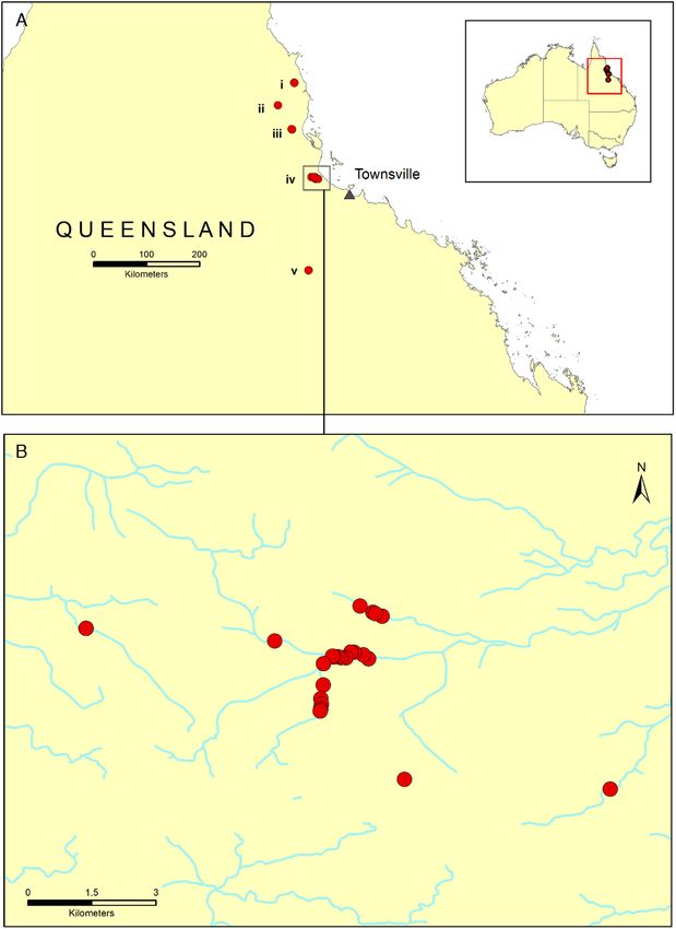
Fig. 1. (A) Map of northern Queensland (insert—Australian continent) with the five locations where wild lizards
were sampled: (i) Wooroonooran National Park, (ii) Tully Gorge National Park, (iii) Girringun National Park,
(iv) Paluma Range National Park, and (v) Wambiana Cattle Station. (B) GPS locations of samples collected from
wild lizards in Paluma Range National Park. Map created using ArcMap 10.3.0. Data sources: Australia and
State boundaries from Australian Bureau of Statistics 2016. Queensland Waterways from Queensland Spatial
Catalogue—Department of Agriculture and Fisheries.
FACETS | 2020 | 5: 758–768 | DOI: 10.1139/facets-2020-0011 762
facetsjournal.comMaclaine et al.
Viral isolation
Viral isolation was attempted by The OIE Reference Laboratory, University of Sydney, on PCR
positive samples as follows: unfiltered material was inoculated, in duplicate, into bluegill fry (BF-2)
cell suspension and incubated for 9 d at 22 °C during which the cells were examined for cytopathic
effect (Passage 1). Cells went through a freeze–thaw cycle once at −20 °C. Duplicate cultures were
pooled and filtered (0.45 μM) before being inoculated into fresh BF-2 cell suspensions and incubated
and observed for a further 9 d (Passage 2). This process was repeated in a third blind passage. An
aliquot of pooled filtered culture supernatant was tested by conventional PCR to detect two different
regions of the ranavirus Major Capsid Protein gene using the OIE Manual of Diagnostic Tests for
Aquatic Animals (OIE 2009).
Results
All lizards sampled in this study were apparently healthy and in good body condition. Although two
of the captive collections (collections 1 and 3) reported experiencing mortality events within the
previous five years. The causes of these mortalities were either unexplored or unknown.
In total, 125 oral-cloacal swabs were collected from 83 captive and 42 wild lizards. Ranaviral DNA was
detected in samples collected from 30/83 (36.1%) captive lizards belonging to collections 1 and 2, and
in 33/42 (78.6%) from wild lizards sampled at Wambiana Cattle Station, Paluma Range National
Park, Girringun National Park, Tully Gorge National Park, and Wooroonooran National Park
(see Table 2).
Ranavirus could not be confirmed by PCR in the original sample material submitted to The OIE
Reference Laboratory, University of Sydney, and viral isolation was not successful.
Discussion
This study detected ranaviral DNA in wild lizards in far north Queensland and in captive lizards in
three states and territories. The molecular detection of ranavirus in asymptomatic lizards supports
the notion that ranavirus circulates naturally within the wild Australian herpetofauna. This is
supported by sero-surveillance for ranaviral antibodies in freshwater turtles and crocodiles, and in
snake populations in northern Queensland, which revealed evidence of previous exposure in several
locations (Ariel et al. 2017).
Although bearded dragons are endemic to Australia, ranaviral infection in this species was first
reported in Germany and Japan (Stöhr et al. 2013; Tamukai et al. 2016). Reports of ranaviral
infections in Australia were previously limited to captive and wild amphibians, farmed and wild fish,
and illegally imported green pythons (Langdon et al. 1986, 1988; Langdon and Humphrey 1987;
Speare and Smith 1992; Whittington et al. 1996, 2010; Hyatt et al. 2002; Weir et al. 2012).
Combined oral-cloacal swab samples collected in this study were tested using PCR. This method is
commonly used to evaluate blood, oral-cloacal swabs, and tissues for ranaviral DNA (Allender et al.
2011; Butkus et al. 2017; Price et al. 2017b). The sensitivity of PCR to detect viral DNA in these sample
types allows researchers to use nonlethal sampling techniques when surveying populations for disease.
This is particularly important when threatened species are involved. However, PCR-based surveys
target the pathogen and therefore can only detect a current infection. An alternative method is
indirect enzyme-linked immunosorbent assays, which has previously been used in tortoises, alligators,
crocodiles, turtles, and snakes to detect antibodies to specific pathogens including iridovirus
(Schumacher et al. 1993; Brown et al. 2001; Origgi et al. 2001; Jacobson et al. 2005; Johnson et al.
2010; Ariel et al. 2017).
FACETS | 2020 | 5: 758–768 | DOI: 10.1139/facets-2020-0011 763
facetsjournal.comMaclaine et al.
Many factors must be considered when conducting molecular or sero-surveys of reptiles such as the
window of detection, sensitivity of the test, carrier states, and nonconverters. It is difficult to
determine the true prevalence of disease in a population as the duration of the infection, antibody
response and survival rate is unknown (Johnson et al. 2010; Ariel et al. 2017). Additionally, it is not
known how long ranaviral antibodies or DNA remain at detectable levels, or if all infected individuals
mount an adaptive immune response (Ariel et al. 2017). Prevalence in wild populations can also be
underestimated if low sero-prevalence is reported in species known to be highly susceptible to
ranavirus under experimental conditions as they would simply die (Johnson et al. 2010; Ariel et al.
2017). Therefore, we recommend conducting molecular and serum surveys simultaneously and at
regular intervals to determine the presence and prevalence of ranavirus in targeted populations.
Additionally, viral isolation would allow for characterisation of any detected isolates and would be
useful for challenge trials.
The examination of samples by The OIE Reference Laboratory did not confirm our results via viral
isolation or PCR. This could reflect a difference in their extraction protocol, amplification protocol,
or degradation of the samples during multiple freeze–thaw cycles or during transport. Additionally,
the primer set used by The Reference Laboratory, and described by Jaramillo et al. (2012), when
overlaid against other ranaviral strains such as BIV, has mismatches on the 3-prime end of the reverse
primer suggesting that it is mostly suitable for samples where EHNV is suspected. This demonstrates
the importance of selecting assays suitable to your sample type, species, and suspected ranavirus.
Similar to PCR, viral isolation is sensitive to sample changes and degradation that may have resulted
in the inability to isolate the viral agent in cell culture. Future systematic surveys of wild Australian
lizards in northern Queensland should aim to collect samples for molecular analysis and use primer
sets that are not only suitable but also that would produce larger amplicons to characterise the
ranaviral status of wild lizard populations.
At the time of sampling all lizards were clinically healthy and had no apparent signs of disease
(e.g., skin lesions or lethargy). This differs from previous reports of diagnostic cases in captive lizards
where the ranaviral infection was associated with clinical signs such as inappetence, lethargy, and skin
lesions (Marschang et al. 2005; Behncke et al. 2013; Stöhr et al. 2013; Tamukai et al. 2016). There are,
however, two previous reports of ranavirus in an asymptomatic host, a wild-caught Iberian mountain
lizard (Iberolacerta monticola) in Portugal and wild eastern fence lizards (Sceloporus undulates) (Alves
De Matos et al. 2011; Goodman et al. 2018). Despite our lizards being asymptomatic at the time of
sampling, the PCR positive samples from 29 captive lizards belonged to a private collection that had
experienced mortality events from unknown causes within the previous five years.
The PCR-positive samples from asymptomatic captive lizards introduces the possibility of carrier
lizards, unbeknown to the keeper, that can infect and kill naïve animals within the collection. A study
using juvenile eastern water dragons has shown that BIV can be transmitted to naïve animals through
direct contact causing disease and mortality (Maclaine et al. 2018). Carrier animals may remain
asymptomatic until times of stress such as breeding or introduction of new animals. This highlights
the importance of educating reptile owners and keepers about viral diseases, basic quarantine and
hygiene practices, as well as the importance of investigating moralities.
The detection of ranaviral antibodies in previous studies and the molecular detection of ranaviral
DNA in asymptomatic hosts in this study suggest that ranavirus may be endemic in Australian
reptiles without necessarily causing disease.
FACETS | 2020 | 5: 758–768 | DOI: 10.1139/facets-2020-0011 764
facetsjournal.comMaclaine et al.
Acknowledgements
We would like to thank Professor R. Whittington, Dr. P. Hick, and A. Tweedie of Sydney University,
Australia, for providing BIV control DNA and for attempting to isolate and detect ranavirus in our
samples; E. Shum of James Cook University, Townsville, for helping with GIS mapping and figures;
Dr. E. Nordberg and R. Diggins of James Cook University, Townsville, and M. McDonald and
R. Maclaine for helping to collect samples; and finally we would like to thank the owners of the captive
collections who participated in this study.
Author contributions
AM and EA conceived and designed the study. AM, WTW, and DTM performed the experiments/
collected the data. AM, WTW, GWB, and EA analyzed and interpreted the data. AM, WTW, DTM,
and EA drafted or revised the manuscript.
Competing interests
Ellen Ariel is a Guest Editor and a co-author.
Data availability statement
All relevant data are within the paper.
References
Allender MC, Abd-Eldaim M, Schumacher J, Mcruer D, Christian LS, and Kennedy M. 2011. PCR
prevalence of ranavirus in free-ranging eastern box turtles (Terrapene carolina carolina) at rehabilita-
tion centers in three southeastern US states. Journal of Wildlife Diseases, 47: 759–764. PMID:
21719848 DOI: 10.7589/0090-3558-47.3.759
Alves De Matos AP, Caeiro MF, Papp T, Matos BA, Correia AC, and Marschang RE. 2011.
New viruses from Lacerta monticola (Serra da Estrela, Portugal): further evidence for a new group
of nucleo-cytoplasmic large deoxyriboviruses. Microscopy and Microanalysis, 17: 101–108. PMID:
21138619 DOI: 10.1017/S143192761009433X
Ariel E, Wirth W, Burgess G, Scott J, and Owens L. 2015. Pathogenicity in six Australian reptile
species following experimental inoculation with Bohle iridovirus. Diseases of Aquatic Organisms,
115: 203–212. PMID: 26290505 DOI: 10.3354/dao02889
Ariel E, Elliott E, Meddings JI, Miller J, Santos MB, and Owens L. 2017. Serological survey of
Australian native reptiles for exposure to ranavirus. Diseases of Aquatic Organisms, 126: 173–183.
PMID: 29160216 DOI: 10.3354/dao03172
Bayley AE, Hill BJ, and Feist SW. 2013. Susceptibility of the European common frog Rana temporaria
to a panel of ranavirus isolates from fish and amphibian hosts. Diseases of Aquatic Organisms, 103:
171–183. PMID: 23574703 DOI: 10.3354/dao02574
Becker JA, Tweedie A, Gilligan D, Asmus M, and Whittington RJ. 2013. Experimental infection of
Australian freshwater fish with epizootic haematopoietic necrosis virus (EHNV). Journal of Aquatic
Animal Health, 25: 66–76. PMID: 23339340 DOI: 10.1080/08997659.2012.747451
Becker JA, Tweedie A, Gilligan D, Asmus M, and Whittington RJ. 2016. Susceptibility of Australian
redfin perch Perca fluviatilis experimentally challenged with epizootic hematopoietic necrosis virus
FACETS | 2020 | 5: 758–768 | DOI: 10.1139/facets-2020-0011 765
facetsjournal.comMaclaine et al.
(EHNV). Journal of Aquatic Animal Health, 28: 122–130. PMID: 27229663 DOI: 10.1080/
08997659.2016.1159621
Behncke H, Stöhr AC, Heckers KO, Ball I, and Marschang RE. 2013. Mass-mortality in green striped
tree dragons (Japalura splendida) associated with multiple viral infections. Veterinary Record, 173:
248. PMID: 23976785 DOI: 10.1136/vr.101545
Bigarré L, Cabon J, Baud M, Pozet F, and Castric J. 2008. Ranaviruses associated with high mortalities
in catfish in France. Bulletin of the European Association of Fish Pathologists, 28: 163–168.
Brenes R, Gray MJ, Waltzek TB, Wilkes RP, and Miller DL. 2014. Transmission of ranavirus between
ectothermic vertebrate hosts. PLoS ONE, 9: e92476. PMID: 24667325 DOI: 10.1371/
journal.pone.0092476
Brown D, Schumacher I, Nogueira M, Richey L, Zacher L, Schoeb T, et al. 2001. Detection of antibod-
ies to a pathogenic mycoplasma in American alligators (Alligator mississippiensis), broad-nosed
caimans (Caiman latirostris), and Siamese crocodiles (Crocodylus siamensis). Journal of Clinical
Microbiology, 39: 285–292. PMID: 11136785 DOI: 10.1128/JCM.39.1.285-292.2001
Butkus CE, Allender MC, Phillips CA, and Adamovicz LA. 2017. Detection of ranavirus using
bone marrow harvested from mortality events in eastern box turtles (Terrapene carolina
carolina). Journal of Zoo and Wildlife Medicine, 48: 1210–1214. PMID: 29297832 DOI: 10.1638/
2017-0098.1
Cullen BR, and Owens L. 2002. Experimental challenge and clinical cases of Bohle iridovirus (BIV) in
native Australian anurans. Diseases of Aquatic Organisms, 49: 83–92. PMID: 12078986 DOI: 10.3354/
dao049083
Cullen BR, Owens L, and Whittington RJ. 1995. Experimental-infection of Australian anurans
(Limnodynastes terraereginae and Litoria latopalmata) with Bohle iridovirus. Diseases of Aquatic
Organisms, 23: 83–92. DOI: 10.3354/dao023083
Goodman RM, Hargadon KM, and Davis Carter E. 2018. Detection of ranavirus in eastern fence
lizards and eastern box turtles in Central Virginia. Northeastern Naturalist, 25: 391–398. DOI:
10.1656/045.025.0306
Hyatt AD, Williamson M, Coupar BE, Middleton D, Hengstberger SG, Gould AR, et al. 2002. First
identification of a ranavirus from green pythons (Chondropython viridis). Journal of Wildlife
Diseases, 38: 239–252. PMID: 12038121 DOI: 10.7589/0090-3558-38.2.239
Jacobson ER, Johnson AJ, Hernandez JA, Tucker SJ, Dupuis AP, Stevens R, et al. 2005. Validation and
use of an indirect enzyme-linked immunosorbent assay for detection of antibodies to West Nile virus
in American alligators (Alligator mississippiensis) in Florida. Journal of Wildlife Diseases, 41: 107–114.
PMID: 15827216 DOI: 10.7589/0090-3558-41.1.107
Jaramillo D, Tweedie A, Becker JA, Hyatt A, Crameri S, and Whittington RJ. 2012. A validated quan-
titative polymerase chain reaction assay for the detection of ranaviruses (Family Iridoviridae) in fish
tissue and cell cultures, using EHNV as a model. Aquaculture, 356: 186–192. DOI: 10.1016/
j.aquaculture.2012.05.017
Jensen BB, Ersboll AK, and Ariel E. 2009. Susceptibility of pike Esox lucius to a panel of ranavirus
isolates. Diseases of Aquatic Organisms, 83: 169–179. PMID: 19402450 DOI: 10.3354/dao02021
FACETS | 2020 | 5: 758–768 | DOI: 10.1139/facets-2020-0011 766
facetsjournal.comMaclaine et al.
Jensen BB, Holopainen R, Tapiovaara H, and Ariel E. 2011. Susceptibility of pike-perch Sander
lucioperca to a panel of ranavirus isolates. Aquaculture, 313: 24–30. DOI: 10.1016/j.aquaculture.
2011.01.036
Johnson AJ, Wendland L, Norton TM, Belzer B, and Jacobson ER. 2010. Development and use of an
indirect enzyme-linked immunosorbent assay for detection of iridovirus exposure in gopher tortoises
(Gopherus polyphemus) and eastern box turtles (Terrapene carolina carolina). Veterinary
Microbiology, 142: 160–167. PMID: 19931321 DOI: 10.1016/j.vetmic.2009.09.059
Kik M, Martel A, Sluijs AS, Pasmans F, Wohlsein P, Gröne A, et al. 2011. Ranavirus-associated
mass mortality in wild amphibians, the Netherlands, 2010: a first report. The Veterinary Journal,
190: 284–286. PMID: 21955440 DOI: 10.1016/j.tvjl.2011.08.031
Langdon JS. 1989. Experimental transmission and pathogenicity of epizootic haematopoietic necrosis
virus (EHNV) in redfin perch, Perca fluviatilis L., and 11 other teleosts. Journal of Fish Diseases, 12:
295–310. DOI: 10.1111/j.1365-2761.1989.tb00318.x
Langdon JS, and Humphrey JD. 1987. Epizootic haematopoietic necrosis, a new viral disease in redfin
perch, Perca fluviatilis L., in Australia. Journal of Fish Diseases, 10: 289–297. DOI: 10.1111/j.1365-
2761.1987.tb01073.x
Langdon JS, Humphrey JD, Williams LM, Hyatt AD, and Westbury HA. 1986. First virus isolation
from Australian fish: an iridovirus-like pathogen from redfin perch, Perca fluviatilis L. Journal of
Fish Diseases, 9: 263–268. DOI: 10.1111/j.1365-2761.1986.tb01011.x
Langdon JS, Humphrey JD, and Williams LM. 1988. Outbreaks of an EHNV-like iridovirus in
cultured rainbow trout, Salmo gairdneri Richardson, in Australia. Journal of Fish Diseases, 11:
93–96. DOI: 10.1111/j.1365-2761.1988.tb00527.x
Leung WTM, Thomas-Walters L, Garner TWJ, Balloux F, Durrant C, and Price SJ. 2017. A
quantitative-PCR based method to estimate ranavirus viral load following normalisation by reference
to an ultraconserved vertebrate target. Journal of Virological Methods, 249: 147–155. PMID:
28844932 DOI: 10.1016/j.jviromet.2017.08.016
Maclaine A, Mashkour N, Scott J, and Ariel E. 2018. Susceptibility of eastern water dragons
Intellagama lesueurii lesueurii to Bohle iridovirus. Disease of Aquatic Organisms, 127: 97–105.
PMID: 29384479 DOI: 10.3354/dao03193
Marschang RE, Braun S, and Becher P. 2005. Isolation of a ranavirus from a gecko (Uroplatus
fimbriatus). Journal of Zoo and Wildlife Medicine, 36: 295–300. PMID: 17323572 DOI: 10.1638/04-008.1
Miller DL, Gray MJ, and Storfer A. 2011. Ecopathology of ranaviruses infecting amphibians. Viruses,
3: 2351–2373. PMID: 22163349 DOI: 10.3390/v3112351
Moody NJG, and Owens L. 1994. Experimental demonstration of the pathogenicity of a frog virus,
Bohle iridovirus, for a fish species, barramundi Lates calcarifer. Diseases of Aquatic Organisms, 18:
95–102. DOI: 10.3354/dao018095
OIE. 2009. Manual of diagnostic tests for aquatic animals. Office International des Epizooties (OIE),
Paris, France.
OIE. 2018. OIE-listed diseases, infections and infestations in force in 2018 [online]: Available from
oie.int/animal-health-in-the-world/oie-listed-diseases-2018/.
FACETS | 2020 | 5: 758–768 | DOI: 10.1139/facets-2020-0011 767
facetsjournal.comMaclaine et al.
Origgi F, Klein P, Mathes K, Blahak S, Marschang R, Tucker S, et al. 2001. Enzyme-linked immuno-
sorbent assay for detecting herpesvirus exposure in Mediterranean tortoises (spur-thighed tortoise
(Testudo graeca) and Hermann’s tortoise (Testudo hermanni)). Journal of Clinical Microbiology, 39:
3156–3163. PMID: 11526144 DOI: 10.1128/JCM.39.9.3156-3163.2001
Price SJ, Ariel E, Maclaine A, Rosa GM, Gray MJ, Brunner JL, et al. 2017a. From fish to frogs and
beyond: impact and host range of emergent ranaviruses. Virology, 511: 272–279. PMID: 28860047
DOI: 10.1016/j.virol.2017.08.001
Price SJ, Wadia A, Wright ON, Leung WTM, Cunningham AA, and Lawson B. 2017b. Screening of a
long-term sample set reveals two Ranavirus lineages in British herpetofauna. PLoS ONE, 12:
e0184768. PMID: 28931029 DOI: 10.1371/journal.pone.0184768
Schumacher IM, Brown MB, Jacobson ER, Collins BR, and Klein PA. 1993. Detection of antibodies to
a pathogenic mycoplasma in desert tortoises (Gopherus agassizii) with upper respiratory tract disease.
Journal of Clinical Microbiology, 31: 1454–1460. PMID: 8314986 DOI: 10.1128/JCM.31.6.1454-
1460.1993
Speare R, and Smith JR. 1992. An iridovirus-like agent isolated from the ornate burrowing frog
Limnodynastes ornatus in northern Australia. Diseases of Aquatic Organisms, 14: 51–57. DOI:
10.3354/dao014051
Stanton JP, and Stanton D. 2005. Vegetation of the wet tropics bioregion of Queensland. Wet Tropics
Management Authority, Cairns, Australia.
Stöhr AC, Blahak S, Heckers KO, Wiechert J, Behncke H, Mathes K, et al. 2013. Ranavirus infections
associated with skin lesions in lizards. Veterinary Research, 44: 84. PMID: 24073785 DOI: 10.1186/
1297-9716-44-84
Tamukai K, Tokiwa T, Kobayashi H, and Une Y. 2016. Ranavirus in an outbreak of dermatophilosis
in captive inland bearded dragons (Pogona vitticeps). Veterinary Dermatology, 27: 99–105e28.
PMID: 26940568 DOI: 10.1111/vde.12288
Weir RP, Moody NJ, Hyatt AD, Crameri S, Voysey R, Pallister J, et al. 2012. Isolation and character-
isation of a novel Bohle-like virus from two frog species in the Darwin rural area, Australia. Diseases
of Aquatic Organisms, 99: 169–177. PMID: 22832715 DOI: 10.3354/dao02472
Whittington RJ, Kearns C, Hyatt AD, Hengstberger S, and Rutzou T. 1996. Spread of epizootic hae-
matopoietic necrosis virus (EHNV) in redfin perch (Perca fluviatilis) in southern Australia.
Australian Veterinary Journal, 73: 112–114. PMID: 8660213 DOI: 10.1111/j.1751-
0813.1996.tb09992.x
Whittington RJ, Kearns C, and Speare R. 1997. Detection of antibodies against iridoviruses in the
serum of the amphibian Bufo marinus. Journal of Virological Methods, 68: 105–108. PMID:
9395145 DOI: 10.1016/S0166-0934(97)00104-3
Whittington RJ, Becker JA, and Dennis MM. 2010. Iridovirus infections in finfish—critical review
with emphasis on ranaviruses. Journal of Fish Diseases, 33: 95–122. PMID: 20050967 DOI: 10.1111/
j.1365-2761.2009.01110.x
Zupanovic Z, Lopez G, Hyatt AD, Green B, Bartran G, Parkes H, et al. 1998. Giant toads Bufo marinus
in Australia and Venezuela have antibodies against ‘ranaviruses’. Diseases of Aquatic Organisms, 32:
1–8. PMID: 9676257 DOI: 10.3354/dao032001
FACETS | 2020 | 5: 758–768 | DOI: 10.1139/facets-2020-0011 768
facetsjournal.comYou can also read