The role of prostaglandin F2α on ovulation and LH release in cows
←
→
Page content transcription
If your browser does not render page correctly, please read the page content below

FULL ARTICLE
ISSN Online 1678-4456
The role of prostaglandin F2α on ovulation and LH release in cows
A função da prostaglandina F2α na ovulação e na liberação de LH em vacas
Natália Ávila de Castro1 ; Carlos Eduardo Porciuncula Leonardi2; Jaswant Singh2;
Augusto Schneider1; Paulo Bayard Gonçalves3; Fernando Caetano Oliveira1; Camila Amaral D’Ávila1;
Rogério Ferreira4; Bernardo Garziera Gasperin1; Elizângela Mírian Moreira5; Jéssica de Souza Andrade6;
Luiz Francisco Machado Pfeifer5
1
Universidade Federal de Pelotas, Pelotas – RS, Brazil
2
University of Saskatchewan, Saskatoon – SK, Canada
3
Universidade Federal de Santa Maria, Santa Maria – RS, Brazil
4
Universidade Estadual de Santa Catarina, Chapecó – SC, Brazil
5
Embrapa-Empresa Brasileira de Pesquisa Agropecuária, Porto Velho – RO, Brazil
6
Bionorte-Rede de Biodiversidade e Biotecnologia da Amazônia Legal, Programa de Pós-graduação em Biodiversidade e Biotecnologia da
Amazônia Legal, Porto Velho – RO, Brazil
ABSTRACT
This study aimed to evaluate the role of prostaglandin F2α (PGF) on ovulation. In Experiment 1, cows were randomly
allocated to two treatments to receive 150 μg of d-Cloprostenol (PGF Group, n = 12) or 2 mL of NaCl 0.9% (Control
Group, n = 11) and CIDRs, were removed 4 days later. No cow ovulated in Control and PGF groups. In Experiment 2,
cows were randomly separated into two experimental groups to receive 4 injections of 150 μg of d-Cloprostenol (n = 9) or
2 mL of NaCL 0.9% (n = 9). In this experiment, ovulation was not observed in any cows. In Experiment 3, ovariectomized
cows receive three injections of 300μg of PGF analog (PGF Group, n = 5), 100μg of Lecirelin (GnRH Group, n = 5)
or 2 mL of PBS (Control Group, n = 4). The LH concentration was higher (P 11.5 mm) were treated with
Saline (Control Group, n = 6); Lecirelin (GnRH Group, n = 7) or Cloprostenol Sodium (PGF Group, n = 6). There was
a significant increase in the vascular area of follicles from 0 to 24 h in GnRH and PGF treatments. In conclusion, PGF
was not able to induce ovulation in cows with high or low plasma progesterone concentration. Additionally, PGF alone
was not able to induce LH release and follicle luteinization, but increased follicular vascularization.
Keywords: Cow. Ovary. Pituitary. Prostaglandin. Reproduction.
RESUMO
O objetivo deste estudo foi avaliar o papel da prostaglandina F2α (PGF) na ovulação. No Experimento 1, as vacas foram
alocadas aleatoriamente em dois tratamentos para receber 150 μg de d-Cloprostenol (Grupo PGF, n = 12) ou 2 mL de
NaCl 0,9% (Grupo Controle, n = 11) e os CIDR, foram removidos 4 dias depois. Nenhuma vaca ovulou nos grupos
Controle e PGF. No Experimento 2, as vacas foram separadas aleatoriamente em dois grupos experimentais para
receber 4 injeções de 150 μg de d-Cloprostenol (n = 9) ou 2 mL de NaCL 0,9% (n = 9). Não foi observada ovulação em
nenhum dos animais deste experimento. No Experimento 3, vacas ovariectomizadas receberam três injeções de 300μg
de análogo de PGF (Grupo PGF, n = 5), 100μg de Lecirelina (Grupo GnRH, n = 5) ou 2 mL de PBS (Grupo Controle,
n = 4). A concentração de LH foi maior (P 11,5 mm) foram tratadas com solução salina (Grupo
Controle, n = 6), Lecirelina (Grupo GnRH, n = 7) ou Cloprostenol Sódico (Grupo PGF, n = 6). Houve um aumento
significativo na área vascular dos folículos de 0 a 24h nos tratamentos com GnRH e PGF. Em conclusão, a PGF não foi
capaz de induzir ovulação em vacas com alta ou baixa concentração plasmática de progesterona. Além disso, a PGF
sozinha não foi capaz de induzir a liberação de LH e a luteinização do folículo, mas aumentou a vascularização folicular.
Palavras-chave: Vaca. Ovário. Pituitária. Prostaglandina. Reprodução.
Braz J Vet Res Anim Sci. 2021;58:e175001
https://doi.org/10.11606/issn.1678-4456.bjvras.2021.175001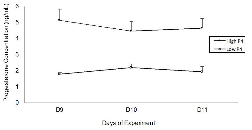
2/10
(Murdoch et al., 1993) and the presence of both receptors
Correspondence to:
Luiz Francisco Machado Pfeifer
was demonstrated in the pituitary gland (Naor et al., 2007).
Embrapa Rondônia Based on these considerations, we hypothesize that
BR 364 - Km 5,5 - Zona Rural PGF treatment affects the hypothalamic-pituitary level to
Caixa postal: 127 CEP: 76815-800, Porto Velho – RO, Brazil
e-mail: luiz.pfeifer@embrapa.br induce ovulation and luteinization in cattle. Therefore, this
study aimed to evaluate: 1) the effect of PGF administered
Received: September 22, 2020 in different time points and doses on ovulation; 2) whether
Approved: December 23, 2020
PGF can induce ovulation in cows with high and low serum
progesterone concentrations; 3) the serum concentration
of LH after PGF treatment and 4) the effect of PGF on
How to cite: Castro NA, Leonardi CEP, Singh J, Schneider
steroidogenesis and vascularization of preovulatory follicles.
A, Gonçalves PB, Oliveira FC, D’Ávila CA, Ferreira R,
Gasperin BG, Moreira EM, Andrade JS, Pfeifer LFM. The
role of prostaglandin F2α on ovulation and LH release in Materials and Methods
cows. Braz J Vet Res Anim Sci. 2021;58:e175001. https:// The Committee for Ethics in Animal Experimentation
doi.org/10.11606/issn.1678-4456.bjvras.2021.175001 from the Brazilian Agricultural Research Corporation
(Embrapa – Rondônia) approved all procedures performed
Introduction in this experiment (Number F.02/2014).
Prostaglandin F2α (PGF) analogs are commonly used
to induce luteolysis in estrous synchronization programs Experiment 1. Effect of a single PGF dose in a high
in cattle (Pursley et al., 1995; Weems et al., 2006; Colazo progesterone environment
& Mapletoft, 2014). However, PGF is also able to hasten Nonlactating crossbred dairy cows (Gyr x Holstein; n = 23)
and synchronize ovulation in cattle (Pfeifer et al., 2014; between 3 and 6 years old, parity between 2 and 4, body
Pfeifer et al., 2016; Castro et al., 2017) independent of its condition score (BCS) between 2.5 and 3.5 (1 = emaciated,
luteolytic action (Leonardi et al., 2012). Previously, we 5 = obese) were used. Cows were maintained in a Brachiaria
observed that d-Cloprostenol (PGF analog) had a similar brizantha pasture, with free access to water and mineral
ovulatory effect to that of estradiol benzoate in timed artificial supplement. Before the experiment, all cows were examined
insemination (TAI) protocols, resulting in similar ovulation by transrectal ultrasonography (SIUI CTS-900 equipped
and pregnancy rates in dairy (Pfeifer et al., 2016; Castro et al., with 5 MHZ linear probe, Guangdong, China) twice, 11 days
2017) and beef cows (Pfeifer et al., 2014; Pfeifer et al., 2018). apart, to confirm that none had a uterine infection as well
Despite this, the mechanism of action for injectable PGF as to evaluate the presence of follicles and corpus luteum
to induce ovulation in cattle is still unknown. (CL). All cows presented a CL and/or a dominant follicle
During a natural estrous cycle, the ovarian dominant (DF; follicle ≥ 8mm) and were included in the study.
follicle reaches ovulatory capacity after luteolysis and a Cows were submitted to a pre-synchronization treatment
consequent decrease in progesterone concentrations (Colazo as shown in Figure 1A. On Day 0, approximately 6 to 7 d after
& Mapletoft, 2014). In a TAI program, however, the use ovulation, cows were treated with two progesterone-releasing
of GnRH can induce ovulation even in the presence of intravaginal implants devices (1.9 g progesterone each, CIDR,
progesterone (Colazo et al., 2008), as it directly stimulates Zoetis, Campinas, Brazil), plus 2 mg of estradiol benzoate
pituitary LH release (Halász et al., 1989). Furthermore, (EB, Bioestrogen, Biogénesis-Bagó, Curitiba, Brazil) i.m.
it was shown that estradiol can induce an LH surge in On Day 8, cows received 300 IU of eCG (Novormon, Zoetis,
ovariectomized cows under high progesterone concentrations Campinas, Brazil) i.m., and 24 h later were randomly separated
(Martínez et al., 2007). Although PGF injection has been into one of two experimental treatments to receive: 1) 150 μg
associated with ovulation after progesterone insert removal of d-Cloprostenol (Croniben, Curitiba, Brazil; PGF Group,
during TAI, little is known about its hypothalamic effects n = 12), or 2) 2 mL of NaCl 0.9% (Control Group, n = 11).
and ability to trigger LH release. The CIDR devices were removed on Day 13.
The administration of PGF associated with GnRH can All cows were evaluated by transrectal ultrasonography
induce an increase in plasma LH levels in postpartum cows, once a day from Day 6 to Day 9 and every 12 h from Day 9
suggesting that PGF increases the responsiveness to GnRH until ovulation, or five days after treatments in the absence of
(Randel et al., 1996). Besides, in mice, prostaglandin E (PGE) ovulation. Also, color Doppler ultrasonography (Mindray
is known to stimulate LH secretion while PGF inhibits it M5 VET equipped with 5 MHZ linear probe) was used
Braz J Vet Res Anim Sci. 2021;58:e1750013/10
Figure 1 - Ovulation synchronization protocol used for cows in (A) Experiment 1 and (B) Experiment 2. *Blood collections
for analysis of serum progesterone concentration. Abbreviations: EB: estradiol benzoate, PGF: prostaglandin F2α,
ECP: estradiol cypionate, eCG: equine chorionic gonadotropin, US: ultrasound examination.
to evaluate the blood flow of the DF. Color gain settings
were kept constant for all ultrasonic assessments. Images
of the follicles were obtained from ultrasound assessments
with maximum color intensity, focusing on the widest
diameter of the follicle. When consistent Doppler signals
were detected in the follicular wall, it was considered that
the follicle had detectable blood flow. The intensity of
follicular vascularization was expressed subjectively as the
percentage of peri-follicular circumference enriched with
Color Doppler signals as described elsewhere (Ginther, Figure 2 - Serum progesterone concentrations in cows exposed
2007; Satheshkumar, 2018). to high (Exp. 1 – two new CIDR®,) and low (Exp. 2 –
one twice used CIDR®,) progesterone concentrations.
Blood samples were collected on Days 9, 10, and 11 of the
experiment, from the coccygeal vein, in vacuum tubes, without
anticoagulant. Immediately after collection, samples were of eCG (Novormon, Zoetis, Campinas, São Paulo, Brazil)
refrigerated at 4 °C, then centrifuged (3000 × g for 15 min) i.m. On Day 9, cows were randomly separated into two
and stored at −20 °C. Serum P4 concentrations were experimental treatments to receive: 1) 4 injections, once
analyzed on D9, D10, and D11 of the experiment (Figure 2) a day, of 150 μg of d-Cloprostenol (4xPGF Group, n = 9),
assessed by chemiluminescence (ADVIA Centaur; Siemens; or 2) 4 injections of 2 mL of NaCl (0.9%, Control Group,
Ref. 01586287; sensitivity of 0.21 ng/mL), with intra- and n = 9). The progesterone inserts were removed on Day 13.
inter-assay values lower than 12%. All cows were evaluated by transrectal ultrasonography,
every 24 h from Day 9 until ovulation, or five days after
Experiment 2. Effect of multiple PGF doses in a low CIDR removal in the absence of ovulation. Blood collection
progesterone environment and serum progesterone concentrations were performed as
Nonlactating crossbred dairy cows (Gyr x Holstein; n = 18) described for Experiment 1.
were given PGF i.m. twice, 11 days apart. Ten days after the
second PGF (Day 0), approximately 5 to 8 days after ovulation, Experiment 3. LH release after PGF treatment
all cows received a hormonal treatment (Figure 1B). On Day 0, Ovariectomized crossbred cows (B. taurus x B. indicus;
cows were treated with a twice-used CIDR device (CIDR, n = 14) were randomly allocated to receive three i.m. injection
Zoetis, Campinas, Brazil), 2 mg of EB i.m. and 150 μg of a (Hour 0, 1 and 2) of one of the following treatments: 300μg
PGF analog (d-Cloprostenol, Croniben, Biogénesis-Bagó, of d-Cloprostenol (PGF analog; Croniben, Curitiba,
Curitiba, Brazil) to induce luteolysis. On Day 6, cows received Brazil; PGF Group, n = 5), 100μg of Lecirelin (GnRH
150 μg of d-Cloprostenol i.m., and 48 hours later, 300 IU analog, Gestran plus, Tecnopec, São Paulo, Brazil; GnRH
Braz J Vet Res Anim Sci. 2021;58:e1750014/10
Group, n = 5) or 2 mL of Saline (Control Group, n = 4). the linear vascularized surface (LVS). The images were
To determine the serum concentration of LH, blood samples overlaid to obtain one single image and then, the LVS was
were collected from all cows, as follows: 2 h before the calculated using the ImageJ software. To determine the
injections, hourly from the moment of injections (Hour 0) proportion of colored pixels surrounding the DF, the LVS
to 6 h, and every 6 h from 6 h to 36 h after injections. Serum was divided by CDF.
LH concentrations were analyzed by radioimmunoassay, Pre-ovulatory follicles were aspirated 24 h after
adapted from Bolt & Rollins (1983) and Bolt et al. (1990). treatments under caudal epidural anesthesia with 80 mg
The minimum detection limit was 0.06 ng/mL. The intra- Lidocaine Chlorhydrate (Anestex Fagra - Vétoquinol,
assay CVs were 4.9 and 7.4% for low- and high- reference Brazil). Follicular aspiration was guided by transvaginal
samples, whereas interassay CVs were 21.9% and 7.2% for ultrasonography (PieMedical Esaote AguilaVet, equipped
low- and high- reference samples. with a 6 MHZ microconvex probe) using an ovum pick up
the system with a 16G catheter (Jelco) attached to a silicon
Experiment 4. Effect of PGF on steroidogenesis and tube and a 5 mL syringe. The follicular fluid samples were
vascularization of pre-ovulatory follicles centrifuged and preserved in liquid nitrogen until estradiol
Nonlactating dairy cows (Jersey and Holstein; n = and progesterone concentration analysis. Follicular fluid
19) with a BCS between 2.5 and 3.5 were maintained in progesterone (P4) and estradiol (E2) concentrations were
a natural pasture with free access to water and mineral assessed by chemiluminescence (ADVIA Centaur; Siemens),
supplement. On Day 0, cows were treated with 2 mg of EB as described in Experiment 1.
i.m. (Gonadiol, Zoetis, Brazil) and 241µg Cloprostenol i.m.
(Estron, Agener, Brazil) simultaneously to the insertion of a Statistical analysis
progesterone-releasing intravaginal implant (1g progesterone; All statistical analyses were performed using SAS 9.0 (SAS,
Sincrogest, Ourofino, Brazil). Furthermore, all follicles > Cary, NC, USA). Continuous variables (diameter of the
8mm were aspirated on Day 0. On Day 8, 241µg cloprostenol dominant follicle, time of ovulation and concentration of
i.m. (Estron, Agener, Brazil) was administered. All cows LH, follicular fluid estradiol and progesterone) were analyzed
were evaluated by transrectal ultrasonography (SonoScape by one-way ANOVA, and means were compared between
A6V equipped with 7.5 MHZ linear probe), every 24 h from groups by the post-hoc Tukey test. Analyses involving
Day 7 to Day 11. On Day 9, the progesterone inserts were repeated measures over time were compared by analysis of
removed and, 12 h later, the cows were randomly allocated variance for repeated measures using the MIXED procedure
into three treatments: 2 mL of Saline i.m. (Control Group, to evaluate the main effects of treatment, time (sampling
n = 6); 100μg of Lecirelin i.m. (GnRH analog, Gestran period), and their interaction (treatment vs. time). When the
plus, Tecnopec, São Paulo, Brazil; GnRH Group, n = 7) or interaction was significant, means were compared among
500 µg Cloprostenol Sodium i.m. (Ciosin - MSD, Brazil; treatments using the Tukey post-hoc test. Ovulation rate
PGF Group, n = 6). was analyzed by the Chi-square test. Data from follicular
All cows had a single follicle with a diameter greater than vascularization had normal distribution according to the
11.5 mm at the moment of the treatment. Color Doppler Shapiro-Wilk’s test and were evaluated using paired data
ultrasonography (Mindray M5 VET equipped with design into a parametric mixed model. Pairwise comparisons
5 MHZ linear probe) was used to evaluate blood flow of were performed using least square corrected means.
the pre-ovulatory follicle 0 and 24 h after treatment. Both Differences between groups were considered significant
ovaries were scanned, and the location of the dominant when the P-value was less or equal to 0.05.
follicle was recorded for each cow. Then, B-mode and color-
flow mode short videoclips (7s duration) were recorded. Results
The intensity of follicular vascularization was blindly
evaluated and objectively expressed as the percentage of the Experiment 1
peripolar circumference that had signs of color Doppler. No cow ovulated in Control and PGF groups when
Thus, the circumference of the DF (CDF) was calculated exposed to high progesterone concentrations. The average
using the internal calipers of the machine. To calculate serum progesterone concentration was 4.7 ± 0.3 ng/mL
the proportion of the peripolar circumference of the DF (Figure 2). Follicular growth and follicular blood flow scores
vascularized, three to four images that better represented the were shown in Figures 3A and 3B, respectively. Follicular
colored area surrounding the DF were chosen to calculate growth was not different between groups (P = 0.32) from
Braz J Vet Res Anim Sci. 2021;58:e1750015/10
Figure 4 - Diameter (Mean ± SEM) of the dominant follicle in
cows treated with four doses of 150µg of PGF analog
(PGF Group, n = 12) or NaCL (Control Group, n = 11)
on Days 9 to 12 of the estradiol-low progesterone-based
protocol (Experiment 2). All cows were synchronized
with a twice-used CIDR®, device, 2 mg of EB i.m.,
and 150 μg of a PGF analog on Day 0. On Day 6,
cows received 150 μg of d-Cloprostenol, and 48 h
later, 300 IU of eCG. CIDR®, devices were removed
on Day 13 of the protocol. *Time points relative to
the first injection of the treatments.
Figure 3 - (A) Diameter (Mean ± SEM) of the dominant follicle
and (B) blood flow score in cows treated with a single
dose of 150µg of PGF analog (PGF Group, n = 12) or
NaCL (Control Group, n = 11) on Day 9 of the estradiol-
high progesterone-based protocol (Experiment 1). All
cows received 2 new CIDR®, devices, and 2 mg of
EB i.m., in a presence of a CL on Day 0. On Day 8,
cows received 300 IU of eCG. CIDR®, devices were
removed on Day 13 of the protocol.
day 6 to 13. However, there was an increase in the blood
flow of the DF after PGF treatment (P = 0.03).
Experiment 2
Figure 5 - Serum LH concentrations after injections of d-Cloprostenol
No cow exposed to low progesterone ovulated in Control (PGF Group, n = 5), Lecirelin (GnRH Group, n = 5),
and 4xPGF groups. The average serum progesterone or Saline (Control Group, n = 4) in ovariectomized
concentration was 1.9 ± 0.1 ng/mL (Figure 2). However, cows from Experiment 3.
after CIDR removal, all cows (9/9) from the Control group
ovulated, and 78% (7/9) of the cows treated with PGF injections, the LH concentration was higher (P 0.05) among
CIDR removal to ovulation was 54.6 ± 3.5 h in Control groups. The LH secretion pattern was not different between
and 42.6 ± 6.5 h in PGF group (P = 0.12). PGF and Control groups (P > 0.05) and was maintained
between 3 and 8 ng/mL throughout the study.
Experiment 3
Serum LH concentrations after saline, GnRH, or PGF Experiment 4
injection are shown in Figure 5. Prior to the injections Follicular fluid progesterone and estradiol concentrations
(Hour 0), LH concentration was similar among groups in Control, GnRH, and PGF treatments are shown in Figure 6.
(P = 0.97; 4.5 ± 0.3 ng/mL). However, 1 h after the first Cows treated with GnRH had a significant decrease in follicular
Braz J Vet Res Anim Sci. 2021;58:e1750016/10
Figure 6 - Follicular fluid estradiol (A) and progesterone (B) concentration in preovulatory follicles from cows 24 h after treatment
with Saline (Control Group, n=6); Lecirelin (GnRH Group, n = 7) or Cloprostenol Sodium (PGF Group, n=6). All the
cows had a single follicle with a diameter greater than 11.5 mm at the moment of treatment. A, B Indicate significant
difference among groups (P7/10
several species (Evans et al., 1983; Dhaliwal et al., 1991; Differently from observed in the cows of our experiments,
Murdoch et al., 1993; Duffy & Stouffer, 2001; Bridges & exogenous PGF induced ovulation in anestrous ewes under
Fortune, 2007). However, most of the reports about the action the effect of medroxyprogesterone (Davies et al., 2006),
of endogenous PGF on periovulatory events in cattle are suggesting a direct effect on the follicle, since circulating
focused on its local action in the dominant follicle (Bridges progesterone concentrations did not change and ovulation
& Fortune, 2007; Fortune et al., 2009; Willis et al., 2017). was not preceded by an LH or FSH surge. In our study,
In response to GnRH, there is an increase in the PGF and progesterone concentrations were high in Exp. 1 (around
PGE receptor expression in theca and granulosa cells of 5 ng/mL), and low in Exp. 2 (around 2 ng/mL), similar to
preovulatory follicles in cattle (Bridges & Fortune, 2007). those previously reported by Pereira et al. (2017), which
Also, the pre-ovulatory LH surge induces granulosa cells induced endocrine profiles of high and low progesterone,
cyclooxygenase-2 activity, which converts arachidonic respectively. It is known that serum progesterone concentrations
acid to PGH2, the precursor of prostaglandins E2 and around 2ng/mL can prevent the LH release necessary for
F22α (Sirois, 1994). The presence of these prostaglandins ovulation of the dominant follicle in cattle (Kojima et al.,
stimulates the activation of proteases in the granulosa 2003). It is possible, therefore, that even the low progesterone
(Fortune et al., 2009) and theca interna cells (Willis et al., concentration can prevent the PGF stimulus on ovulation.
2017), promoting follicular rupture and extracellular This suggests that other periovulatory events need to be
matrix remodeling (Richards et al., 2005; Shozu et al., triggered to PGF have a stimulatory effect.
2005). These reports demonstrated the importance of The results from our study also demonstrate that PGF
endogenous PGF in ovulation and support the hypothesis alone is not able to stimulate the LH surge in cows, as low
that PGF administration may directly alter the follicular levels of LH were maintained even after three injections of
environment. In our study, although PGF was not able to PGF in ovariectomized cows. In postpartum cows, it was
induce ovulation in cows with high progesterone levels, previously demonstrated that PGF increases LH secretion
it improved follicular vascularization, suggesting a local after GnRH injection (Randel et al., 1996), suggesting a
action on the preovulatory follicle. Initially, we tested the direct effect of PGF on the anterior pituitary (Weems et al.,
effect of PGF in a high progesterone environment, and 2006). However, the effect of PGF on LH secretion suggested
since PGF did not induce ovulation, we attempted to test by those authors was not confirmed in our current study
multiple PGF injections in a low progesterone environment. using PGF alone. Besides inducing ovulation, the LH
The presence of progesterone, even at low levels as surge induced by GnRH promotes dramatic effects on
observed in experiment 2, blocked the ovulatory action of follicular steroidogenesis, which initiates before follicular
PGF. However, after the progesterone source removal, almost rupture and culminates with luteinization of the follicle
60% of PGF treated cows ovulated until 36 h, while only (Komar et al., 2001).
22% of control cows ovulated in the same interval. In rhesus Thus, we investigated if PGF administration in cows
monkeys, LH increased PGE and PGF concentrations in with preovulatory follicles during proestrus/estrus would
follicular fluid about 10 h before ovulatory follicle rupture induce such effects. As expected, GnRH treatment decreased
(Duffy & Stouffer, 2001). This suggests that PGs may modulate estradiol synthesis and increased progesterone concentration
the effects of the LH surge in ovarian tissue remodeling, (Komar et al., 2001; Santos et al., 2012), validating the
which is essential for follicle rupture and luteinization. luteinization model. However, PGF treatment did not promote
These results, associated with our findings, suggest that luteinization, as estradiol and progesterone concentrations
PGF could have a local effect in the pre-ovulatory follicle. were similar to those observed in non-treated control cows,
In previous studies, we verified the ability of PGF to further confirming the absence of an indirect effect of PGF
hasten ovulation when progesterone sources were removed mediated by GnRH/LH. Interestingly, the evaluation of the
before PGF treatment to induce ovulation (Leonardi et al., preovulatory follicles by Color Doppler ultrasonography
2012; Pfeifer et al., 2014; Pfeifer et al., 2016; Castro et al., revealed that PGF treatment induced a significant increase
2017). In the current study, to evaluate possible mechanisms in follicular vascularization, as observed in GnRH-treated,
of action, we maintained the source of progesterone during but not in control cows.
PGF injection. With this, we aimed to determine if, while It was previously demonstrated that PGF induces a
pituitary gonadotropin release is inhibited by progesterone, significant increase in vascularization at the periphery of
PGF would still be able to induce ovulation. However, our the CL during both spontaneous and induced luteolysis,
results demonstrated that this did not happen, PGF did not through inducing the expression of endothelial nitric
induce ovulation in the presence of any level of progesterone. oxide synthase (eNOS) (Shirasuna et al., 2008). However,
Braz J Vet Res Anim Sci. 2021;58:e1750018/10
that effect seems to last only for 6 h, and in the present Ethics Statement
study, the evaluation was performed 24 h after treatment. The Committee for Ethics in Animal Experimentation
Considering the short half-life of PGF, further studies are from the Brazilian Agricultural Research Corporation
necessary to identify the mechanisms involved in the local (Embrapa – Rondônia) approved all procedures performed
action of PGF during ovulation. in this experiment (Number F.02/2014).
Conclusion Acknowledgments
Our results show that PGF did not induce ovulation under
any serum progesterone level. Furthermore, PGF did not The authors are thankful to the National Council for Scientific
trigger an LH surge in ovariectomized cows and PGF alone was and Technological Development (CNPq), Foundation
not able to induce follicle luteinization, although it increased of Research Support of the State of Rio Grande do Sul
follicular vascularization. Together, the results suggest that (Project n: 16/2551-0000494-3). This study was financed
PGF may act locally to induce ovulation in cattle. Further in part by the Coordenação de Aperfeiçoamento de Pessoal
studies are needed to understand its mechanism of action. de Nível Superior - Brasil (CAPES) - Finance Code 001.
This research was also supported by CNPq (Universal
Conflict of interest Project n: 407307/2016-8) and EMBRAPA (Project MP2
The authors declare no conflict of interest. n 02.12.01.021.00.00).
References
Bolt DJ, Rollins R. Development and application of a Dhaliwal GS, Sharma RD, Prabhakar S. Ovarian changes
radioimmunoassay for bovine follicle-stimulating hormone. in buffaloes following PGF 2 administration using two
J Anim Sci. 1983;56(1):146-54. http://dx.doi.org/10.2527/ routes. Buffalo Bull. 1991;10:32-7.
jas1983.561146x. PMid:6402479.
Davies KL, Bartlewski PM, Epp T, Duggavathi R, Barrett
Bolt DJ, Scott V, Kiracofe GH. Plasma LH and FSH after
estradiol, norgestomet and Gn-RH treatment in ovariectomized DMW, Bagu ET, Cook SJ, Rawlings NC. Does injection of
beef heifers. Anim Reprod Sci. 1990;23(4):263-71. http:// prostaglandin F2α (PGF2α) cause ovulation in anestrous
dx.doi.org/10.1016/0378-4320(90)90040-M. Western White Face ewes? Theriogenology. 2006;66(2):251-
9. http://dx.doi.org/10.1016/j.theriogenology.2005.08.027.
Bridges PJ, Fortune JE. Regulation, action and transport of PMid:16336995.
prostaglandins during the periovulatory period in cattle.
Mol Cell Endocrinol. 2007;263(1–2):1-9. http://dx.doi. Duffy DM, Stouffer RL. The ovulatory gonadotrophin surge
org/10.1016/j.mce.2006.08.002. PMid:17064845.
stimulates cyclooxygenase expression and prostaglandin
production by the monkey follicle. Mol Hum Reprod.
Castro NA, Neves PMA, Cestaro JP, Melo VTO, Schneider
2001;7(8):731-9. http://dx.doi.org/10.1093/molehr/7.8.731.
A, Pfeifer LFM. Use of prostaglandin F2α as ovulatory
stimulus for synchronizing dairy cattle. Res Vet Sci. PMid:11470860.
2017;2018(118):151-4.
Evans G, Dobias M, King GJ, Armstrong DT. Production
Colazo MG, Mapletoft RJ. A review of current timed- of Prostaglandins by Porcine Preovulatory Follicular
AI (TAI) programs for beef and dairy cattle. Can Vet J. Tissues and Their Roles in Intrafollicular Function 1.
2014;55(8):772-80. PMid:25082993. Biol Reprod. 1983;28(2):322-8. http://dx.doi.org/10.1095/
biolreprod28.2.322. PMid:6404317.
Colazo MG, Kastelic JP, Davis H, Rutledge MD, Martinez
MF, Small JA, Mapletoft RJ. Effects of plasma progesterone
Fortune JE, Willis EL, Bridges PJ, Yang CS. The periovulatory
concentrations on LH release and ovulation in beef cattle
given GnRH. Domest Anim Endocrinol. 2008;34(1):109- period in cattle: progesterone, prostaglandins, oxytocin
17. http://dx.doi.org/10.1016/j.domaniend.2006.11.004. and ADAMTS proteases. Anim Reprod. 2009;6(1):60-71.
PMid:17210239. PMid:20390049.
Braz J Vet Res Anim Sci. 2021;58:e1750019/10
Ginther OJ. Ultrasonic Imaging and Animal Reproduction: 1993;46(8):85-115. http://dx.doi.org/10.1016/0090-
color-Doppler Ultrasonography. 1. ed. Cross Plains: 6980(93)90037-8. PMid:8210447.
Equiservices Publishing; 2007.
Naor Z, Jabbour HN, Naidich M, Pawson AJ, Morgan K,
Halász B, Kiss J, Molnár J. Regulation of the gonadotropin- Battersby S, Millar MR, Brown P, Millar RP. Reciprocal cross
releasing hormone (GnRH) neuronal system: morphological talk between gonadotropin-releasing hormone (GnRH)
aspects. J Steroid Biochem. 1989;33(4 Part 2):663-8. http:// and prostaglandin receptors regulates GnRH receptor
dx.doi.org/10.1016/0022-4731(89)90475-5. PMid:2513450. expression and differential gonadotropin secretion. Mol
Endocrinol. 2007;21(2):524-37. http://dx.doi.org/10.1210/
Jaeger JR, Turner HA, Stormshak F. Gonadotropin releasing me.2006-0253. PMid:17138645.
hormone-induced secretion of luteinizing hormone during
the milk-ejection reflex in the postpartum Beef Cow2. J Pereira MHC, Sanches CP Jr, Guida TG, Wiltbank MC,
Anim Sci. 1987;65(2):543-7. http://dx.doi.org/10.2527/ Vasconcelos JLM. Comparison of fertility following use of one
jas1987.652543x. PMid:3305457. versus two intravaginal progesterone inserts in dairy cows
without a CL during a synchronization protocol before timed
Kojima FN, Bergfeld EGM, Wehrman ME, Cupp AS, AI or timed embryo transfer. Theriogenology. 2017;89:72-
Fike KE, Mariscal-Aguayo DV, Sanchez-Torres T, Garcia- 8. http://dx.doi.org/10.1016/j.theriogenology.2016.10.006.
Winder M, Clopton DT, Roberts AJ, Kinder JE. Frequency PMid:28043373.
of luteinizing hormone pulses in cattle influences duration
of persistence of dominant ovarian follicles, follicular fluid Pfeifer LFM, Leonardi CEP, Castro NA, Viana JHM, Siqueira
concentrations of steroids, and activity of insulin-like growth LGB, Castilho EM, Singh J, Krusser RH, Rubin MIB. The
factor binding proteins. Anim Reprod Sci. 2003;77(3-4):187- use of PGF2α as ovulatory stimulus for timed artificial
211. http://dx.doi.org/10.1016/S0378-4320(03)00038-1. insemination in cattle. Theriogenology. 2014;81(5):689-95.
PMid:12695054. http://dx.doi.org/10.1016/j.theriogenology.2013.11.016.
PMid:24412682.
Komar CM, Berndtson AK, Evans ACO, Fortune JE. Decline
in circulating estradiol during the periovulatory period Pfeifer LFM, Siqueira LGB, Arashiro EKN, Castro NÁ,
is correlated with decreases in estradiol and androgen, Viana JHM. Prostaglandin F2α or estradiol benzoate to
and in messenger RNA for P450 aromatase and P450 induce ovulation in timed artificially inseminated dairy
17α-hydroxylase, in bovine preovulatory follicles. Biol cows. Pesqui Agropecu Bras. 2016;51(6):738-44. http://
Reprod. 2001;64(6):1797-805. http://dx.doi.org/10.1095/ dx.doi.org/10.1590/S0100-204X2016000600005.
biolreprod64.6.1797. PMid:11369611.
Pfeifer LFM, Rodrigues WB, Silva KC, Anache NA, Castro
Lamb GC, Dahlen CR, Larson JE, Marquezini G, Stevenson NA, Castilho EM, Nogueira E. Different protocols using
JS. Control of the estrous cycle to improve fertility for PGF2α as ovulation inducer in Nelore cows subjected
fixed-time artificial insemination in beef cattle: a review. to estradiol-progesterone timed AI based protocols.
J Anim Sci. 2010;88(13, Suppl.):E181-92. http://dx.doi. Theriogenology. 2018;120:56-60.http://dx.doi.org/10.1016/j.
org/10.2527/jas.2009-2349. PMid:19783709. theriogenology.2018.06.030. PMid: 30092375.
Leonardi CEP, Pfeifer LFM, Rubin MIB, Singh J, Mapletoft Pursley JR, Mee MO, Wiltbank MC. Synchronization
RJ, Pessoa GA, Bainy AM, Silva CAM. Prostaglandin F2α of ovulation in dairy cows using PGF2α and GnRH.
promotes ovulation in prepubertal heifers. Theriogenology. Theriogenology. 1995;44(7):915-23. http://dx.doi.
2012;78(7):1578-82. http://dx.doi.org/10.1016/j. org/10.1016/0093-691X(95)00279-H. PMid:16727787.
theriogenology.2012.06.030. PMid:22925644.
Randel RD, Lammoglia MA, Lewis AW, Neuendorff DA,
Martínez MF, Kastelic JP, Colazo MG, Mapletoft RJ. Guthrie MJ. Exogenous PGF2α enhanced GnRH-induced LH
Effects of estradiol on gonadotrophin release, estrus and release in postpartum cows. Theriogenology. 1996;45(3):643-
ovulation in CIDR-treated beef cattle. Domest Anim 54. http://dx.doi.org/10.1016/0093-691X(95)00410-A.
Endocrinol. 2007;33(1):77-90. http://dx.doi.org/10.1016/j. PMid:16727826.
domaniend.2006.04.009. PMid:16797154.
Richards JAS, Hernandez-Gonzalez I, Gonzalez-Robayna
Murdoch WJ, Hansen TR, McPherson LA. A review - Role I, Teuling E, Lo Y, Boerboom D, Falender AE, Doyle KH,
of eicosanoids in vertebrate ovulation. Prostaglandins. LeBaron RG, Thompson V, Sandy JD. Regulated expression
Braz J Vet Res Anim Sci. 2021;58:e17500110/10
of ADAMTS family members in follicles and cumulus oocyte Sirois J. Induction of prostaglandin endoperoxide synthase-2
complexes: evidence for specific and redundant patterns by human chorionic gonadotropin in bovine preovulatory
during ovulation. Biol Reprod. 2005;72(5):1241-55. http:// follicles in vivo. Endocrinology. 1994;135(3):841-8. http://
dx.doi.org/10.1095/biolreprod.104.038083. PMid:15659705. dx.doi.org/10.1210/endo.135.3.8070377. PMid:8070377.
Salasel B, Mokhtari A. Effect of early postpartum PGF2α Shozu M, Minami N, Yokoyama H, Inoue M, Kurihara H,
treatment on reproductive performance in dairy cows with Matsushima K, Kuno K. ADAMTS-1 is involved in normal
calving and puerperal traits. Theriogenology. 2011;76(9):1723- follicular development, ovulatory process and organization
9. http://dx.doi.org/10.1016/j.theriogenology.2011.07.004. of the medullary vascular network in the ovary. J Mol
PMid:21872307. Endocrinol. 2005;35(2):343-55. http://dx.doi.org/10.1677/
jme.1.01735. PMid:16216914.
Satheshkumar S. Peri-follicular blood flow in the
follicle from which ovulation occurs in cows. Anim
Weems CW, Weems YS, Randel RD. Prostaglandins and
Reprod Sci. 2018;198:154-9. http://dx.doi.org/10.1016/j.
reproduction in female farm animals. Vet J. 2006;171(2):206-28.
anireprosci.2018.09.014. PMid:30279026.
http://dx.doi.org/10.1016/j.tvjl.2004.11.014. PMid:16490704.
Santos JT, Ferreira R, Gasperin BG, Siqueira LC, de
Willis EL, Bridges PJ, Fortune JE. Progesterone receptor
Oliveira JF, Santos RA, Reis AM, Gonçalves PB. Molecular
characterization and regulation of the angiotensin-con- and prostaglandins mediate luteinizing hormone-induced
verting enzyme type 2/Angiotensin-(1-7)/MAS recep- changes in messenger RNAs for ADAMTS proteases in theca
tor axis during the ovulation process in cattle. J Renin cells of bovine periovulatory follicles. Mol Reprod Dev.
Angiotensin Aldosterone Syst. 2012;13(1):91-8. http:// 2017;84(1):55-66. http://dx.doi.org/10.1002/mrd.22761.
dx.doi.org/10.1177/1470320311417273. PMid:21824993. PMid:27879029.
Shirasuna K, Watanabe S, Asahi T, Wijayagunawardane MPB,
Financial Support: National Council for Scientific
Sasahara K, Jiang C, Matsui M, Sasaki M, Shimizu T, Davis
and Technological Development (CNPq), Foundation
JS, Miyamoto A. Prostaglandin F2α increases endothelial
of Research Support of the State of Rio Grande do Sul
nitric oxide synthase in the periphery of the bovine corpus
(Project n: 16/2551-0000494-3). This research was also
luteum: the possible regulation of blood flow at an early
stage of luteolysis. Reproduction. 2008;135(4):527-39. supported by CNPq (Universal Project n: 407307/2016-8)
http://dx.doi.org/10.1530/REP-07-0496. PMid:18296510. and EMBRAPA (Project MP2 n 02.12.01.021.00.00).
Braz J Vet Res Anim Sci. 2021;58:e175001You can also read


