Trends of Human Plague, Madagascar, 1998-2016 - CDC
←
→
Page content transcription
If your browser does not render page correctly, please read the page content below
SYNOPSIS

                    Trends of Human Plague,
                    Madagascar, 1998–2016
                  Voahangy Andrianaivoarimanana,1 Patrice Piola,1,2 David M. Wagner,
               Fanjasoa Rakotomanana, Viviane Maheriniaina, Samuel Andrianalimanana,
           Suzanne Chanteau,3 Lila Rahalison,4 Maherisoa Ratsitorahina, Minoarisoa Rajerison

Madagascar is more seriously affected by plague, a zoo-              Restriction of plague primarily to the highlands is probably
nosis caused by Yersinia pestis, than any other country.             caused by the absence of S. fonquerniei fleas at elevations
The Plague National Control Program was established in               90% of global human            Data Analysis
plague cases, and most cases are reported from Madagascar            We analyzed data for human plague surveillance activities
and the Democratic Republic of the Congo (1).                        (Appendix, https://wwwnc.cdc.gov/EID/article/25/2/17-
     Plague was introduced to Madagascar in 1898 at the              1974-App1.pdf) by using Stata 14 (StataCorp LLC, https://
port of Toamasina and reached the capital (Antananarivo)             www.stata.com). We also analyzed epidemiologic trends
in 1921. It is endemic to the central and northern highlands         of calculated suspected, presumptive, and confirmed cases.
(elevation >800 m). The main reservoir is the black rat              Cases were considered confirmed if Y. pestis was isolated
(Rattus rattus), and 2 main flea species (Xenopsylla cheo-           by culture or mouse inoculation, presumptive if positive
pis, a cosmopolitan species, and Synopsyllus fonquerniei,            results were obtained by F1RDT or microscopy (i.e., gram-
an endemic species) are involved in transmission. S. fon-            negative coccobacilli showing bipolar staining) without
querniei fleas show a greater transmission efficiency (2,3).         isolation of Y. pestis, or suspected if no samples were avail-
                                                                     able for testing or all test results were negative (9).
Author affiliations: Institut Pasteur, Antananarivo, Madagascar           Changes in screening tests resulted in different
(V. Andrianaivoarimanana, P. Piola, F. Rakotomanana,                 case definitions: before 2002, the screening test was
S. Chanteau, L. Rahalison, M. Rajerison); Northern Arizona
University, Flagstaff, Arizona, USA (D.M. Wagner); Ministry of
                                                                     1
                                                                         These authors contributed equally to this article.
Public Health, Antananarivo (V. Maheriniaina, S. Andrianalimanana,
                                                                     2
                                                                         Current affiliation: Institut Pasteur, Phnom Penh, Cambodia.
M. Ratsitorahina)
                                                                     3
                                                                         Retired.
                                                                     Current affiliation: Centers for Disease Control and Prevention,
                                                                     4

DOI: https://doi.org/10.3201/eid2502.171974                          Atlanta, Georgia, USA.

220                         Emerging Infectious Diseases • www.cdc.gov/eid • Vol. 25, No. 2, February 2019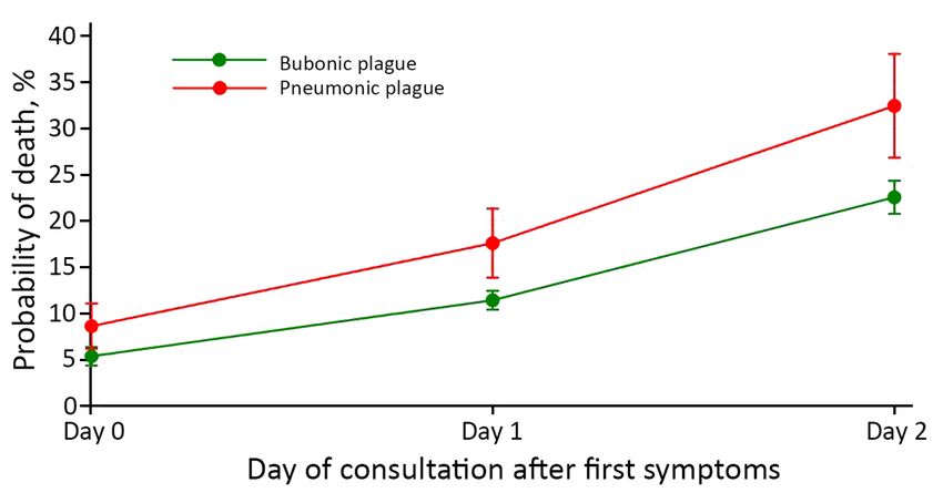
Human Plague in Madagascar, 1998–2016

microscopy, but during 2002–2008, F1RDT replaced                  (yes/no); month of plague reporting; and mean elevation of
microscopy. We estimated performance of microscopy                district residence. Clinical form was defined on the basis
(1998–2001) and F1RDT (2002–2007) by measuring                    of when the case was initially examined. Thus, PP cases
sensitivity and specificity compared with bacteriological         included persons with primary PP and secondary PP arising
culture; from 2008 on, only positive F1RDT results at the         from BP. Recent travel was defined as traveling in a plague-
Central Laboratory for Plague were confirmed by culture.          endemic focus area within 10 days before disease onset.
Cases for which samples were not available for testing            Elevation was included because it has been described as a
are listed as unknown (Figure 1) and were excluded from           risk factor for death from pneumonia (10).
sensitivity and specificity analyses.                                  To assess death risk factors, we also analyzed all
     A retrospective case–control study identified death          variables associated with death in univariate analysis at
risk factors for confirmed plus presumptive cases. Case-          the p2 days); clini-     description of infection clusters (groups of >2 confirmed
cal form (BP or PP); time period (before 2009 or from 2009        case-patients or presumptive case-patients who reported
on); contact with another confirmed plus presumptive case;        physical contact, close proximity, or other possible infec-
reporting presence of dead rats (yes/no); age (
SYNOPSIS General Trends 1998–2001) and a specificity of 96% (3,123/3,253). During January 1, 1998–December 31, 2016, a total of F1RDT sensitivity was 100% (1,411/1,411; samples 13,234 clinically suspected plague cases were reported to from 2002–2007) and specificity was 59% (1,420/2,399); the PNCP. Of these cases, 3,628 (27%) were classified as F1RDT specificity was higher for BP (60.5%; samples confirmed and 2,191 (17%) as presumptive (Figures 1, 2); from 2002–2007) than for PP (44.7%; p
Human Plague in Madagascar, 1998–2016

Figure 3. Yearly (A) and monthly (B) distributions of bubonic and pneumonic plague and their CFR, Madagascar, 1998–2016. CFR,
case-fatality rate.

     We found by univariate analysis that death was associ-                    PP increased from 9% (95% CI 6.2%–11.1%) when con-
ated more with PP (odds ratio [OR] 2.0, 95% CI 1.57–2.53)                      sulting on first day of symptoms to 32% when consulting
than with BP. For PP case-patients, death was associated                       after the second day (95% CI 26.8%–38.1%); these values
with delayed consultations (OR 2.7, 95% CI 1.70–4.37),                         for BP were 5% (95% CI 4.3%–6.4%) and 23% (95% CI
occurrence after 2009 (OR 1.60, 95% CI 1.02–2.52), and                         20.8%–24.4%) (Figure 5).
age group (OR  0.50, 95% CI 0.29–0.85). For BP case-
patients, death was associated with delayed consultations                      Discussion
(OR 3.44, 95% CI 2.92–4.04), contact with another case-                        Analysis of 19 years of human plague surveillance in Mad-
patient (OR 1.86, 95% CI 1.45–2.37), occurrence after                          agascar showed a distinct peak of confirmed plus presump-
2009 (OR 1.56, 95% CI 1.33–1.83), and not reporting dead                       tive cases during 2003 and 2004. This peak, which might
rats (OR 0.67, 95% CI 0.54–0.85).                                              have been influenced by global climate phenomena, such
     When we combined BP and PP case-patients (Table 3),                       as the El Niño Southern Oscillation and Indian Ocean Di-
we found that death was associated with age, sex, and recent                   pole (11,12), was partly caused by an increase in plague
travel. However, month and mean elevation of district of res-                  cases reported primarily from districts in rural areas of
idence were not associated with death, except for the month                    middle-west (including Ambohidratrimo, Miarinarivo, and
of August. In the multivariate model (Table 3), we found                       Tsiroanomandidy) and central (Antananarivo-Renivohitra)
that the remaining factors with an association after adjust-                   Madagascar. The first 3 districts have long been consid-
ments were delayed consultation (from OR 3.2 at day 2 up to                    ered plague hotspots (13); they reported at least twice the
OR 5.0 at day 4), pneumonic plague (OR 1.6), contact with                      mean confirmed plus presumptive reports during the 5
another plague case (OR 1.9), year 2009 or after (OR 1.6),                     years before 2004. In 2004, Antananarivo-Renivohitra had
and not reporting dead rats (OR  0.7) (all p values 35 years of age had a moderately higher risk for                      ment II of the capital (Antananarivo). This unusual event
death than did persons
SYNOPSIS
Table 2. Sociodemographic and epidemiologic characteristics of persons with confirmed, presumptive, and suspected plague cases,
Madagascar, 1998–2016*
Bubonic plague case status, no. patients Pneumonic plague case status, no. patients
Confirmed and Confirmed and
Characteristic Suspected, n = 6,454 presumptive, n = 5,132 Suspected, n = 579 presumptive, n = 409
Median age, y (IQR) 11 (6–20) 13 (8–24) 26 (17–40) 29 (20–42)
Sex ratio (M:F) 1.44 (3,803:2,639) 1.38 (2,972:2,157) 1.08 (300:278) 1.28 (230:179)
Presence of rats, % 15.1 18.6 11.3 9.3
Recent trip, % 6.4 6.9 7.0 13.6
Mean (SE) days to care 2.0 (0.05) 1.7 (0.03) 2.61 (0.15) 2.21 (0.18)
Median elevation, m (IQR) 1,262 (1,111–1,384) 1,275 (1,063–1,409) 1,228 (921–1,333) 1,284 (1,111–1,355)
Contact with other plague cases, % 7 11 21 23
*IQR, interquartile range.
A similar situation occurred in 1994, when a suspected weather patterns (16). In more recent years, overall confirmed
plague outbreak affected the cities of Surat and Beed in In- plus presumptive cases and CFR have both increased in Au-
dia and caused 54 deaths in 876 reported case-patients (9). gust, during what is typically low season for human plague
However, Y. pestis was not isolated from this outbreak, and in Madagascar. Increased CFR in August might have been
many patients were subsequently given a diagnosis of fatal caused by misdiagnosis by health workers unaccustomed to
illnesses resembling acute pneumonia and not plague (14). suspecting plague cases in this month or delayed treatment.
In our study, total annual cases peaked in 2004 and de- Relatively low specificity (59% overall, 44.7% for
creased to the lowest value during 2016 (Figure 2). This re- PP) of the F1RDT compared with that for culture might be
duction occurred primarily in suspected cases and might be caused by several factors, including badly preserved and
caused by improved adherence to case criteria after train- contaminated field samples and long delays in transport to
ing and education that occurred during widespread national laboratory; both factors could lead to negative results from
implementation of the F1RDT starting in 2002. culture even if the cultured material originated from a true
Monthly distribution of human plague cases in Madagas- plague case. In addition, negative culture results might have
car in this study was consistent with previous findings, except resulted from administration of antimicrobial drugs by lo-
for a troubling recent trend of the season starting earlier dur- cal health officials before sampling (17), whereas F1RDT
ing the year (Figure 3). In general, the human plague season results remained positive >3 weeks after treatment initia-
in Madagascar started with early recrudescence in Septem- tion (18). Moreover, pain, fever, and cough are often treat-
ber and peaked in November (5,15); this pattern is associated ed with self-medication in Madagascar (19). Thus, specific-
with temperature effects on flea abundance and climate and ity of the F1RDT test is almost certainly underestimated by
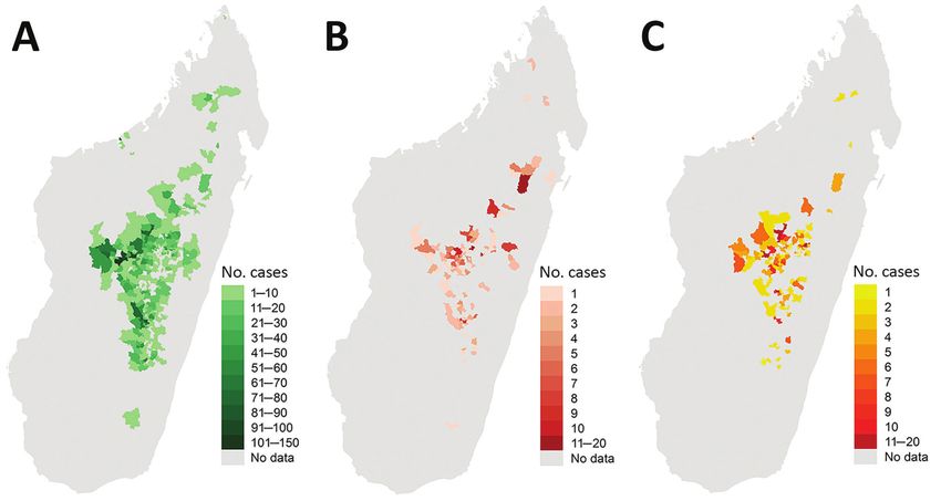
Figure 4. Geographic distributions of bubonic plague (A), pneumonic plague (B), and infection clusters (C), Madagascar, 1998–2016.
224 Emerging Infectious Diseases • www.cdc.gov/eid • Vol. 25, No. 2, February 2019Human Plague in Madagascar, 1998–2016
Table 3. Risk factors for death from plague, Madagascar, 1998–2016*
Univariate analysis Multivariate analysis
Factor No. patients OR (95% CI) p value OR (95% CI) p value
Delay to seek healthcare, d
0 (same day) 1,142 1 1
1 2,426 0.91 (0.73–1.14) 0.42
2 1,059 2.51 (2.00–3.16)SYNOPSIS
Figure 5. Predictive mortality rate
according to clinical form and
day of consultation for cases of
plague, Madagascar, 1998–2016.
Error bars indicate 95% CI.
Spatially, most confirmed plus presumptive cases dur- locations in Madagascar, clinicians and authorities outside
ing 1998–2016 were confined to the Central Highlands, but traditional plague foci need to be aware of this risk.
some occurred in other regions where plague has been his- It has been long speculated that plague epidemics in
torically absent (Figure 4). Concentration of human plague Madagascar occur in 4–5-year cycles; however, no clear
in the Central Highlands, consistent with past trends in periodicity could be defined at the level of rural districts
Madagascar (5), is likely caused by long-term persistence (15), which could be attributed to dynamics of plague in
of plague in this region within rodent reservoirs. A recent rodents and fleas. BP outbreaks are preceded by rodent epi-
phylogeographic analysis showed that different and distinct zootics (3), which reduce local rodent populations; it is un-
Y. pestis subpopulations largely persist at the same geo- known how long it takes for rodent populations to recover
graphic locations in the Central Highlands over time, sug- sufficiently to support another epizootic. Fluctuation in ro-
gesting that plague is constantly and stably maintained lo- dent abundance caused by plague might vary depending on
cally in rodent populations in this region and not regularly species, fecundity, availability of food, or lifespan, or larg-
reintroduced from other locations (13). During this period, er factors, such as weather and climate. High abundance
BP cases occurred throughout the Central Highlands, es- of rodents might lead to more contact between humans
pecially in the central and western portions (Figure 4). PP and animals and, therefore, to outbreaks (31). In Himach-
cases were scattered throughout the Central Highlands but al Pradesh, India, examination of past plague epidemics
concentrated in the northeastern portion in areas that typi- showed that outbreaks occur in cycles of 10–15 years (28).
cally report few human plague cases (Figure 4). The rela- Tools for early detection and treatment, as well as
tively high number of PP cases in these areas was caused properly trained health workers, are critical to reducing
by misdiagnosis and ignorance of plague by health workers overall plague deaths and progression of BP to PP. Just
and the local population, which delayed treatment and en- a 2-day delay in treatment dramatically increased death
abled BP to progress to PP. rates for BP and PP (Figure 5); these national findings for
Human plague is now occurring in some regions of PP are similar to those from a single PP plague outbreak
Madagascar where it was historically absent. An outbreak in Moramanga in 2015 (26). That study highlighted the
of BP emerged in southeast Madagascar (Befotaka and need for education and rapid clinical decision-making for
Iakora Districts) in November 2016, outside previously reducing deaths because the average duration from full
known limits of plague-endemic foci. It is not clear wheth- onset of PP to death was 1.9 days. Because it is highly
er plague has always been present in these regions and not sensitive and can be used in remote settings, F1RDT is
previously detected or was recently transported to these a proven preliminary diagnosis tool that has led to ear-
regions. Imported plague caused by human movements lier detection of outbreaks and rapid implementation of
or transportation has been documented for Mahajanga, a control measures in multiple countries in Africa (32–34).
coastal location outside the traditional regions of plague in This more local and focused diagnostic capability pro-
Madagascar, resulting in reintroduction and establishment vides considerable savings in terms of treatment, che-
of plague in this port city. It then cycled in local rodent and moprophylaxis, and pesticide treatment; in Madagascar,
flea populations for several years before apparently disap- these activities are conducted systematically and free of
pearing (30). Because plague can suddenly occur in new charge when a plague case is suspected.
226 Emerging Infectious Diseases • www.cdc.gov/eid • Vol. 25, No. 2, February 2019Human Plague in Madagascar, 1998–2016
An increase in the proportion of PP cases over time plague had not previously been reported or not reported
(Figure 3) was caused by untreated BP cases progressing for several years, a limitation evident for the reemerg-
to PP and is symptomatic of the deteriorating health sys- ing focus in Moramanga (26) and the recent outbreak in
tem in Madagascar, a result of sociopolitical and economic southeastern Madagascar, where many deaths were re-
crises of 2002 and 2009 (1). Specifically, this deteriora- ported but samples were not always available for confir-
tion is likely caused by fast turnover of health workers in mation. Thus, risk factors associated with complications
plague-endemic regions, especially after changes in the were not analyzed for these cases.
government in Madagascar or political crises; new work- In summary, a seasonal pattern of plague was observed
ers were not always properly trained for plague diagnosis. in Madagascar during September–March. Annual cases of
Surveillance and control activities throughout Madagascar plague peaked in 2004 and decreased in 2016. This reduc-
are also affected by lack of financial support. Discontinua- tion might be caused by improved adherence to case crite-
tion of regular plague surveillance in Antananarivo during ria during implementation of the F1 rapid diagnostic test in
2006–2018, caused by financial shortages, likely contrib- 2002. Because plague can suddenly occur in new locations
uted to the reappearance of plague in its suburbs 6 years in Madagascar, clinicians and authorities outside tradition-
after the previous last reported case. In 2011, two human al plague foci need to be aware of this risk.
cases were confirmed there outside the typical season, and
Y. pestis was also isolated from the spleen of an R. rattus rat Acknowledgments
(2). More concerning, in 2017 a large PP outbreak occurred We thank Mamy Ratsimba for data collection and Bienvenue
in Madagascar, with many cases in Antananarivo (35). A S. Rahoilijaona and the Clinical Research Unit of the Institut
similar overall situation has transpired in the Democratic Pasteur de Madagascar for plague mapping.
Republic of the Congo, where civil war and political crisis,
leading to a lack of resources, has adversely affected plague About the Author
control activities (9). Dr. Andrianaivoarimanana is a researcher at Rahoilijaoana–
Control and prevention of human plague cases in Mad- Epidemiology and in the Plague Unit at the Institut Pasteur de
agascar faces challenges. BP acquired from a Y. pestis–in- Madagascar, Antananarivo, Madagascar. Her research interests
fected flea is the most common form and the ultimate source are immune response between Y. pestis and its hosts and plague
of PP cases and outbreaks. Thus, prevention strategies are epidemiology.
focused on rodent hosts and flea vectors. Unfortunately, in-
appropriate use of chemical insecticides, contrary to PNCP
recommendations, has promoted emergence in Madagascar References
1. Bertherat E. Plague around the world, 2010–2015. Wkly Epidemiol
of fleas resistant to multiple classes of insecticides, lead-
Rec. 2016;91:89–93.
ing to survival of Y. pestis–infected fleas (36,37). Human 2. Andrianaivoarimanana V, Kreppel K, Elissa N, Duplantier JM,
contact with rodent hosts, and consequently flea vectors, Carniel E, Rajerison M, et al. Understanding the persistence of
is increased by common practices of storing food and dis- plague foci in Madagascar. PLoS Negl Trop Dis. 2013;7:e2382.
http://dx.doi.org/10.1371/journal.pntd.0002382
posing of garbage near households (21) and close proxim-
3. Duplantier JM, Duchemin JB, Chanteau S, Carniel E. From the
ity of households in many locations. Fortunately, control recent lessons of the Malagasy foci towards a global understanding
of human plague cases is still possible by use of standard of the factors involved in plague reemergence. Vet Res.
antimicrobial drugs (assuming treatment is started in time) 2005;36:437–53. http://dx.doi.org/10.1051/vetres:2005007
4. Brygoo ER. Epidemiology of plague in Madagascar [in French].
because only a few Y. pestis strains of >4,000 strains exam-
Arch Inst Pasteur Madagascar. 1966;35:9–147.
ined have been resistant to major antimicrobial drugs, such 5. Migliani R, Chanteau S, Rahalison L, Ratsitorahina M,
as streptomycin and doxycycline (38–40). Also, plague di- Boutin JP, Ratsifasoamanana L, et al. Epidemiological trends for
agnosis by using F1RDT on dead rodent samples can also human plague in Madagascar during the second half of the 20th
century: a survey of 20,900 notified cases. Trop Med Int Health.
contribute to efficient plague prevention (8).
2006; 11:1228–37. http://dx.doi.org/10.1111/j.1365-3156.2006.01677.x
This study has major strengths and potential limita- 6. Neerinckx S, Bertherat E, Leirs H. Human plague occurrences in
tions. Strengths include the length of the study and large Africa: an overview from 1877 to 2008. Trans R Soc Trop Med Hyg.
sample sizes. These sample sizes benefit from the fact 2010; 104:97–103. http://dx.doi.org/10.1016/j.trstmh.2009.07.028
7. Pollitzer R. Plague. World Health Organization Monograph Series
that all suspected plague in Madagascar must be reported
22. Geneva: The Organization; 1954.
with standard epidemiologic information. One possible 8. Chanteau S, Rahalison L, Ralafiarisoa L, Foulon J, Ratsitorahina M,
limitation is that diagnostic tools used for case defini- Ratsifasoamanana L, et al. Development and testing of a rapid
tion evolved during the study. Also, only confirmed plus diagnostic test for bubonic and pneumonic plague. Lancet.
2003;361:211–6. http://dx.doi.org/10.1016/S0140-6736(03)12270-2
presumptive cases were analyzed, which might not fully
9. World Health Organization. International meeting on preventing
represent the national situation. Furthermore, samples for and controlling plague: the old calamity still has a future. Wkly
confirmation were often unavailable from regions where Epidemiol Rec. 2006;81:278–84.
Emerging Infectious Diseases • www.cdc.gov/eid • Vol. 25, No. 2, February 2019 227SYNOPSIS
10. Pérez-Padilla R, Franco-Marina F. The impact of altitude on of pneumonic plague in Madagascar. Lancet. 2000;355:111–3.
mortality from tuberculosis and pneumonia. Int J Tuberc Lung Dis. http://dx.doi.org/10.1016/S0140-6736(99)05163-6
2004;8:1315–20. 26. Ramasindrazana B, Andrianaivoarimanana V, Rakotondramanga JM,
11. Kreppel KS, Caminade C, Telfer S, Rajerison M, Rahalison L, Birdsell DN, Ratsitorahina M, Rajerison M. Pneumonic plague
Morse A, et al. A non-stationary relationship between global transmission, Moramanga, Madagascar, 2015. Emerg Infect Dis.
climate phenomena and human plague incidence in Madagascar. 2017;23:521–4. http://dx.doi.org/10.3201/eid2303.161406
PLoS Negl Trop Dis. 2014;8:e3155. http://dx.doi.org/10.1371/ 27. Gupta ML, Sharma A. Pneumonic plague, northern India, 2002.
journal.pntd.0003155 Emerg Infect Dis. 2007;13:664–6. http://dx.doi.org/10.3201/
12. Ben-Ari T, Neerinckx S, Gage KL, Kreppel K, Laudisoit A, eid1304.051105
Leirs H, et al. Plague and climate: scales matter. PLoS Pathog. 28. Joshi K, Thakur JS, Kumar R, Singh AJ, Ray P, Jain S, et al.
2011;7:e1002160. http://dx.doi.org/10.1371/journal.ppat.1002160 Epidemiological features of pneumonic plague outbreak in
13. Vogler AJ, Andrianaivoarimanana V, Telfer S, Hall CM, Sahl JW, Himachal Pradesh, India. Trans R Soc Trop Med Hyg.
Hepp CM, et al. Temporal phylogeography of Yersinia pestis in 2009;103:455–60. http://dx.doi.org/10.1016/j.trstmh.2008.11.026
Madagascar: Insights into the long-term maintenance of plague. 29. Bertherat E, Lamine KM, Formenty P, Thuier P, Mondonge V,
PLoS Negl Trop Dis. 2017;11:e0005887. http://dx.doi.org/10.1371/ Mitifu A, et al. Major pulmonary plague outbreak in a mining
journal.pntd.0005887 camp in the Democratic Republic of Congo: brutal awakening
14. Ramalingaswami V. Plague in India. Nat Med. 1995;1:1237–9. dof an old scourge [in French]. Med Trop (Mars). 2005;65:511–4.
http://dx.doi.org/10.1038/nm1295-1237 30. Vogler AJ, Chan F, Wagner DM, Roumagnac P, Lee J, Nera R,
15. Chanteau S, Ratsitorahina M, Rahalison L, Rasoamanana B, et al. Phylogeography and molecular epidemiology of Yersinia
Chan F, Boisier P, et al. Current epidemiology of human plague pestis in Madagascar. PLoS Negl Trop Dis. 2011;5:e1319.
in Madagascar. Microbes Infect. 2000;2:25–31. http://dx.doi.org/ http://dx.doi.org/10.1371/journal.pntd.0001319
10.1016/S1286-4579(00)00289-6 31. Davis S, Calvet E, Leirs H. Fluctuating rodent populations and risk
16. Kreppel KS, Telfer S, Rajerison M, Morse A, Baylis M. Effect of to humans from rodent-borne zoonoses. Vector Borne Zoonotic
temperature and relative humidity on the development times and Dis. 2005;5:305–14. http://dx.doi.org/10.1089/vbz.2005.5.305
survival of Synopsyllus fonquerniei and Xenopsylla cheopis, the 32. Bertherat E, Bekhoucha S, Chougrani S, Razik F, Duchemin JB,
flea vectors of plague in Madagascar. Parasit Vectors. 2016;9:82. Houti L, et al. Plague reappearance in Algeria after 50 years, 2003.
http://dx.doi.org/10.1186/s13071-016-1366-z Emerg Infect Dis. 2007;13:1459–62. http://dx.doi.org/10.3201/
17. Forrester JD, Apangu T, Griffith K, Acayo S, Yockey B, eid1310.070284
Kaggwa J, et al. Patterns of human plague in Uganda, 2008-2016. 33. Cabanel N, Leclercq A, Chenal-Francisque V, Annajar B,
Emerg Infect Dis. 2017;23:1517–21. http://dx.doi.org/10.3201/ Rajerison M, Bekkhoucha S, et al. Plague outbreak in Libya, 2009,
eid2309.170789 unrelated to plague in Algeria. Emerg Infect Dis. 2013;19:230–6.
18. Andrianaivoarimanana V, Bertherat E, Rajaonarison R, http://dx.doi.org/10.3201/eid1902.121031
Rakotondramaro T, Rogier C, Rajerison M. Mixed pneumonic 34. Bertherat E, Thullier P, Shako JC, England K, Koné ML,
plague and nosocomial MDR-bacterial infection of lung: a rare case Arntzen L, et al. Lessons learned about pneumonic plague
report. BMC Pulm Med. 2018;18:92. http://dx.doi.org/10.1186/ diagnosis from two outbreaks, Democratic Republic of the Congo.
s12890-018-0656-y Emerg Infect Dis. 2011;17:778–84. http://dx.doi.org/10.3201/
19. Sendrasoa FA, Razanakoto NH, Ranaivo IM, Andrianasolo RL, eid1705.100029
Randria MJDD, Rakotoarivelo RA. Antibiotic and antimalarial 35. Roberts L. Echoes of Ebola as plague hits Madagascar. Science.
self-medication in Antananarivo, Madagascar. International Journal 2017;358:430–1. http://dx.doi.org/10.1126/science.358.6362.430
of Infectious Diseases and Therapy. 2016;1:1–5. 10.11648/ 36. Boyer S, Miarinjara A, Elissa N. Xenopsylla cheopis (Siphonaptera:
j.ijidt.20160101.11 Pulicidae) susceptibility to Deltamethrin in Madagascar. PLoS One.
20. Boisier P, Rahalison L, Rasolomaharo M, Ratsitorahina M, 2014;9:e111998. http://dx.doi.org/10.1371/journal.pone.0111998
Mahafaly M, Razafimahefa M, et al. Epidemiologic features 37. Miarinjara A, Boyer S. Current perspectives on plague vector
of four successive annual outbreaks of bubonic plague in control in Madagascar: susceptibility status of Xenopsylla cheopis
Mahajanga, Madagascar. Emerg Infect Dis. 2002;8:311–6. to 12 insecticides. PLoS Negl Trop Dis. 2016;10:e0004414.
http://dx.doi.org/10.3201/eid0803.010250 http://dx.doi.org/10.1371/journal.pntd.0004414
21. Kilonzo BS, Mvena ZS, Machangu RS, Mbise TJ. Preliminary 38. Galimand M, Guiyoule A, Gerbaud G, Rasoamanana B,
observations on factors responsible for long persistence and continued Chanteau S, Carniel E, et al. Multidrug resistance in Yersinia pestis
outbreaks of plague in Lushoto district, Tanzania. Acta Trop. mediated by a transferable plasmid. N Engl J Med. 1997;337:677–
1997;68:215–27. http://dx.doi.org/10.1016/S0001-706X(97)00096-X 80. http://dx.doi.org/10.1056/NEJM199709043371004
22. Barreto A, Aragon M, Epstein PR. Bubonic plague outbreak in 39. Guiyoule A, Gerbaud G, Buchrieser C, Galimand M,
Mozambique, 1994. Lancet. 1995;345:983–4. http://dx.doi.org/ Rahalison L, Chanteau S, et al. Transferable plasmid-mediated
10.1016/S0140-6736(95)90730-0 resistance to streptomycin in a clinical isolate of Yersinia pestis.
23. Eisen RJ, MacMillan K, Atiku LA, Mpanga JT, Emerg Infect Dis. 2001;7:43–8. http://dx.doi.org/10.3201/
Zielinski-Gutierrez E, Graham CB, et al. Identification of risk eid0701.010106
factors for plague in the West Nile Region of Uganda. Am J 40. Cabanel N, Bouchier C, Rajerison M, Carniel E. Plasmid-mediated
Trop Med Hyg. 2014;90:1047–58. http://dx.doi.org/10.4269/ doxycycline resistance in a Yersinia pestis strain isolated from a
ajtmh.14-0035 rat. Int J Antimicrob Agents. 2018;51:249–54. http://dx.doi.org/
24. Richard V, Riehm JM, Herindrainy P, Soanandrasana R, 10.1016/j.ijantimicag.2017.09.015
Ratsitoharina M, Rakotomanana F, et al. Pneumonic plague
outbreak, Northern Madagascar, 2011. Emerg Infect Dis. Address for correspondence: Minoarisoa Rajerison, Plague Unit, Institut
2015;21:8–15. http://dx.doi.org/10.3201/eid2101.131828
Pasteur de Madagascar, BP 1274 Ambatofotsikely, Antananarivo 101,
25. Ratsitorahina M, Chanteau S, Rahalison L, Ratsifasoamanana L,
Boisier P. Epidemiological and diagnostic aspects of the outbreak Madagascar; email: mino@pasteur.mg
228 Emerging Infectious Diseases • www.cdc.gov/eid • Vol. 25, No. 2, February 2019You can also read


